Chronic pancreatitis
Progressive irreversible inflammation and fibrosis of the pancreas causing pain, exocrine insufficiency and diabetes; causes, diagnosis, and treatment.
- Chronic pancreatitis is a progressive, irreversible inflammation and fibrosis of the pancreas that destroys both exocrine and endocrine tissue.
- Alcohol and tobacco are the dominant environmental causes, with alcohol alone behind roughly 60 to 70 percent of cases in many published series; genetic, autoimmune, obstructive and metabolic causes account for much of the rest.
- Patients present with recurrent or persistent upper abdominal pain, weight loss, fatty stools, deficiency of the vitamins A, D, E and K and, later, diabetes secondary to pancreatic disease (type 3c diabetes).
- Diagnosis rests mainly on imaging: computed tomography for calcifications and ductal dilation, magnetic resonance cholangiopancreatography (often with secretin) and endoscopic ultrasound; faecal elastase under 200 micrograms per gram supports exocrine insufficiency.
- Treatment combines removing the cause, stepwise pain relief, pancreatic enzyme replacement with meals, and surgery or endoscopy for ductal obstruction; good nutrition care and bone monitoring are part of long-term care.
By the end of this page you should be able to:
- Define chronic pancreatitis and contrast it with acute pancreatitis.
- List the major causes and risk factors, including the hereditary mutations.
- Explain the clinical features from the underlying mechanism.
- Choose and interpret the first-line investigations and diagnostic clues.
- Plan stepwise management, including enzyme dosing and timing, analgesia, endoscopy and surgery.
- Recognise and manage complications, including type 3c diabetes and pancreatic cancer risk.
- Adapt care in low-resource settings and tropical calcific pancreatitis.
DefinitionChronic pancreatitis
A progressive, irreversible inflammatory condition of the pancreas that leads to fibrosis and loss of both exocrine and endocrine function. It is now also described as a fibroinflammatory syndrome in people with genetic, environmental or other risk factors who develop a persistent pathological response to pancreatic injury.[1]
Chronic pancreatitis is usually diagnosed from imaging rather than from symptoms alone, and its severity ranges from painful but functional early disease to calcific, atrophic, insufficiency disease. It is worth distinguishing from acute pancreatitis at the outset: acute pancreatitis is a sudden inflammatory illness that may settle, whereas chronic pancreatitis leaves permanent structural change.
Chronic pancreatitis is not rare in gastroenterology practice, but its measured incidence and prevalence vary widely with alcohol intake, smoking, and access to imaging. Aetiology differs by region: alcoholic disease predominates in many Western series, whereas tropical calcific pancreatitis is common in parts of Africa and Asia.[6][8]
Alcohol accounts for a large share of cases, about 60 to 70 percent in many published series, though some adult cohorts report a lower share.[6] Tobacco is an independent and additive risk factor, and smokers with chronic pancreatitis progress faster and have more pain.[6] In practice the two exposures are common together, and simply separating them is difficult.
Other recognised causes include:
- Hereditary pancreatitis from mutations in PRSS1, SPINK1, CFTR and CTRC, which are major risk factors; hereditary disease runs in families with early-onset recurrent attacks and a high lifetime risk of pancreatic cancer.[6][7]
- Autoimmune pancreatitis: type 1, an IgG4-related disease that often involves the bile ducts and other organs, and type 2, which is linked to granulocytic epithelial lesions and lacks IgG4 elevation.[9]
- Obstructive disease from main pancreatic duct stricture, ampullary obstruction or pancreas divisum.
- Metabolic causes such as hypertriglyceridaemia and hypercalcaemia.
- Tropical calcific pancreatitis in Africa and Asia.
- Idiopathic disease, which is increasingly relabelled as late-onset or risk-factor-negative rather than truly unexplained.
Clinical pearlA 55-year-old vignette
A 55-year-old man who drinks heavily and smokes presents with repeated epigastric pain. Computed tomography shows multiple pancreatic calcifications and a dilated, irregular pancreatic duct. Before assuming alcohol is the sole culprit, ask about family history and smoking, and look for features of autoimmune IgG4-related disease.
The modern mechanistic view is that chronic pancreatitis is a fibroinflammatory syndrome rather than a simple post-inflammatory scar.[1] The sequence, from cause to signs, is roughly:
- Initial injury damages acinar cells. Triggers include alcohol metabolites, smoking compounds, ductal obstruction, genetic trypsin-related defects, autoimmune attack or high triglyceride levels.
- Premature trypsin activation and sustained protease activity in hereditary and some idiopathic disease convert the gland into its own aggressor. Mutations in PRSS1, SPINK1, CFTR and CTRC disturb the balance between proteases and their inhibitors.[6][7]
- Inflammasome and cytokine signalling perpetuate inflammation rather than resolving it, so macrophages and lymphocytes remain in the interstitium.
- Pancreatic stellate cells activate and deposit collagen, producing the dense fibrosis that defines the disease and stiffens the gland.
- Ductal obstruction and hypertension follow from strictures, protein plugs and stones, which raise intraductal pressure and worsen pain.
- Loss of acinar mass reduces digestive enzyme output, so malabsorption and steatorrhoea appear.
- Islet loss and fibrosis around islets impair insulin secretion, producing type 3c diabetes rather than insulin resistance.
- Neuroplastic and neuropathic changes in the pancreatic nerves amplify pain, often before gross structural change is visible.
From cause to complications in chronic pancreatitis
- Toxins, genes, obstruction or autoimmunity injure the pancreasAlcohol, smoking, hereditary mutations, stones, strictures, IgG4 disease, metabolic causes
- Persistent inflammationFailed resolution; cytokines; activated pancreatic stellate cells
- Fibrosis and ductal obstructionCalcification, strictures, protein plugs, widespread scarring
- Exocrine insufficiencyLow enzyme output, malabsorption, steatorrhoea, deficiency of the vitamins A, D, E and K
- Endocrine insufficiencyType 3c diabetes from loss of insulin-secreting islets
- Pain sensitisationNeuropathic pain, opioid dependence, reduced quality of life
Grossly the gland becomes hard, fibrotic and shrunken, with white calcified deposits in dilated ducts and common bile duct narrowing. Microscopically one sees dense interlobular and intralobular fibrosis, atrophy of acini, protein plugs and calculi, ductal squamous metaplasia and loss of islets. In type 1 autoimmune disease there is a dense lymphoplasmacytic infiltrate with many IgG4-positive plasma cells and storiform fibrosis; type 2 shows granulocytic epithelial lesions. These changes explain why damage is irreversible and why resection or decompression, rather than anti-inflammatory drugs, is usually needed for established pain.
The hallmark is recurrent or persistent epigastric pain, often radiating to the back, worsened by meals, sometimes colicky or boring. Pain may have started as attacks indistinguishable from acute pancreatitis and become continuous; early on the pancreas may look normal on imaging despite severe pain. Patients lose weight, and may describe pale, bulky, offensive stools (steatorrhoea) despite a reasonable appetite. Nausea, bloating and early satiety come from maldigestion. Late disease may be almost painless, with malnutrition and diabetes dominating.[1]
A shortage of vitamins A, D, E and K can present as bleeding tendency, night blindness, bone pain or osteoporotic fracture. Opioid dependence is common in longstanding disease and must be assessed honestly and managed with a multidisciplinary team rather than simply escalating doses.[3]
Examination can be surprisingly unrewarding. Epigastric tenderness is common; a mass suggests a pseudocyst; jaundice suggests common bile duct stricture; splenomegaly suggests splenic vein thrombosis. Evidence of malnutrition, peripheral neuropathy or peripheral oedema may be present. Look also for features of autoimmune disease such as sicca symptoms, or of genetic syndromes.
Red flagRed flags in chronic pancreatitis
Weight loss with steatorrhoea, jaundice, a new mass, gastrointestinal obstruction, or rapidly worsening pain each demand urgent reassessment, because they may signal complications or pancreatic malignancy rather than routine disease.[3]
Several conditions can mimic chronic pancreatitis or coexist with it.
| Presentation or finding | Chronic pancreatitis | Main alternatives to consider |
|---|---|---|
| Recurrent upper abdominal pain | Epigastric, often radiates to back, worse after meals; imaging shows calcification or ductal change | Acute recurrent pancreatitis, peptic disease, biliary colic, mesenteric ischaemia |
| Steatorrhoea | Low faecal elastase; responds to pancreatic enzymes | Coeliac disease, small-bowel disease, bile salt malabsorption, cystic fibrosis |
| Jaundice | Common bile duct stricture from fibrosis | Cholangiocarcinoma, primary sclerosing cholangitis, IgG4-related sclerosing cholangitis |
| Pancreatic mass or cyst | Pseudocyst, calcified gland | Neuroendocrine tumour, cystic neoplasm, pancreatic adenocarcinoma |
| Diabetes | Type 3c diabetes, low or absent insulin with often low glucagon | Type 1 and type 2 diabetes mellitus |
Investigations answer three questions: is this chronic pancreatitis, how much function is lost, and what complication or cause needs treating.
- Transabdominal ultrasound may show calcification and ductal change but is often limited by bowel gas.
- Computed tomography is the usual first cross-sectional test. It demonstrates calcifications, glandular atrophy, ductal dilation and complications such as pseudocyst or vascular thrombosis.[1]
- Magnetic resonance cholangiopancreatography, especially with secretin stimulation, displays the ductal system and minor-duct variants such as pancreas divisum, and can show the "chain-of-lakes" irregular ductal dilation typical of established disease.
- Endoscopic ultrasound is highly sensitive for early parenchymal and ductal changes and is preferred when initial imaging is unrevealing. It also guides biopsy and cyst drainage.[2]
Figure 1Contrast CT of chronic pancreatitis
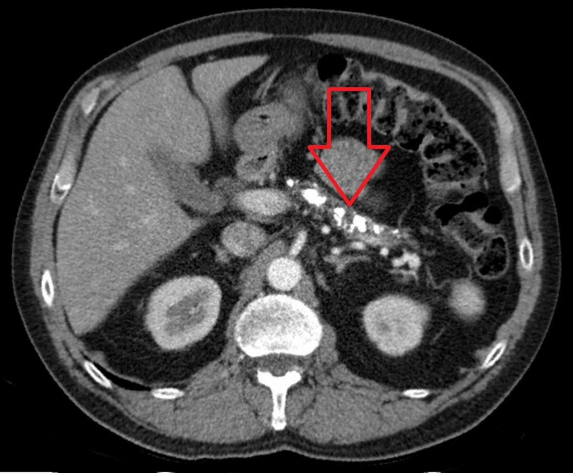
Long description
Axial contrast-enhanced CT. Dense calcifications run through the body and tail of the pancreas, which is the hallmark of chronic pancreatitis, with duct dilatation and a shrunken gland.

- Faecal elastase-1 under 200 micrograms per gram supports pancreatic exocrine insufficiency.[5]
- Serum amylase and lipase are often normal or low in chronic disease, which is an important contrast with acute pancreatitis and can be misleading.
- Faecal fat estimation and targeted vitamin levels assess nutritional impact.
- Glucose and HbA1c should be checked because diabetes may be present at diagnosis.
- Serum IgG4 supports type 1 autoimmune pancreatitis; raised levels are characteristic but not diagnostic on their own.
- Genetic testing for PRSS1, SPINK1, CFTR and CTRC is appropriate with a strong family history, young age or hereditary features.[7]
- Lipid profile and calcium assess metabolic causes.
- Autoimmune panel and bile duct imaging help with IgG4 disease.
Approach to suspected chronic pancreatitis
- Suspect chronic pancreatitisRecurrent pain, calcification on X-ray/ultrasound, steatorrhoea, diabetes
- Cross-sectional imaging (CT first)Calcification, atrophy, ductal dilation, pseudocyst, splenic vein thrombosis
- Question: Diagnosis clear?
Yes
- Assess function and complicationsFaecal elastase, vitamins, HbA1c, nutrition review
- Start cause-directed and symptomatic management
No, but suspicion remains
- MRCP with secretin, then EUSDuctal anatomy, early parenchymal change, pancreas divisum
- Add function tests and aetiology screenIgG4, genetics, lipids, calcium
There is no single, universal histological or imaging score that grades every patient neatly. In practice the diagnosis is made when imaging shows permanent structural change, such as calcification, ductal irregularity or atrophy, in a compatible clinical setting.[1] Severity is better judged by what the patient has lost: pain burden, exocrine insufficiency, endocrine insufficiency and the presence of complications. Because endoscopic ultrasound, secretin-stimulated magnetic resonance cholangiopancreatography and pancreatic function tests can be abnormal before computed tomography, early disease can be missed if only a late structural test is used. For staging discussions, describe disease as early, established or advanced in terms of pain, nutrition and endocrine status; this is often more clinically useful than a radiological score alone.
Management should not be reduced to painkillers. It has four parallel aims: remove the trigger, control pain, replace missing enzymes and vitamins, and treat complications. Care is shared between gastroenterology, surgery, dietetics, pain services and diabetes teams.[3]
- Analgesia for active pain, with a clear plan to avoid long-term opioid escalation.
- Nutritional support: small, frequent meals rather than prolonged fasting; many patients need oral supplements. Enteral feeding may be needed during severe flares.
- Treat infection when a complication such as infected pseudocyst or cholangitis is present.
- Biliary drainage if obstructive jaundice is present.
- Alcohol cessation is strongly advised; ongoing drinking accelerates damage.[3]
- Smoking cessation is equally important and is often harder to achieve.[3]
- Diet should be a normal or slightly high energy intake in small, frequent meals. A blanket low-fat diet is not recommended, because it worsens malnutrition; fat is allowed alongside adequate enzyme replacement.[4][3]
- Psychological and social support, occupational help and pain-coping strategies reduce disability.
- Non-opioid analgesia and non-drug measures, including heat, positioning and activity modification.
- Neuropathic pain agents such as pregabalin or gabapentin where neuropathic pain dominates, following standard neuropathic pain guidance.[3]
- Antioxidants, which may reduce pain in some patients.[10]
- Opioids, used cautiously and reviewed regularly, with a plan to stop if they do not help.
- Celiac plexus block, which should not be routine but may be considered case by case when other measures fail, after discussing uncertain benefit and procedural risk.[2]
- Endoscopic therapy for ductal obstruction, stones or strictures.
- Surgery for selected patients.
Endoscopic treatment is appropriate for painful obstructive chronic pancreatitis and for local complications, but surgery is generally the more durable long-term option.[2]
- Small (up to 5 millimetre) main pancreatic duct stones can be treated with pancreatography and conventional extraction.
- Larger stones may require extracorporeal shockwave lithotripsy, with or without pancreatoscopy and intraductal lithotripsy.[2][3]
- Prolonged stent therapy of 6 to 12 months, by sequential placement of multiple plastic stents in parallel, can remodel main pancreatic duct strictures and relieve symptoms.[2]
- Endoscopic retrograde cholangiopancreatography with stent insertion is the preferred treatment for benign biliary stricture caused by chronic pancreatitis; fully covered self-expanding metal stents are favoured over multiple plastic stents when feasible.[2]
- Endoscopic ultrasound-guided or transpapillary drainage is offered for symptomatic pseudocysts, and considered for non-symptomatic pseudocysts with duct disruption, vascular or diaphragmatic pressure, rupture risk or suspected infection.[3]
When endoscopy is pursued for pancreas divisum, its value is debated; minor papilla endotherapy may be considered only when there are objective signs of outflow obstruction, and there is no role for treating pain alone. For unexplained recurrent acute pancreatitis with normal ductal anatomy, the benefits of endoscopic retrograde cholangiopancreatography are uncertain and should be discussed in detail before proceeding.[2]
Surgery should be considered first-line for painful chronic pancreatitis that causes main pancreatic duct obstruction.[3] Procedures in common use include:
- Lateral pancreaticojejunostomy (Puestow procedure) for a dilated duct with multiple stones, to decompress the duct and relieve pain.
- Frey procedure for a dilated duct combined with cored excavation of the pancreatic head.
- Pancreaticoduodenectomy (Whipple procedure) for disease predominantly in the head, suspected malignancy or failed decompression.
- Total pancreatectomy with islet autotransplantation for selected patients with diffuse small-duct disease, in centres with appropriate expertise.
Endoscopic shockwave lithotripsy remains an alternative when surgery is unsuitable. Combined surgical and endoscopic approaches are common in specialist centres. Total pancreatectomy is the only known cure for chronic pancreatitis, but difficult patient selection and operative complications mean it is usually unattractive except in carefully chosen patients.[1][3]
All patients with proven or strongly suspected exocrine insufficiency should be offered pancreatic enzyme replacement.[3][4]
- Starting dose: 25,000 to 40,000 units of lipase per meal.[4]
- Maximum: 75,000 to 80,000 units of lipase per meal if standard doses fail.[4]
- Timing: take capsules with the first mouthful of food and spread further capsules through the meal; enzymes must be present when food enters the duodenum. Enteric-coated preparations should not be crushed or chewed. Acid-suppressing therapy can be a useful adjunct if symptoms persist on high doses.[4]
- Monitoring: titrate to stools, weight and vitamin levels; faecal elastase confirms the diagnosis but is not a good tool for dose titration.
- Dietetic support from a specialist dietitian is recommended for all patients.[3]
- Monitor for pancreatic exocrine insufficiency and malnutrition at least every 12 months, and every 6 months in patients under 16 years; adjust vitamin and mineral replacement accordingly.[3]
- Patients have a high risk of osteoporosis, osteopenia and fracture, so offer a bone density assessment every 2 years in adults and replace vitamin D.[3][10]
- Ensure vitamin and mineral replacement is guided by measured levels rather than by symptoms alone.
Diabetes caused by chronic pancreatitis (type 3c diabetes) is a state of insulin deficiency rather than insulin resistance, and it is often brittle.[3][10]
- Assessment: check HbA1c at least every 6 months and formally screen for diabetes.[3]
- Non-insulin therapy: follow standard type 2 diabetes guidance where insulin is not yet needed.
- Insulin therapy: when insulin is required, follow type 1 diabetes guidance on injection technique and blood glucose monitoring; assess every 6 months for the benefit of insulin.[3]
- Education: patients need specific advice about hypoglycaemia, variable absorption, rotating injection sites and the possibility of concurrent glucagon deficiency.
Refer to a specialist pancreatic centre for complex pain, ductal obstruction, pseudocyst, biliary or duodenal obstruction, splenic vein thrombosis, suspected cancer, hereditary disease and all surgical or advanced endoscopic care.[3]
- Monitor HbA1c for diabetes at least every 6 months.[3]
- Monitor pancreatic exocrine insufficiency and malnutrition at least annually.[3]
- Offer bone density assessment every 2 years in adults.[3]
- Be vigilant for pancreatic cancer, especially in hereditary disease, for which the lifetime risk is around 40 percent; annual monitoring may be considered in hereditary pancreatitis.[3]
- Pseudocyst may cause pain, gastric or duodenal obstruction, infection or haemorrhage.
- Common bile duct stricture causes conjugated hyperbilirubinaemia and cholangitis.
- Duodenal obstruction from inflammation and fibrosis or pseudocyst.
- Splenic vein thrombosis with gastric varices and upper gastrointestinal bleeding.
- Pancreatic fistula, ascites and pleural effusion after ductal disruption.
- Pancreatic cancer, whose risk is increased in chronic pancreatitis and is highest (around 40 percent lifetime) in hereditary disease.[3]
- Opioid dependence and chronic pain syndrome, a major determinant of quality of life.
- Nutritional failure with loss of fat and fat-soluble vitamins.
Prognosis depends on cause, smoking and alcohol status, pain severity and the development of exocrine and endocrine insufficiency. Alcoholic patients who continue to drink have worse pain and faster progression. Patients who stop smoking and drinking, and who receive proper enzyme and nutritional care, can do much better. Type 3c diabetes is a marker of more advanced disease. Overall survival is reduced, mainly through malignancy, cardiovascular disease and infection.
- Prevent new damage by treating hypertriglyceridaemia, hypercalcaemia and other underlying causes.
- Support abstinence from alcohol and smoking for life.[3]
- Screen for diabetes regularly and replace vitamins proactively.[3]
- Screen for osteoporosis with bone density assessment every 2 years in adults.[3]
- In hereditary pancreatitis, consider annual surveillance for pancreatic cancer in specialist programmes.[3]
Children. Hereditary pancreatitis is the leading genetic consideration; more than half of children with chronic pancreatitis carry one of the known variants in some series. Management balances growth, nutrition and pain, and genetic counselling is offered to families.[7]
Pregnancy. Management focuses on nutrition, pain control with drugs chosen for pregnancy, and correction of vitamin deficiency; advanced endoscopy and surgery are reserved for complications and undertaken in specialist centres.
Older adults. Malnutrition, osteoporosis and frailty dominate; enzyme doses may need individual titration and diabetes management should avoid hypoglycaemia.
Immunocompromised patients need prompt treatment of infected complications and careful antibiotic choice.
In settings where advanced imaging or specialist care is limited, the key is to recognise the disease early from clinical features and basic tests, and to treat what matters most.
- Transabdominal ultrasound is often the only imaging available; look for calcification and ductal change.
- Faecal fat and clinical assessment can substitute for faecal elastase where the latter is unavailable, and an empirical trial of pancreatic enzymes is reasonable in a malnourished patient with steatorrhoea.
- Pain control may need strong opioids; use the simplest effective regimen and reassess regularly.
- Insulin for type 3c diabetes and basic metabolic monitoring remain essential; oral hypoglycaemic agents are less useful once insulin deficiency is established.
- Vitamin D and calcium intake, together with sunlight exposure, support bone health.
- In tropical calcific pancreatitis, seen in Africa and Asia, patients are often malnourished adolescents or young adults with abdominal pain, large pancreatic calculi and a ketosis-resistant but insulin-requiring diabetes. Pancreatic cancer complicates up to 10 percent of cases. Management follows the same principles: analgesia, pancreatic enzymes, diabetes control and, when feasible, endoscopic or surgical decompression with stone removal.[8]
- Chronic pancreatitis is irreversible fibroinflammatory damage to both the exocrine and endocrine pancreas, most often beginning with repeated parenchymal injury.
- Alcohol accounts for 60 to 70 percent of cases in many series, and smoking is an independent, additive risk factor.
- Hereditary forms involve mutations in PRSS1, SPINK1, CFTR and CTRC.
- Type 1 (IgG4-related) and type 2 autoimmune pancreatitis both respond to steroids, but type 1 relapses in 30 to 50 percent of cases within 6 to 12 months.
- Patients present with epigastric pain, weight loss, steatorrhoea, deficiency of the vitamins A, D, E and K and, later, type 3c diabetes.
- Computed tomography shows calcification and ductal change; MRCP (especially with secretin) shows ductal anatomy and the chain-of-lakes appearance; endoscopic ultrasound is the most sensitive early test.
- Faecal elastase under 200 micrograms per gram supports exocrine insufficiency; serum amylase and lipase are often normal.
- Enzyme replacement starts at 25,000 to 40,000 units lipase per meal, maximum 75,000 to 80,000, taken with the first mouthful.
- Surgery is first-line for painful duct-obstructive disease; endoscopic therapy is valuable but less durable.
- Type 3c diabetes is insulin deficiency from islet loss, with up to 80 percent lifetime risk.
- Hereditary disease carries up to about 40 percent lifetime risk of pancreatic cancer.
- Antioxidants may reduce pain; celiac plexus block should not be routine.
- Osteoporosis, malnutrition and opioid dependence need active, long-term management.
Answer each one in your head first, then open the answer.
A 42-year-old man has recurrent epigastric pain radiating to the back, worse after meals, with weight loss and pale bulky stools. Computed tomography shows pancreatic calcifications and a dilated pancreatic duct. Which single test best supports exocrine insufficiency, and what result would you expect?
Show answerHide answer
Faecal elastase-1 under 200 micrograms per gram. In this patient with structural disease, a low value confirms pancreatic exocrine insufficiency and should prompt enzyme replacement and dietetic review.
Which four genes are most commonly implicated in hereditary chronic pancreatitis, and why does an early-onset case in a young patient matter beyond pain control?
Show answerHide answer
PRSS1, SPINK1, CFTR and CTRC. It matters because hereditary disease progresses faster, may need family counselling and genetic testing, and carries a substantially higher lifetime risk of pancreatic cancer.
How should pancreatic enzyme capsules be taken, and what is the usual adult starting and maximum lipase dose per meal?
Show answerHide answer
Start with the first mouthful and continue through the meal; do not crush or chew enteric-coated capsules. Start at 25,000 to 40,000 units lipase per meal and increase up to 75,000 to 80,000 units if needed.
What distinguishes type 3c diabetes from type 1 and type 2 diabetes, and what monitoring is required in chronic pancreatitis?
Show answerHide answer
It is diabetes from insulin deficiency caused by destruction of pancreatic islets, not autoimmune type 1 or insulin-resistant type 2 disease. Monitor HbA1c at least every 6 months and assess for insulin therapy benefit.
Chronic pancreatitis is discussed alongside acute pancreatitis, exocrine pancreatic insufficiency, type 3c diabetes mellitus, pancreatic cancer, chronic abdominal pain, and tropical calcific pancreatitis (plain text; no topic links are used on this page).
- Timothy B. Gardner, Douglas G. Adler, Chris E. Forsmark, Bryan G. Sauer, Jason R. Taylor, David C. Whitcomb. ACG Clinical Guideline: Chronic Pancreatitis. American Journal of Gastroenterology 2020;115(3):322–339. doi:10.14309/ajg.0000000000000535. Accessed .
- Daniel S. Strand, Ryan J. Law, Dennis Yang, B. Joseph Elmunzer. AGA Clinical Practice Update on the Endoscopic Approach to Recurrent Acute and Chronic Pancreatitis: Expert Review. Gastroenterology 2022;163(4):1107–1114. doi:10.1053/j.gastro.2022.07.079. Accessed .
- National Institute for Health and Care Excellence. Pancreatitis (NICE guideline NG104). NICE guideline, published 2018, last updated 2020. Accessed .
- Mehrdad Nikfarjam, Jeremy S. Wilson, Ross C. Smith; Australasian Pancreatic Club Pancreatic Enzyme Replacement Therapy Guidelines Working Group. Diagnosis and management of pancreatic exocrine insufficiency. Medical Journal of Australia 2017;207(4):161–165. doi:10.5694/mja16.00851. Accessed .
- Sudipta Dhar Chowdhury, Reuben Thomas Kurien, Anup Ramachandran, et al. Pancreatic exocrine insufficiency: Comparing fecal elastase 1 with 72-h stool for fecal fat estimation. Indian Journal of Gastroenterology 2016;35(6):441–444. doi:10.1007/s12664-016-0714-4. Accessed .
- Eva C. Vaquero Raya. [Chronic pancreatitis: beyond alcohol]. Gastroenterologia y Hepatologia 2012;35(8):577–584. doi:10.1016/j.gastrohep.2012.03.002. Accessed .
- Arvind Vasant Panchoo, Grant H. VanNess, Edgardo Rivera-Rivera, Trevor J. Laborda. Hereditary pancreatitis: An updated review in pediatrics. World Journal of Clinical Pediatrics 2022;11(1):27–37. doi:10.5409/wjcp.v11.i1.27. Accessed .
- Rakesh K. Tandon. Tropical calcific pancreatitis. Journal of Gastroenterology and Hepatology 1998;13(S3):S284–S288. doi:10.1111/j.1440-1746.1998.tb01894.x. Accessed .
- Amir Mari, Anas Kadah, Mahmud Mahamid, Wisam Sbeit, Tawfik Khoury. IgG4 Related Autoimmune Pancreatitis: An Overview and the Emerging Role of Serum Eotaxin as a Potential Treatment Target. Israel Medical Association Journal 2019;21(9):620–623. Accessed .
- Anand R. Gupte, Chris E. Forsmark. Chronic pancreatitis. Current Opinion in Gastroenterology 2014;30(5):500–505. doi:10.1097/MOG.0000000000000094. Accessed .
Figure credits
Educational content for medical students and doctors. It is not a substitute for clinical judgement or local guidelines.