Acute appendicitis
A common surgical emergency: how obstruction of the appendix leads to pain, perforation and abscess, and how it is diagnosed and treated.
- Acute appendicitis is inflammation of the appendix, usually starting when its narrow lumen becomes blocked by a faecal stone (faecolith) or swollen lymphoid tissue.
- It is one of the commonest reasons for abdominal surgery in young people, with a lifetime risk of about 7 to 8 percent and a peak between the ages of 10 and 30 years.
- The classic story is vague periumbilical colicky pain that steadily intensifies and moves to the right iliac fossa, with anorexia, nausea, vomiting and a low fever; focal tenderness at McBurney's point is the hallmark sign.
- Diagnosis is clinical but imaging helps: graded-compression ultrasound first in children and in pregnancy, and computed tomography in most adults when the diagnosis is uncertain.
- The definitive treatment is appendectomy, now almost always laparoscopic; selected uncomplicated cases can be treated with antibiotics alone, but about 3 in 10 people treated this way need appendectomy within 90 days and about 4 in 10 have a recurrence within 5 years.
By the end of this page you should be able to:
- Trace how luminal obstruction of the appendix produces pain, infection, gangrene and perforation, and explain the positions that change the clinical picture.
- Recognise the classical presentation of acute appendicitis and the atypical forms seen in children, older adults, pregnant women and retrocaecal or pelvic appendices.
- Use the Alvarado, Appendicitis Inflammatory Response (AIR) and Pediatric Appendicitis Score (PAS) scores to stratify risk and decide when imaging is needed.
- Choose and interpret first-line investigations (full blood count, C-reactive protein, urinalysis, pregnancy test, ultrasound, computed tomography, magnetic resonance imaging) for each patient group.
- Compare the differential diagnoses of right iliac fossa pain and state the feature that separates each.
- Plan management from resuscitation through antibiotics, appendectomy, antibiotics-only non-operative treatment, drainage of an abscess and follow-up, and mention appendiceal tumours found incidentally, complications, mortality and low-resource practice.
DefinitionAcute appendicitis
Acute appendicitis is inflammation of the vermiform appendix, the small blind-ended tube attached to the caecum. It begins as mucosal and wall inflammation, can progress through gangrenous change to perforation, and is the most common abdominal surgical emergency in young patients. The diagnosis is clinical and is confirmed by imaging and, ultimately, by histopathology of the removed appendix.
The appendix is a narrow (average resting diameter well under a centimetre) tubular out-pouching of the caecum with a small and end-artery blood supply from the appendicular branch of the ileocolic artery, and it contains abundant lymphoid tissue. That narrow lumen, end-arterial supply and lymphoid-rich wall explain almost everything about how appendicitis behaves.
The appendix lies at the point where the three taeniae coli of the caecum converge. Its tip is mobile, so its position in the abdomen varies:
- Retrocaecal (behind the caecum) or retrocolic is the commonest position.
- Pelvic (descending into the true pelvis) is next in frequency.
- Subcaecal, pre-ileal and post-ileal positions are less common.
Position decides the pain. Early appendicitis distends the appendix; this activates visceral afferent nerves at the level of the tenth thoracic segment, producing the midline, poorly localised, colicky periumbilical or epigastric discomfort that patients describe as "a griping around the navel". Once inflammation reaches the serosa and the adjacent parietal peritoneum, somatic nerves take over and the pain sharpens and "moves" to whichever part of the abdominal wall overlies the inflamed appendix. That is why pain moves to the right iliac fossa in the common positions, to the right flank or lumbar region in a retrocaecal appendix, and lower down, sometimes with urinary frequency or diarrhoea, in a pelvic one.
The classic McBurney point, the spot of maximal tenderness, is about 1.5 to 2 inches (roughly 4 to 5 cm) along a line from the right anterior superior iliac spine to the umbilicus. [2]
Acute appendicitis is, in most cases, a problem of drainage. The sequence is:
- Obstruction of the lumen. In adults the common obstructing objects are a faecolith (a stony, calcified mass of stool) or a foreign body; in children and adolescents lymphoid hyperplasia after a viral infection or gastroenteritis is the more usual cause. A caecal or appendiceal tumour can obstruct in older patients. The appendix keeps secreting mucus, but the mucus now has nowhere to go, so intraluminal pressure rises.
- Impaired drainage and bacterial overgrowth. Rising pressure exceeds venous pressure first; mucosal blood flow falls, the mucosal barrier fails, and the normal mixed gut flora (the usual enterobacteria and anaerobes of the colon) multiply and invade the wall. Low-grade inflammation at this stage is "catarrhal" enteritis of the appendix.
- Progression to gangrene. Sustained ischaemia makes the whole wall necrotic (transmural necrosis, i.e. gangrenous appendicitis). Thrombosis of the small appendicular vessels and microscopic perforations follow. This stage can be surprisingly fast, often within 24 to 48 hours of symptom onset.
- Perforation. Gangrene gives way to free perforation into the peritoneal cavity, with consequences that depend on how the body walls off the leak:
- free perforation causes generalised peritonitis;
- localised containment produces an appendiceal phlegmon (an inflammatory mass) or an abscess;
- perforation of an appendix carrying a mucinous neoplasm (see below) can shower mucin throughout the abdomen and create pseudomyxoma peritonei.
- Resolution. Not every inflamed appendix perforates. Some cases, particularly early and uncomplicated ones, resolve spontaneously, and this is one reason the distinction between uncomplicated and complicated disease now matters so much.
The severity spectrum matters clinically. Uncomplicated appendicitis shows suppurative (phlegmonous) inflammation only. Complicated appendicitis means gangrenous change, perforation, an abscess or phlegmon, or, in some classifications, the presence of an appendicolith. [3] [7]
A higher temperature, a very raised white cell count or markers of severe sepsis at presentation are clues that the patient may already be in the gangrenous or perforated stage, and they change how quickly source control is needed.
Acute appendicitis is a worldwide problem with a strong age and geography pattern.
- Lifetime risk is about 7 to 8 percent overall, and higher in males than females in most series. One US review quotes 8.6 percent for males and 6.7 percent for females. [2] [3]
- In developed countries the incidence is reported at 5.7 to 50 new cases per 100,000 inhabitants per year, and the rate has fallen since the mid-twentieth century; conversely it is rising in South America, Asia and the Middle East. [1] [3]
- Peak incidence is between the ages of 10 and 30 years, with the highest burden in adolescents (about 15 to 19 years in global burden data) and a slight male preponderance. Some series report a male-to-female ratio of about 1.4 to 1, while global burden analyses find no statistically significant difference between sexes; this is probably because both sex-specific estimates and age distributions differ between populations. [1] [2] [3]
- Lifetime risk by region is given by the WSES as 9 percent in the USA, 8 percent in Europe and 2 percent in Africa, reflecting the impact of diet, environment and access to care. [1]
- Perforation is commoner than many expect. The perforation rate varies from 16 to 40 percent, rising to roughly 40 to 57 percent in the youngest patients and to roughly 55 to 70 percent in patients over 50 years, partly because diagnosis is delayed and partly because the infection may be more virulent in those groups. [1]
- Among people over 50 who attend an emergency department with acute abdominal pain, about 15 percent have acute appendicitis, and it is the second commonest acute surgical condition in that age group. [3]
- Children under 4 years are uncommonly affected (roughly 1 to 2 cases per 10,000 children under 4, rising to 25 per 10,000 by the second decade), which is why their disease is often recognised late and perforated. [4]
- The initiating event is obstruction, usually idiopathic. The most frequent identifiable causes are faecoliths, especially in adults, and lymphoid hyperplasia, especially in children and adolescents.
- Regional differences in incidence (and the historical decline in developed countries) suggest a role for diet, childhood environment and hygiene, but no single modifiable risk factor has been proven.
- Infectious triggers for lymphoid hyperplasia, such as viral enteritis, are sometimes identifiable; parasitic infestation can cause obstruction in endemic areas.
- Female sex is protective in most series but the reason is not established.
- Age changes the risk of complication far more than the risk of disease: perforation and tumour discovery rise markedly after 40 to 50 years.
- A past episode of appendicitis can recur, and an untreated or incompletely treated first episode leaves the same anatomy at risk.
Acute appendicitis classically unfolds in a recognisable order. The patient's own words are often more reliable than any score.
- Pain. A vague, colicky pain around the navel or epigastrium begins first. Over 12 to 24 hours it becomes continuous, sharp and localised in the right iliac fossa, usually somewhere near McBurney's point. Movement, coughing and deep breathing worsen it; the patient often lies still.
- Anorexia is marked and almost constant; many patients turn away food and drink.
- Nausea follows the pain; vomiting, when it occurs, is usually once or twice and is not projectile. Vomiting that starts before the pain and is repeated points toward gastroenteritis or an obstructed intestinal loop rather than appendicitis.
- Fever is low grade (often about 37.5 to 38.0 degrees Celsius) and appears after the pain.
- Bowel habit. Most patients are constipated; diarrhoea occurs with a pelvic appendix because it irritates the rectum.
- Urinary frequency or dysuria can occur if an inflamed pelvic appendix lies near the bladder or ureter.
- Menstrual history is essential in anyone who could be pregnant.
Clinical pearlThe order of symptoms
In appendicitis, pain comes first, then anorexia and nausea. In gastroenteritis and food poisoning, nausea and vomiting usually come first, with crampy periumbilical pain following. This single observation separates many cases in the emergency department.
- General. The patient looks unwell, lies still with the knees slightly flexed, and is tender. Bowel sounds may be reduced. A low-grade tachycardia and fever are common.
- Inspection and palpation. Look for guarding and rebound tenderness in the right iliac fossa. Press gently first; do not start by jabbing the tender spot.
- McBurney point tenderness is the hallmark.
- Guarding (voluntary or involuntary rigidity) and rebound tenderness (pain as the hand is released) indicate parietal peritoneal involvement.
- Rovsing's sign (pain in the right iliac fossa when the left iliac fossa is pressed) is present in about 3.5 percent of cases; it is specific enough to be useful when found, but many patients do not have it.
- Neurodynamic signs help with an atypically placed appendix:
- the psoas sign (pain on extending the right hip, the hip then being pushed back) suggests a retrocaecal appendix lying on psoas major;
- the obturator sign (pain on internal rotation of a flexed right hip) suggests a pelvic appendix.
- Rectal or (in women) vaginal examination can find tenderness or a mass on the right in pelvic appendicitis; it is used selectively.
- Peritonitis. Generalised tenderness, rigidity, absent bowel sounds and tachycardia indicate that the appendix has perforated: this is now an operative emergency.
- Retrocaecal. The pain is duller and higher, in the right flank or lumbar region, and classical right iliac fossa tenderness may be absent; the psoas sign is the useful clue.
- Pelvic. Pain is suprapubic or worsens on defaecation; there may be diarrhoea, tenesmus or urinary symptoms, and rectal tenderness on the right.
- Children. Communication is harder and perforation rates are higher. Fever may be higher; diarrhoea and cough-like pain can dominate; generalised abdominal tenderness is common. About 28 to 57 percent of children under 12 are misdiagnosed on the first assessment in some series, and misdiagnosis is more frequent still in children under 2. [1] (These figures are quoted from the cited literature; local rates vary with the availability of imaging.)
- Older adults. Pain is often less severe and less well localised; fever is blunted (about 20 to 30 percent have little or no fever response); anorexia and malaise may dominate. Perforation, morbidity (19 to 46 percent versus 5 to 9 percent in younger patients) and mortality (0.7 to 6 percent versus less than 0.3 percent in the general population) are all higher. [3]
- Pregnant women. The gravid uterus displaces the caecum and appendix upward and outward, so pain may be higher, nearer the right hypochondrium or flank, and the appendix is not always in the right iliac fossa. Leucocytosis is also a normal feature of pregnancy, which blunts laboratory interpretation. Because untreated appendicitis in pregnancy carries a serious risk for the fetus, a low threshold for imaging and for surgical consultation is justified. [1]
Right iliac fossa pain has a broad list of causes. The table does not list every possibility, but it covers those that most commonly mimic appendicitis.
| Condition | What points away from appendicitis |
|---|---|
| Gastroenteritis | Vomiting before pain, profuse watery diarrhoea, no focal guarding, pain crampy and central; usually resolves within 24 hours |
| Mesenteric adenitis | Often after a viral illness; tender but less toxic; ultrasound shows enlarged mesenteric nodes; appendix is normal |
| Crohn disease (terminal ileitis) | Recurrent abdominal pain and loose stools, weight loss, oral ulcers; imaging shows thickened terminal ileum |
| Meckel diverticulitis | Identical presentation possible; often indistinguishable pre-operatively; found at laparoscopy |
| Caecal or colonic diverticulitis (right-sided) | Older patient, tenderness a little higher or more medial; CT shows an inflamed diverticulum |
| Gynaecological disease (ovarian cyst, torsion, ectopic pregnancy, pelvic inflammatory disease) | Sudden or cyclical pain, vaginal discharge or bleeding, cervical excitation; pregnancy test and ultrasound are decisive |
| Ureteric colic / pyelonephritis | Colicky loin-to-groin pain, haematuria, urinary infection; urinalysis and renal imaging |
| Psoas or retrocaecal abscess, psoas haematoma | Back or flank pain, limited hip movement, fever |
| Caecal carcinoma / appendiceal tumour | Older patient, weight loss, anaemia, atypical recurrence; imaging and histology settle it |
| Inguinal or femoral hernia strangulation | A groin lump, irreducible and tender, bowel obstruction |
| Rectus sheath haematoma | Localised wall plaque, no deeper tenderness, often after cough or anticoagulation |
Two practical rules. First, in a woman of childbearing age, exclude pregnancy early: an ectopic pregnancy can look exactly like appendicitis and is dangerous to miss. Second, in a patient over about 50 with new or atypical appendicitis, keep the possibility of a tumour in mind until histology is available. [2] [3]
- Full blood count. A neutrophil leucocytosis supports appendicitis; a normal white cell count does not exclude it. In children the combination of white cells at least 16,000 per microlitre and a raised C-reactive protein is strongly predictive. [1]
- C-reactive protein. A value of 10 mg/L or more is a strong supportive finding, especially in children, and rising serial values suggest progression. It is normal early on; a single normal value in the first 12 hours is not reassuring. [1] [2]
- Urinalysis. Should be normal in appendicitis (leucocytes or nitrites point to infection, haematuria to a stone). A few red or white cells can appear if an inflamed appendix irritates the ureter or bladder, so do not dismiss the diagnosis on a single dipstick.
- Pregnancy test (beta-human chorionic gonadotrophin) in every girl or woman of childbearing age before surgery.
- Liver function and bilirubin may help when biliary or hepatic causes are in the differential; a raised bilirubin has been studied as a marker of perforation but is not diagnostic on its own.
- Blood cultures if sepsis or high fever is present.
Figure 1CT scan of the abdomen showing acute appendicitis
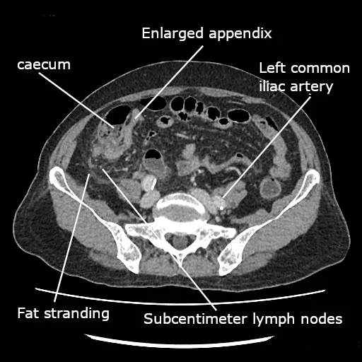
Long description
CT scan of the abdomen showing an enlarged appendix, suggestive of acute appendicitis.

The aim of imaging is to confirm appendicitis, judge whether it is complicated, and answer the surgical questions, not to replace clinical judgement.
- Ultrasound (graded compression) of the right iliac fossa is the safest first test, avoids radiation and is operator-dependent. It should be the first imaging in children and in pregnancy, and a reasonable first step in adults when it is readily available. It shows the classic picture of a non-compressible tubular structure more than 6 mm in outer-to-outer diameter with wall thickening, and secondary signs such as periappendiceal fat change or free fluid. [2] [4]
- Computed tomography (CT) is the most definitive cross-sectional test in adults and is widely used when ultrasound is inconclusive, particularly in older or obese patients, where it also helps in differentiating uncomplicated from complicated disease and in excluding tumours. [2] [7]
- When a scan is needed in adolescents and young adults, a contrast-enhanced low-dose protocol has accuracy not inferior to a standard-dose scan and saves radiation. [1]
- In adults, second-line CT after an ultrasound has sensitivity and specificity around 90 to 94 percent in meta-analyses. [3]
- CT is not first-line in children: clinical scores plus ultrasound (and MRI where available) should limit its use, because radiation matters in a population that often undergoes repeat scanning. [1]
- Magnetic resonance imaging (MRI) is highly accurate (reported sensitivity roughly 96 to 97 percent and specificity about 96 to 97 percent in recent studies) and involves no radiation. [2] It should be the second-line test in pregnancy after an inconclusive ultrasound, and it is a good second-line test in children where it is available; in adults it is more expensive and less immediately available. [1]
- Plain abdominal radiograph is not diagnostic. It can occasionally show a faecolith (which is radiologically visible in a minority of cases) and is useful to exclude free air or obstruction when perforation is suspected, but it should not delay adequate imaging and treatment.
A right-iliac-fossa pregnancy is still possible: in pregnant patients, graded compression ultrasound followed by MRI if it is inconclusive is recommended. [1]
A score alone does not diagnose appendicitis; it estimates risk and decides who needs imaging. The three most used are below, taken from published tables; thresholds vary slightly with the series, and local validation matters.
| Feature | Points |
|---|---|
| Migratory right iliac fossa pain | 1 |
| Nausea or vomiting | 1 |
| Anorexia | 1 |
| Tenderness in the right iliac fossa | 2 |
| Rebound tenderness in the right iliac fossa | 1 |
| Elevated temperature | 1 |
| Leucocytosis | 2 |
| Shift to the left of neutrophils | 1 |
Score 1 to 4 suggests low risk and the patient can be discharged with safety netting; 5 to 6, observation and reassessment; 7 to 10, high probability and surgical review. [2] [1] The WSES 2020 guideline recommends that the Alvarado score not be used to confirm the diagnosis in adults, because specificity is limited. [1]
| Feature | Score |
|---|---|
| Vomiting | 1 |
| Pain in the right inferior fossa | 1 |
| Rebound tenderness or muscular defence - light | 1 |
| Rebound tenderness or muscular defence - moderate | 2 |
| Rebound tenderness or muscular defence - strong | 3 |
| Body temperature over 38.5 degrees Celsius | 1 |
| Polymorphonuclear leucocytes 70 to 84 percent | 1 |
| Polymorphonuclear leucocytes over 85 percent | 2 |
| White cell count 10.0 to 14.9 times 10^9 per litre | 1 |
| White cell count 15.0 or more times 10^9 per litre | 2 |
| C-reactive protein 10 to 49 mg per litre | 1 |
| C-reactive protein over 50 mg per litre | 2 |
Scores of 0 to 4 suggest low risk and discharge; 5 to 8, active observation and imaging; 9 to 12, strong probability of appendicitis and a surgical plan. The AIR score and the Adult Appendicitis Score (AAS) have the highest discriminating power among adult scores and reduce negative appendectomy and imaging in both low- and intermediate-risk groups. [2] [1]
| Feature | Points |
|---|---|
| Anorexia | 1 |
| Nausea or vomiting | 1 |
| Fever (over 38 degrees Celsius) | 1 |
| Migration of pain to the right lower quadrant | 1 |
| Tenderness in the right lower quadrant | 2 |
| Cough, percussion or hopping tenderness | 2 |
| Leucocytosis (white cells over 10,000 per microlitre) | 1 |
| Neutrophilia (neutrophils over 75 percent) | 1 |
Risk bands quoted in validation studies are low risk below 3, intermediate 4 to 6, and high risk above 7. [4] The WSES recommends that appendicitis in children is not diagnosed on a score alone, and that both biomarkers and scoring be used to predict severity and the need for imaging. [1]
How to use scores in practice. Estimate pre-test probability clinically, then use the score: a low score supports discharge with safety-netting and no imaging in a well, improving child or adult; an intermediate score is the group that benefits from systematic imaging (and, in children, ultrasound first); a high score in an adult under 40 with an AIR score of 9 to 12, an Alvarado score of 9 to 10 or an AAS of 16 or more allows proceeding toward diagnostic laparoscopy without a preceding CT, if local practice supports it. Scores combined with ultrasound outperform either alone and reduce the need for CT. [1]
If history and examination suggest appendicitis but ultrasound, and then CT, is negative, the WSES recommends observation with cross-sectional imaging before any operation, and laparoscopy when pain persists or progresses in order to establish or exclude the diagnosis (and to treat its cause). [1]
A practical approach to suspected acute appendicitis
- Patient with right iliac fossa or central migrating painHistory, examination, observations, full blood count, CRP, urinalysis, pregnancy test where relevant
- Stratify risk with a clinical score (Alvarado / AIR in adults; PAS in children)Does not replace judgement; combines with imaging
- Question: Risk band
Low risk
- Reassess, observe and safely discharge if improvingClear return instructions and review plan
Intermediate risk
- Image in a structured wayAdults: ultrasound first, then contrast-enhanced low-dose CT if needed; children: ultrasound first, MRI if available and ultrasound inconclusive; pregnancy: ultrasound then MRI
- Question: Imaging confirms appendicitis?
Yes
- Manage (see below)
No, but pain persists
- Cross-sectional imaging and diagnostic laparoscopy if needed
High risk (adult under 40: AIR 9-12, Alvarado 9-10, AAS 16 or more)
- Proceed toward diagnostic and therapeutic laparoscopy without a preceding CTWhere local practice supports it; consultant involvement
- Suspected perforation or sepsis at any stageResuscitate, start broad antibiotics and expedite source control
- Nothing by mouth, intravenous fluids and correction of electrolytes; analgesia (opiates are safe and do not mask the diagnosis once the decision to operate is made), antiemetics and oxygen if needed.
- Preoperative intravenous antibiotics are recommended for all patients, with spectrum to cover gut enterobacteria and anaerobes. The exact choice follows local resistance patterns and guidelines; confirm against the local formulary and pharmacy advice.
- Consent must include the possibility of finding a normal appendix or a complicated one, the conversion to open surgery, and the small risk of serious complications.
- Calculate a risk score on admission and document it; it helps communicate with the surgical team and will guide postoperative review.
- Laparoscopic appendectomy is the preferred approach over open appendectomy for both uncomplicated and complicated acute appendicitis where equipment and expertise are available, with lower wound infection, shorter stay and faster recovery. [1]
- Delay is safe in uncomplicated disease but has a ceiling. Short in-hospital delay up to 24 hours is safe and does not increase complications or perforation; appendectomy should not be delayed beyond 24 hours from admission, and appendectomy done on hospital day 3 or later carries higher mortality and complications. [1] [7]
- In complicated appendicitis, early appendectomy within about 8 hours is recommended. [1]
- Uncomplicated appendectomy can be a day-case where a clear enhanced-recovery pathway exists. [7]
Selected adults and children with imaging-confirmed uncomplicated appendicitis and no appendicolith can be offered antibiotics as a first-line option after a shared decision.
- Candidacy. Exclusions (from the evidence): an appendicolith on imaging, suspected complicated disease, pregnancy (non-operative management is not recommended in pregnancy until better evidence exists), inability to comply with follow-up, or immunocompromise where surgery is preferable. [1] [7]
- Protocol. An initial intravenous course followed by oral antibiotics to complete a 10-day course; one large pragmatic trial (CODA, 1552 adults in 25 US centres) used 10 days and found antibiotics non-inferior to appendectomy for 30-day general health status, with nearly half of antibiotic patients not needing admission for the index episode. [3]
- Outcomes. In that trial, 29 percent of the antibiotics group had undergone appendectomy by 90 days (41 percent of those with an appendicolith, 25 percent of those without), and non-operative management had more emergency department visits and hospitalisations after the index treatment. [3]
- Longer term. About 39 percent of patients treated with antibiotics alone have a recurrence by 5 years; initial success is high (about 86 percent in pooled analyses), so the trade-off is early avoidance of surgery against later recurrence. [2] [3]
- Guideline position. Overall guidance is graded toward operative management (conditional recommendation), particularly where appendicoliths or recurrent disease are present; antibiotics-first is an option to discuss, not the default. [7] Where non-operative treatment is chosen for complicated disease (abscess, phlegmon), the same guiding principle applies: drainage and antibiotics first, with surgery when it fails.
When imaging or laparoscopy shows perforation, phlegmon or abscess, the first question is whether the patient is stable and the infection is contained.
- Contained disease (phlegmon or abscess) in a stable patient. Non-operative management with broad antibiotics, with percutaneous drainage if a discrete, drainable abscess is present and accessible, succeeds in over 90 percent of cases, with a 7.4 percent risk of recurrence and only about 20 percent of abscesses needing drainage. [1]
- Antibiotics after adequate source control. Postoperative (or post-drainage) antibiotics should be short: a 3 to 5 day course is generally sufficient for adults, and guidance recommends against prolonging them beyond 3 to 5 days once source control is adequate. Early transition to oral antibiotics is safe and cost-effective in children. [1] [7]
- Interval appendectomy (removal weeks later). This is a judgement call rather than a universal rule. The WSES states it is not always indicated after successful non-operative treatment, whereas some surgical guidance suggests it in adults (conditional recommendation); it is reasonable where there is residual faecolith, atypical imaging, symptoms, or an older patient in whom a tumour must be excluded. [1] [7]
- In practice many teams remove the appendix after 6 to 8 weeks, both to prevent recurrence and to send the specimen for histology.
- Operative disease. Early appendectomy within 8 hours is recommended for complicated appendicitis, and more extensive resection (ileocaecal or right-sided colonic) is occasionally needed with a destructive or mass-forming process; but where the inflammation is severe and difficult, initial non-operative treatment may be safer. [1] [7]
Surgical and emergency-medicine societies differ on routine interval appendectomy; local policy should govern individual decisions.
- Surgical technique in complicated disease. Lavage versus suction alone does not clearly change outcomes; both are acceptable. Routine drains are not recommended. [7]
- Antibiotics choice. Confirm route, dose and any renal or hepatic adjustment with the current local guideline or formulary before prescribing.
- Peritoneal contamination raises the risk of intra-abdominal abscess (about 5 to 20 percent after appendectomy for complicated disease), so postoperative temperature, pain and inflammatory markers need monitoring and imaging when recovery stalls. [7]
- Every removed appendix should be examined histologically (see the tumours section); the result is usually available before discharge.
- Wound check, return to activities (most uncomplicated patients recover in 1 to 2 weeks), and a review for complications.
- After an antibiotics-only pathway, the patient should know the recurrence risk and return promptly if symptoms recur.
Appendiceal neoplasms are uncommon, but the diagnosis is usually made only by the pathologist; many are entirely invisible at operation. [5]
- Neuroendocrine (carcinoid) tumours are the commonest appendiceal neoplasm (roughly 30 to 80 percent), with an annual incidence of about 0.15 to 0.6 per 100,000, and are found incidentally in about 3 to 5 per 1000 appendectomies. They peak in young adults (mean age 38 to 51 years) with a female preponderance in some series. Most are under 2 cm and in the tip of the appendix, are clinically silent, are found incidentally at appendectomy, and are cured by simple appendectomy. Tumours over 2 cm, and selected tumours 1 to 2 cm with adverse features (location at the base, lymphovascular invasion, grade 2), are considered for right hemicolectomy because lymph node involvement rates rise. Appendiceal neuroendocrine tumours have an excellent prognosis, with 10-year survival above 90 percent in most series. [5]
- Mucinous neoplasms are the next most frequent. They range from low-grade mucinous neoplasms to invasive mucinous adenocarcinomas; mucinous lesions are reported to account for about 58 percent of malignant appendiceal tumours in a SEER review, and lower-grade mucinous disease combined with perforation can seed the peritoneum, producing pseudomyxoma peritonei, a gelatinous filling of the abdomen. [6]
- Pseudomyxoma peritonei is a clinical syndrome rather than a single tumour, and the appendix is its commonest origin. It presents, not uncommonly, as suspected appendicitis with perforation on CT, and is now managed in specialist centres with cytoreductive surgery and intraperitoneal chemotherapy, but that is beyond the scope of this page.
- Other rare lesions (goblet cell adenocarcinoma, signet-ring and non-mucinous adenocarcinoma, lymphoma, sarcoma) occur; atypical appendicitis in older adults, particularly with a mass, weight loss or anaemia, warrants tumour exclusion and a right hemicolectomy when adenocarcinoma is confirmed.
- The risk of an appendiceal neoplasm is reported as high (3 to 17 percent) in adult patients 40 years and older with appendicitis, one reason to send every specimen for histology and to have a low threshold for interval appendectomy after non-operative treatment in that age group. [1]
- Perforation occurs in roughly 16 to 40 percent of cases, more often at the extremes of age.
- Localized abscess or phlegmon, sometimes needing percutaneous drainage.
- Generalised peritonitis, which is more likely with delayed presentation.
- Wound infection after open surgery is reported at about 11.1 percent overall versus 8.7 percent after laparoscopy; intra-abdominal abscess and ileus also occur. [1]
- Bowel obstruction from adhesions after laparotomy.
- Pylephlebitis (septic thrombophlebitis of the portal vein) is a rare but dangerous complication of perforated appendicitis: fever, rigors and jaundice in a patient recovering from apparently simple appendicitis, or after a pelvic abscess, should raise it.
- Complications of non-operative management: recurrence (about 39 percent by 5 years after antibiotics), undetected malignancy, and the small number of early failures that present with delayed perforation.
- Stump appendicitis, a rare recurrence after incomplete appendectomy, when the remnant stump re-inflames.
- Recovery is usually complete and prompt after uncomplicated appendectomy.
- Mortality is under 0.1 percent for acute but non-gangrenous disease, about 0.6 percent for gangrenous appendicitis and around 5 percent for perforated appendicitis; in the elderly, mortality ranges from about 0.7 to 6 percent. [1] [3]
- About 15 percent of adults over 50 attending emergency departments with abdominal pain will turn out to have appendicitis, and in this group both the death rate and the chance of a complicated course are higher. [3]
- Late after appendicitis, there is a recognised association with colorectal cancer (about 2.9 percent of patients with acute appendicitis in one series had colorectal cancer, compared with 0.1 percent of those without appendicitis, and in patients 55 years or older acute appendicitis was associated with right-sided neoplasm), so follow-up colorectal evaluation is prudent once the acute illness has settled, especially after the age of 50. [2]
- A removed appendix has no measurable long-term physiological consequence and the small bowel generally adapts without symptoms.
There is no population screen for appendicitis. Practical risk reduction is about timely recognition rather than prevention:
- Recognise the typical sequence of symptoms and the danger of ignoring right iliac fossa pain, because delay is what converts uncomplicated into perforated disease.
- Avoid repeated enemas and excessive laxatives in someone with evolving abdominal pain.
- Every specimen goes to histology; atypical pathology is how silent tumours are caught.
- In older adults, colonoscopic evaluation for right-sided neoplasia is sensible once recovery is complete. [2]
Children. Appendicitis is the commonest surgical emergency in children. Scores (PAS in particular) and ultrasound should lead the pathway; first-line CT is discouraged, and MRI is acceptable where available. Perforation and postoperative sepsis are more likely than in adults, and younger children may not communicate pain. A confident diagnosis often needs senior input and, occasionally, honest uncertainty left to be resolved by close reassessment. [4] [1]
Pregnancy. Graded-compression ultrasound first, then MRI if inconclusive. Operating when appendicitis is strongly suspected is safer for the fetus than delay, particularly in severe disease; non-operative treatment is not recommended in pregnancy on current evidence. Laparoscopy is increasingly used in pregnancy and should be planned with obstetric involvement. [1]
Older adults. The presentation is atypical, fever is blunted, and the differential is broader, so liberal CT (non-contrast where possible) is appropriate. Perforation rates (roughly 55 to 70 percent in those over 50), postoperative morbidity and mortality are all higher, and so are the chances of cancer; in patients over 65 who survive an inflammatory mass, the probability of an appendiceal tumour is reported at about 6 to 20 percent. [3]
People with comorbidity or a hostile abdomen. The risks and benefits of surgery must be assessed case by case, and initial non-operative treatment with antibiotics and drainage is sometimes the safer plan for major phlegmons. [7]
Immunosuppressed patients, including those on corticosteroids or biologics, may have a muted inflammatory response and therefore a normal temperature and white cell count despite advanced disease; they need a lower threshold for imaging and more urgent management, because presentation is delayed and complications come earlier. (This follows from the observation that fever is a late and unreliable sign even in immunocompetent older adults. [3])
The WSES draws attention to the wide global variation in presentation, radiological work-up and surgery with country income. [1] Practical points where CT, MRI, laparoscopy or consultant care are limited:
- Clinical scoring (Alvarado is simple, cheap and still widely useful) combined with careful repeated examination, and ultrasound where it is available, should carry the diagnostic weight. [2] [1]
- Lifetime risk is as low as 2 percent in Africa, against 9 percent in the USA, and rates in Asia, South America and the Middle East are rising, so appendicitis will increasingly be seen in settings where it was historically rare. [1]
- Perforation and generalised peritonitis are more common where presentation is delayed, and in many centres the rate of complicated disease is substantially higher than the 16 to 40 percent quoted from developed-world series. [1]
- Open appendectomy is safe and effective; where laparoscopy is unavailable, doing it well and early is more important than the access route.
- Where percutaneous drainage is unavailable, a contained appendiceal abscess can sometimes be managed conservatively with high-dose antibiotics and close observation, with surgical drainage as the fall-back.
- Send every specimen for histology if pathology is available at all, and arrange referral or tele-pathology for older patients or for interval appendectomy specimens.
- Where antibiotics are scarce, a short course of 3 to 5 days after adequate source control is supported; prolonged broad-spectrum empiric therapy is not. [1]
- Teenagers and young adults with atypical or worsening abdominal pain should not be sent home solely because tests are unavailable; a period of observation with re-examination costs little and saves lives.
East Africa
East African practice reflects many of these constraints: late presentation is common, perforation rates are higher, ultrasound may be the only available cross-sectional imaging, open surgery is standard and theatre capacity sets the pace. Early recognition, resuscitation and timely source control produce outcomes comparable with better resourced centres.
- Acute appendicitis is most often caused by luminal obstruction by a faecolith (adults) or lymphoid hyperplasia (children and adolescents), followed by bacterial overgrowth, distension and ischaemia.
- Not all cases progress to perforation, but the transition from uncomplicated to perforated disease is the main source of serious morbidity and death.
- The classic picture is periumbilical pain migrating to the right iliac fossa with anorexia, nausea, vomiting and low fever; pain coming before nausea is the key clue.
- McBurney point tenderness, guarding, rebound, Rovsing, psoas and obturator signs are the examination findings that matter; absent signs do not exclude it.
- Presentations in children, older adults, pregnant women, and retrocaecal or pelvic appendices are commonly atypical and delayed.
- Alvarado, AIR and PAS scores estimate risk but do not diagnose; low risk means discharge and safety netting, intermediate means imaging, and high risk in a young adult means proceeding to laparoscopy.
- Ultrasound first for children and pregnancy and when available; CT for adults when the diagnosis is uncertain; MRI for pregnancy and as a second line in children.
- Laparoscopic appendectomy is preferred; delay up to 24 hours is safe in uncomplicated disease, but not beyond it.
- Non-operative management with antibiotics is reasonable for selected uncomplicated cases without an appendicolith: expect about a 29 percent appendectomy rate by 90 days (41 percent with an appendicolith) and roughly 39 percent recurrence by 5 years.
- Contained abscess or phlegmon responds to antibiotics with or without drainage in over 90 percent of cases; postoperative antibiotics after adequate source control need only 3 to 5 days.
- Mortality is under 0.1 percent without gangrene, about 0.6 percent with gangrene and about 5 percent when perforated.
- Send every specimen for histology: about 3 to 17 percent of adults 40 years or older with appendicitis turn out to have an appendiceal neoplasm.
Answer each one in your head first, then open the answer.
A 17-year-old has 12 hours of colicky periumbilical pain that has moved to the right iliac fossa, with one vomit after the pain began, anorexia and a temperature of 37.7 degrees Celsius. He is tender at McBurney's point with mild guarding. What is the likely diagnosis and what are the key features that point to it?
Show answerHide answer
Acute appendicitis. Pain that began centrally and migrated to the right iliac fossa, then anorexia and nausea after the pain, localised tenderness with guarding at McBurney's point and a low fever all fit appendicitis. The sequence (pain first, then anorexia and vomiting) argues against gastroenteritis, where vomiting usually precedes the pain.
A 32-year-old woman 18 weeks pregnant has right flank and upper abdominal pain, low fever and a raised white cell count. Ultrasound is inconclusive. What should happen next?
Show answerHide answer
Magnetic resonance imaging is recommended when ultrasound is inconclusive in pregnancy, and surgical consultation should be arranged early rather than waiting, because appendicitis in pregnancy carries fetal risk if treatment is delayed. Non-operative management with antibiotics is not recommended in pregnancy on current evidence. [1]
Which scoring system component is worth 2 points in the Alvarado system, and what is the risk band for an Alvarado score of 5 to 6?
Show answerHide answer
Tenderness in the right iliac fossa (2) and leucocytosis (2) are the only items worth 2 points. A score of 5 to 6 falls in the observation band (scores 1 to 4 low risk suggest discharge; 5 to 6 observation; 7 to 10 high probability). [2]
A CT in an adult shows a non-perforated uncomplicated appendicitis with an appendicolith. The patient asks whether antibiotics alone could avoid surgery. What do you say?
Show answerHide answer
You should discuss surgery as standard with antibiotics as a conditional alternative, but flag the higher failure risk in the presence of an appendicolith. In the CODA trial, 41 percent of patients with an appendicolith treated with antibiotics needed appendectomy by 90 days, compared with 25 percent without one; longer-term recurrence is about 39 percent by 5 years. Absolute risk and patient preference both matter, and this decision should be shared. [3] [7]
List the conditions in the differential diagnosis of right iliac fossa pain and state which one observation separates gastroenteritis from appendicitis.
Show answerHide answer
Mesenteric adenitis, Crohn disease, ovarian torsion or cyst, ectopic pregnancy, pelvic inflammatory disease, ureteric colic, Meckel diverticulitis, caecal diverticulitis, gastroenteritis, right-sided colitis and tumours. Gastroenteritis usually starts with vomiting before pain and produces watery diarrhoea, while appendicitis starts with pain and then anorexia and nausea.
A patient treated non-operatively for an appendiceal phlegmon is now well 6 weeks later. What are the arguments for and against routine interval appendectomy?
Show answerHide answer
For: prevention of recurrence (7.4 percent after non-operative management), exclusion of a tumour that is increasingly likely with age, and removal of a residual faecolith. Against: not all societies recommend it (WSES says it is not always indicated, while some surgical guidance leans towards interval appendectomy in adults), and many patients do well without it. Many teams remove the appendix at 6 to 8 weeks, but the decision should be individualised by age, imaging and symptoms. [1] [7]
See also: Crohn disease, mesenteric adenitis, ovarian torsion, ectopic pregnancy, pelvic inflammatory disease, ureteric colic, caecal diverticulitis, Meckel diverticulum, colorectal cancer screening and inflammatory bowel disease.
- Di Saverio S, Podda M, De Simone B, et al. Diagnosis and treatment of acute appendicitis: 2020 update of the WSES Jerusalem guidelines. World Journal of Emergency Surgery 2020;15:27.. Accessed .
- Krzyzak M, Mulrooney SM. Acute Appendicitis Review: Background, Epidemiology, Diagnosis, and Treatment. Cureus 2020;12(6):e8562.. Accessed .
- Lapsa S, Ozolins A, Strumfa I, Gardovskis J. Acute Appendicitis in the Elderly: A Literature Review on an Increasingly Frequent Surgical Problem. Geriatrics (Basel) 2021;6(3):93.. Accessed .
- Simkhada S, Thapa B, Basnet AT, Karki S. Pediatric Appendicitis Score and Ultrasound in children with Acute Appendicitis: An Observational Study. Journal of the Nepal Medical Association 2025;63(282):109-113.. Accessed .
- Andrini E, Lamberti G, Alberici L, Ricci C, Campana D. An Update on Appendiceal Neuroendocrine Tumors. Current Treatment Options in Oncology 2023;24(7):742-756.. Accessed .
- Ramaswamy V. Pathology of Mucinous Appendiceal Tumors and Pseudomyxoma Peritonei. Indian Journal of Surgical Oncology 2016;7(2):258-267.. Accessed .
- Kumar SS, Collings AT, Lamm R, et al. Guideline for the Diagnosis and Treatment of Appendicitis. SAGES (Society of American Gastrointestinal and Endoscopic Surgeons), 2023.. Accessed .
Figure credits
Educational content for medical students and doctors. It is not a substitute for clinical judgement or local guidelines.