Diverticular disease
Diverticulosis and its complications - bleeding, diverticulitis and SCAD - from pathogenesis to management.
- Diverticulosis is the presence of small pouches (diverticula) pushed out through the colon wall; it becomes common with age and is usually silent.
- It turns into disease in three main ways: inflammation of a diverticulum (diverticulitis), bleeding from a vessel at a diverticular mouth (diverticular bleeding), and a chronic, symptomatic state without inflammation.
- Left-sided, sigmoid diverticulitis is typical of Western countries; right-sided disease is much more common in Asian populations and can mimic appendicitis.
- Uncomplicated diverticulitis without red flags is now often managed without antibiotics; complicated disease needs admission, imaging-guided drainage and sometimes surgery.
- Bleeding is usually painless, large-volume and stops by itself, yet it is a major cause of lower gastrointestinal bleeding in older adults.
By the end of this page you should be able to:
- Define diverticulosis, diverticulitis (uncomplicated and complicated), symptomatic uncomplicated diverticular disease, diverticular bleeding, and segmental colitis associated with diverticulosis.
- Explain how a false diverticulum forms and why the sigmoid colon is the usual site in Western patients.
- Describe the risk factors and the factors that protect against diverticulitis.
- Recognise the clinical pictures of diverticulitis and diverticular bleeding and choose the right first investigation.
- Use the Hinchey and modified Hinchey classifications to stage diverticulitis.
- Manage uncomplicated versus complicated diverticulitis, including antibiotics, abscess drainage and surgery.
- Plan colonoscopic follow-up and an individualised discussion of elective colectomy.
- Adapt care for older, immunocompromised and low-resource settings.
DefinitionDiverticular disease of the colon
A spectrum of conditions arising from colonic diverticula: diverticulosis (pouches without symptoms), diverticulitis (inflammation, micro-perforation or perforation of one or a few diverticula), diverticular bleeding (arterial haemorrhage from a vessel at the neck or dome of a diverticulum), and non-inflammatory symptomatic states.
A diverticulum of the colon is an outpouching. Most colonic diverticula are false diverticula: only the mucosa and submucosa push through the muscularis propria, at sites where the small penetrating branches of the colonic vessels (the vasa recta) enter the wall. Because they lack a full muscular coat, they are prone to rupture and to bleeding from the exposed vessel.
Diverticulosis is simply the presence of such pouches, usually without symptoms. Colonic diverticulitis is inflammation of one or a few adjacent diverticula and the surrounding colon and fat. It is categorised as:
- Uncomplicated diverticulitis: thickening of the colon wall with pericolic inflammatory change only.
- Complicated diverticulitis: abscess, perforation, obstruction from stricture, or fistula in addition to the inflammation.
Symptomatic uncomplicated diverticular disease (SUDD), also called symptomatic diverticular disease, describes recurrent lower abdominal pain and altered bowel habit in a person with known diverticulosis, in the absence of evidence of inflammation on imaging, laboratory tests and endoscopy. The symptoms overlap with irritable bowel syndrome and are best understood as visceral hypersensitivity rather than smouldering inflammation.
Diverticular bleeding is bleeding from a single diverticulum into the lumen, usually sudden, painless and large in volume.
Segmental colitis associated with diverticulosis (SCAD) is a rare condition in which colitis is confined to a diverticulum-bearing segment, characteristically sparing the rectum, and is generally considered to sit on the spectrum of inflammatory bowel disease.
Diverticulosis increases steeply with age: it is present in roughly 25 to 35 percent of people under 50 years and up to 60 percent of those older than 60 years, rising to about 70 percent by the age of 85 years. [3] About 2 to 4 percent of people with diverticulosis ever develop diverticulitis, and 20 to 25 percent of those who have had one episode experience at least one further episode. [3] The overall lifetime risk of developing diverticulitis in a person with diverticulosis is about 4 percent, but the figure is about 11 percent in those first found to have diverticulosis between 40 and 49 years of age. [2][3]
In the United States one source reports more than 1.9 million outpatient visits and 208,000 inpatient admissions for diverticulitis each year, at a cost of about 5.5 billion US dollars, and an incidence of about 180 cases per 100,000 persons per year. [1] These figures vary with the year and the source, and all are rising. The relative rise in incidence has been greatest in younger adults: in people aged 40 to 49 years it rose by about 132 percent from 1980 to 2007. [1] In Western countries the incidence rises steadily with age, from about 10 per 100,000 person-years in the second decade of life to about 535 per 100,000 person-years in the ninth decade. [3] Men are slightly more often affected in younger adults, but from the sixth decade onwards diverticulitis becomes more common in women. [3]
About 12 percent of patients present with complicated rather than uncomplicated disease. [1]
Diverticular bleeding is less common than diverticulitis overall but rises even more sharply with age: by the ninth decade it is twice as common as diverticulitis. [3] Diverticular bleeding accounts for an estimated 27 to 64 percent of acute lower gastrointestinal bleeding, and the risk of incident diverticular bleeding increases more than 200-fold between the third and ninth decades of life. [3] Admission rates in one series were about 27 per 100,000 hospitalisations for diverticular bleeding compared with about 92 per 100,000 for diverticulitis. [3]
The causes of diverticula are mechanical, dietary and genetic combined:
- Low-fibre, low-quality diet and a Western dietary pattern high in red meat and refined carbohydrate, versus the protective effect of a high-quality diet rich in fibre, fruit, vegetables and whole grains.
- Obesity, particularly central obesity, and weight gain.
- Smoking.
- Alcoholism (but not modest alcohol consumption by itself).
- Physical inactivity.
- Drugs: regular use of non-steroidal anti-inflammatory drugs, taken 2 or more times a week (aspirin prescribed for secondary cardiovascular prevention excepted), plus opiate analgesics and corticosteroids, all of which raise the risk of diverticulitis and of perforation.
- Female sex hormones: menopausal hormone therapy is associated with increased risk, independent of dose and duration.
- Genetics: twin and sibling studies attribute 40 to 50 percent of the risk of diverticulitis to genetic factors; siblings of affected patients have roughly three times the risk, and more than 30 susceptibility loci have been described. [1]
- Connective tissue disorders such as Ehlers-Danlos and Marfan syndromes (and other conditions with reduced connective-tissue strength), in which multiple, early or unusual diverticula can occur.
- Aging and colonic dysmotility.
Put together, these factors raise intracolonic pressure, lengthen colonic transit and weaken the bowel wall at the points where the vasa recta penetrate it.
Patients should also be told that fear about food is misplaced: consumption of nuts, corn, popcorn and fruits with small seeds such as strawberries and blueberries has not been associated with an increased risk of diverticulitis. [1]
The bowel wall is pierced at many points by the small penetrating vessels that run from the mesenteric circulation into the submucosa. With age, and in the setting of a low-fibre diet producing small, hard stool and segmented high-pressure contractions, the mucosa and submucosa are forced out through these weak points, forming false diverticula. The sigmoid colon, which has the highest pressures and the smallest diameter, is the usual site in Western populations.
Stasis of stool and mucus in a narrow-necked pouch allows bacterial overgrowth. Micro-perforation at the weakest point leads to pericolic inflammation and, when contained, to phlegmon or abscess. Uncontained perforation produces purulent or faecal peritonitis. Repeated inflammation and healing stiffen the wall and can produce stricture, obstruction and fistula into neighbouring organs.
Diverticular bleeding is an arterial problem, not an inflammatory one: an atherosclerotic vasa recta in the wall of or at the neck of a diverticulum ruptures into the pouch and the lumen. It characteristically presents as painless, sudden, large-volume haematochezia, and in the majority of cases it stops spontaneously.
Although diverticulosis itself is found everywhere, the disease phenotype differs. Diverticulitis in Western populations is usually left-sided (sigmoid), whereas right-sided disease (caecum and ascending colon) is much more common in Asian populations and presents more often in younger patients, sometimes mimicking appendicitis. [3][2]
Diverticulitis classically produces a fairly sudden onset of constant pain in the left lower quadrant, worse on movement, with fever, anorexia, nausea without vomiting and a change in bowel habit. [1][3] Older adults, however, may present subtly: less fever, less leukocytosis, more fatigue and even rectal bleeding, and abdominal pain as the sole symptom in up to about 20 percent. [3] By contrast, the pain of uncomplicated diverticular disease (SUDD) is chronic and intermittent, with bloating and irregular bowels but no fever and normal inflammatory markers.
Diverticular bleeding presents as sudden, painless, often large-volume rectal bleeding that can be maroon or bright red depending on its rate and origin. [3] Severe bleeding presents with tachycardia, hypotension, syncope and transfusion need. Patients older than 75 years have a four-fold increased risk of severe diverticular bleeding. [3]
Tenderness is maximal in the left lower quadrant and there may be local guarding. In more severe disease there is generalised guarding, absent bowel sounds and evidence of sepsis or peritonitis. A palpable mass suggests an abscess.
- SUDD: chronic, intermittent left lower-quadrant pain with bowel habit change; normal full blood count, C-reactive protein, imaging and endoscopy. [1][3]
- SCAD: diarrhoea, often with blood and mucus, and pain confined to a diverticulum-bearing segment; the colon is inflamed but the rectum is spared, and the condition behaves more like inflammatory bowel disease than diverticulitis.
The list for left-lower-quadrant pain is wide; imaging is required because clinical diagnosis of diverticulitis is correct in only 40 to 65 percent of patients. [1][2]
| Condition | Typical clinical picture | Point that separates it from diverticulitis |
|---|---|---|
| Colorectal cancer | Progressive change in bowel habit over weeks to months; late pain | Palpable mass; local bowel wall thickening without pericolic fat stranding on CT; raised CEA in some |
| Inflammatory bowel disease | Subacute chronic pain, diarrhoea, weight loss, fever | Endoscopic and histological diagnosis; also common first presentation over 60 years |
| Ischaemic colitis | Acute pain before diarrhoea or bleeding; vascular risk factors | Diffuse rather than focal pain; elevated lactate in advanced disease |
| Infectious colitis | Watery profuse diarrhoea 1–3 days after an exposure | Stool pathogen testing; more continuous bowel wall thickening |
| Symptomatic uncomplicated diverticular disease | Chronic, intermittent left lower-quadrant pain without fever | Normal laboratory, imaging and endoscopy |
| Irritable bowel syndrome | Chronic abdominal pain with altered stools | Diagnosis of exclusion; less likely first diagnosed in older adults |
| Obstruction or volvulus | Constipation and distension over days; vomiting | Dilated bowel with a transition point on imaging; previous surgery or inflammatory bowel disease |
| Acute appendicitis (right-sided disease) | Right lower-quadrant pain, especially in Asian patients | CT signs of appendiceal inflammation |
| Biliary disease | Epigastric or right-upper-quadrant pain after meals, jaundice | Raised liver enzymes; ultrasound or CT biliary abnormality |
| Urogynaecological causes | Suprapubic or pelvic pain, dysuria, gynaecological symptoms | Urinalysis; imaging abnormalities of the uterus or adnexa |
For diverticular bleeding, the main alternatives are post-polypectomy bleeding, haemorrhoids, stercoral ulcers, angiodysplasia (especially in patients over 50 years and those with renal or aortic valve disease), ischaemic colitis and colorectal cancer. [3]
Figure 1CT of acute sigmoid diverticulitis
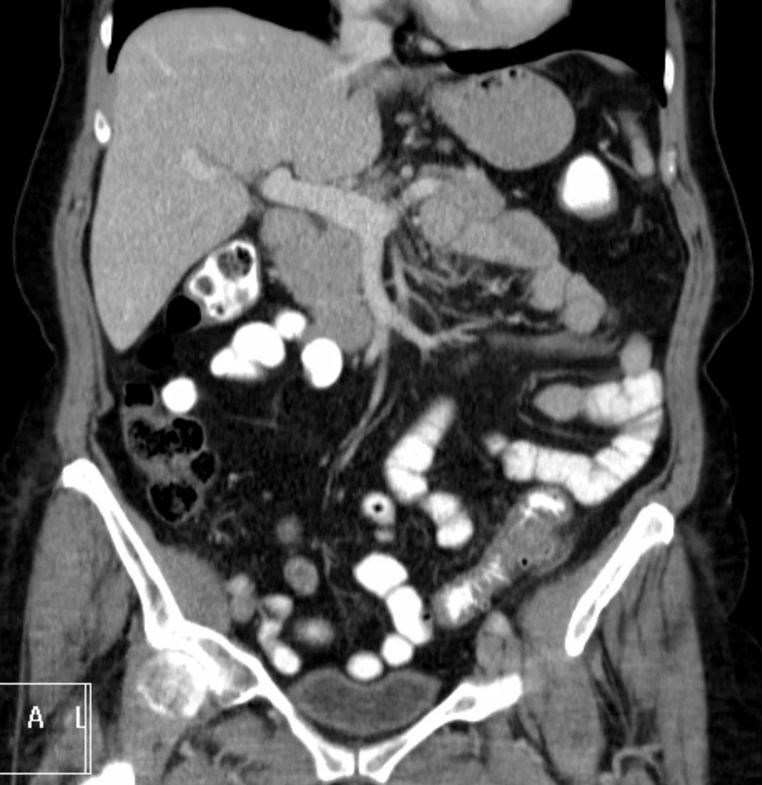
Long description
Coronal CT of the abdomen and pelvis with a thickened sigmoid colon wall, diverticula and surrounding fat stranding. The image comes from a patient with contained perforation and a small abscess, a complicated diverticulitis picture.

Clinical diagnosis alone is correct in only about 40 to 65 percent of patients, so cross-sectional imaging should be obtained in a first or severe episode. [1][2] The first-line test is a CT of the abdomen and pelvis with intravenous contrast (oral contrast according to local practice). It shows the diagnosis with a sensitivity of about 95 percent and a specificity of about 96 percent, better than ultrasound at about 90 percent sensitivity and 90 percent specificity. [2] CT also stages the disease by revealing wall thickening, pericolic fat stranding, fluid, free gas and abscess.
Acute and subacute inflammatory markers help but should not replace imaging. C-reactive protein is a useful marker of severity: values above 150 to 175 mg/L make complicated disease much more likely, and a level above 170 mg/L distinguishes severe from mild diverticulitis in several series. [2] White blood cell count and C-reactive protein are also used to decide whether antibiotics are needed (see below). Patients over 65 years with suspected diverticulitis warrant a particularly low threshold for CT, since older adults more often present atypically. [3]
Plain radiographs do not confirm the diagnosis but are useful for free air or obstruction. Bedside ultrasound is operator dependent and is best used in a step-up approach where CT is unavailable; magnetic resonance imaging is sensitive but less specific and not practical in the acute setting. [1][2] Barium or water-soluble contrast enema and colonoscopy should not be performed during the acute episode.
Patients should be resuscitated first. Once stable, CT angiography is performed when bleeding is brisk, because it can localise the bleeding vessel and guide embolisation. Colonoscopy should follow when the patient is stable. In practice CTA is used for massive ongoing bleeding and colonoscopy for patients who have stopped bleeding or have only modest haemorrhage.
Colonoscopy also has a delayed diagnostic role after an episode of diverticulitis (see follow-up below) to exclude colorectal cancer, which can be misdiagnosed as diverticulitis.
Diverticulitis becomes a diagnosis of uncomplicated or complicated disease on CT, and for many years the Hinchey classification, originally based on operative findings, has been used to describe complicated disease. [2]
- Hinchey I: pericolic abscess.
- Hinchey II: pelvic, intra-abdominal or retroperitoneal abscess.
- Hinchey III: generalized purulent peritonitis.
- Hinchey IV: generalized faecal peritonitis.
Because management is now CT-driven, modified systems are used. The modified (Kaiser) Hinchey classification reads: [2]
| Stage | CT findings |
|---|---|
| 0 | Mild clinical diverticulitis |
| 1a | Confined pericolic inflammation |
| 1b | Confined pericolic abscess |
| 2 | Pelvic or distant intra-abdominal abscess |
| 3 | Generalized purulent peritonitis |
| 4 | Faecal peritonitis |
Stages 0 and 1a represent uncomplicated disease; stages 1b to 4 are complicated. The 2020 World Society of Emergency Surgery guideline uses an even simpler CT-only grouping (uncomplicated; air bubbles or small pericolic fluid; abscess 4 cm or less; abscess larger than 4 cm or distant gas; diffuse fluid with or without free gas), but whatever system is used the management decision rests on whether the disease is uncomplicated and whether an abscess is present and drainable. [2][4]
Initial management is conservative: clear or low-residue diet as tolerated, intravenous or oral hydration, analgesia (paracetamol and antispasmodics are preferred; avoid systemic opioids where possible), and close follow-up. [1][3]
Four well-designed European and Oceania randomised controlled trials in immunocompetent patients with CT-proven uncomplicated diverticulitis found that antibiotics impart no benefit on time to symptom resolution, complications, recurrence or mortality. [3] Consistent with this, the 2020 World Society of Emergency Surgery guideline recommends against antibiotics in uncomplicated diverticulitis without systemic inflammation, and the American Gastroenterological Association advises that antibiotics can be used selectively rather than routinely. [2][1]
Antibiotics are advised when any of the following is present: [1]
- Comorbidity, frailty or immunosuppression.
- Refractory symptoms or vomiting.
- C-reactive protein above 140 mg/L or white blood cell count above 15 times 10 to the 9 per litre.
- CT evidence of a fluid (abscess) collection or a longer segment of inflammation (about 86 mm compared with about 65 mm in patients who did not progress to complicated disease).
When antibiotics are needed in the outpatient setting, the usual choices are oral amoxicillin-clavulanate alone or a fluoroquinolone plus metronidazole. [1][3] Treatment usually lasts 4 to 7 days; one study found similar outcomes with a 4-day course. In older patients, fluoroquinolones carry a higher risk of tendon injury and of Clostridioides difficile infection, so amoxicillin-clavulanate should be preferred where appropriate. [1][3]
Immunocompetent patients with mild uncomplicated disease, adequate support at home and no red flags can be managed as outpatients with close follow up. [1][3] Patients who are immunosuppressed, on chronic corticosteroids, frail or showing systemic inflammatory signs should have CT imaging, antibiotic therapy and early surgical consultation; immunosuppressed patients need broader gram-negative and anaerobic cover, usually for 10 to 14 days. [1][2]
Admit all such patients. The main components are:
- Intravenous broad-spectrum antibiotics covering gram-negative aerobes and anaerobes, intravenous fluids, and bowel rest with a nasogastric tube if obstructed. [1][2]
- Percutaneous drainage for abscess, combined with antibiotics. Many guidelines advise drainage for abscesses of 3 cm or more; the 2020 World Society of Emergency Surgery guideline sets a higher threshold of 4 to 5 cm for a "large" abscess. The evidence for the exact cut-off is of low quality, and practice varies, so the decision should be led by imaging, anatomy and the patient's condition. [4][2]
- Surgery for failure of conservative management or for free perforation with generalised peritonitis. Options include primary resection of the diseased segment, with either a primary anastomosis (with or without a diverting loop ileostomy) or a Hartmann procedure (resection with an end colostomy) depending on contamination, physiology and surgical judgement. Laparoscopic peritoneal lavage is used in selected patients with purulent peritonitis (Hinchey III) without faecal contamination and without septic shock, though it is not universally adopted. [3][1]
- Elective resection after successful non-operative treatment of an abscess is no longer automatic and is individualised (see below).
Fistulas (colovesical, colovaginal, colocutaneous) and strictures leading to obstruction are dealt with on their merits, usually with resection of the diseased segment and closure or repair of the fistula.
A more complete decision pathway is shown below.
Management of CT-confirmed acute colonic diverticulitis
- CT-confirmed diverticulitisClassify as uncomplicated or complicated
- Question: Complicated disease? (abscess, perforation, obstruction, stricture or fistula)
No (uncomplicated)
- Outpatient care if well and supportedLiquid or low-residue diet, analgesia, hydration; antibiotics only when red flags are present: comorbidity or frailty, vomiting, CRP above 140 mg/L or WBC above about 15 x 10^9 per L (cut-offs vary between guidelines), or fluid collection or long inflammation
- Admit if immunosuppressed, frail or systemically unwellBroader, longer (10 to 14 day) antibiotic cover; early surgical review
Yes (complicated)
- AdmitIV broad-spectrum antibiotics covering gram-negative aerobes and anaerobes; fluids; NPO if obstructed
- Question: Abscess present?
Less than 3 cm
- Antibiotics aloneReassess clinically with interval imaging if no improvement
3 cm or more
- Add image-guided percutaneous drainage where anatomically feasibleThresholds vary between 3 cm and 4 to 5 cm among guidelines
- Question: Free perforation with generalised peritonitis?
Yes
- Emergency surgeryResection, with Hartmann procedure or primary anastomosis with or without diverting ileostomy according to contamination and physiology; lavage for selected Hinchey III
No
- Conservative or drainage strategyContinue antibiotics; escalate on failure
- Follow-upColonoscopy at 6 to 8 weeks after complicated disease, or a first episode, to exclude cancer
Cancer can be misdiagnosed as diverticulitis. In a meta-analysis of 31 studies and 50,445 patients, the pooled prevalence of colorectal cancer after diverticulitis was 1.9 percent, higher after complicated disease (7.9 percent) than after uncomplicated disease (1.3 percent), with advanced adenomas in 4.4 percent and adenomas in 14.2 percent. [1] Consistent with this, colonoscopy should be delayed 6 to 8 weeks after an episode, or until symptoms have fully resolved. [1] It is advised after an episode of complicated diverticulitis and after a first episode of uncomplicated diverticulitis, and may be deferred if a high-quality colonoscopy was done within the previous year. Patients with recurrent uncomplicated disease follow routine colorectal cancer screening intervals.
About 8 percent of patients have a recurrence within the first year and about 20 percent within 10 years; after a second episode the figures are 18 percent within 1 year and 55 percent within 10 years, and after a third episode 40 percent by 3 years. [1] Recurrence is about 25 percent within 5 years after an abscess managed conservatively. [1]
Importantly, complicated disease is usually the first presentation rather than a consequence of repeated episodes: 74 percent of patients with complicated diverticulitis had had no earlier episode, and recurrent diverticulitis reduces the odds of complicated diverticulitis (odds ratio about 0.78). [1] Therefore elective colectomy should not be advised on the number of episodes alone. [1] The decision should be individualised, weighing severity, quality of life, comorbidity (notably immune status), patient preference and surgical risk. Elective segmental resection reduces but does not eliminate recurrence: about 15 percent versus 61 percent at 5 years. Chronic gastrointestinal symptoms after recovery do not always improve with surgery in 22 to 25 percent of patients, and 5-year quality of life is better with elective resection when symptoms are marked. [1] Patients with a history of complicated disease managed without surgery remain at higher risk of recurrence and complicated recurrence and should be reviewed by a colorectal surgeon. Chronically immunosuppressed patients should consult a colorectal surgeon after recovery. [1]
Fewer than 10 percent of patients have diverticulitis that fails to settle with treatment or relapses quickly; this requires prolonged antibiotics and, if unsuccessful, segmental resection. Differentiate it from visceral hypersensitivity, where the patient has ongoing lower abdominal pain but normal imaging, laboratory tests and endoscopy and does not respond to antibiotics. [3]
- Abscess (pericolic, pelvic, retroperitoneal).
- Perforation with purulent or faecal peritonitis.
- Fistula, most commonly colovesical (usually in men, more common after previous pelvic surgery is absent) and colovaginal (typically after hysterectomy); also colocutaneous.
- Stricture and obstruction from repeated inflammation and scarring.
Most episodes of uncomplicated diverticulitis settle, although recurrent attacks are common. Smaller subgroups have smouldering disease, chronic pain from visceral hypersensitivity or complicated recurrence. Mortality is low in uncomplicated disease but rises with age, comorbidity, sepsis and emergency surgery, particularly in older adults. [3]
Prevention targets the risk factors rather than the pouches:
- A high-quality, high-fibre diet low in red meat, with adequate fluid and physical activity.
- Normal body weight.
- No smoking; avoidance of heavy alcohol use.
- Avoidance of regular non-steroidal anti-inflammatory drug use where possible (aspirin for secondary cardiovascular prevention is an exception).
- Reasonable caution with opiates and corticosteroids.
The reassuring facts to share are that the overall contribution of diet and lifestyle is substantial (adherence to a low-risk pattern was associated with about 75 percent lower risk of incident diverticulitis in a prospective cohort of men), while about 40 to 50 percent of risk is genetic, and most complications happen on the first episode rather than later ones. [1][3]
Measures that are not recommended for preventing recurrence, because the evidence is weak or negative, include 5-aminosalicylic acid (mesalazine), probiotics and cyclic rifaximin. [1] Nuts, corn, popcorn and small-seeded berries need not be avoided. [1]
Colorectal cancer screening should follow normal population pathways; colonoscopy timing after an episode is given above.
Presentation is often atypical (less fever and leukocytosis, more fatigue, rectal bleeding, abdominal pain alone); the threshold for CT should be low in patients over 65 years, because a competing diagnosis is common. [3] Antibiotic choice should balance effectiveness against the higher risk of Clostridioides difficile infection and tendon injury; frailty, comorbidity and social support determine whether a patient is treated as an outpatient. Emergency or elective surgery carries substantially higher risk of death with advancing age, so preoperative optimisation is important. [3]
These patients have a higher risk of severe and complicated disease, a low threshold is needed for CT imaging and antibiotics, and the regimen should be broader (gram-negative and anaerobic cover) and longer (10 to 14 days); consultation with a colorectal surgeon is advised after recovery. [1][2]
Diverticulitis in pregnancy is uncommon. Imaging should use ultrasound and magnetic resonance imaging first; complicated disease needs obstetric and surgical input. Management of the individual patient should follow local guidance.
- Base the diagnosis on the classic clinical picture plus whatever imaging is available; ultrasound in experienced hands is a reasonable first test, escalating to CT where inconclusive, given its high sensitivity and specificity. [2]
- C-reactive protein and full blood count, where available, help with severity; plain radiographs remain useful for free air and obstruction.
- Use oral amoxicillin-clavulanate or a fluoroquinolone plus metronidazole for outpatient treatment of uncomplicated disease; reserve IV broad-spectrum therapy for inpatients.
- Percutaneous drainage may not be available; larger or non-drainable abscesses and free perforation then require surgery.
- Emphasise dietary fibre from cheap local sources, weight, activity, and avoidance of unnecessary non-steroidal anti-inflammatory drugs.
- Arrange delayed colonoscopy where services allow; otherwise prioritise colorectal cancer pathways for patients with alarm symptoms.
- Diverticulosis is common and rises steeply with age; only 2 to 4 percent of patients develop diverticulitis.
- Colonic diverticula are usually false, pulsion pouches through weak points in the wall where vessels enter.
- Sigmoid disease dominates in Western countries; right-sided disease is common in Asia.
- Uncomplicated diverticulitis in a well immunocompetent patient often needs no antibiotics; complicated disease needs admission, drainage and sometimes surgery.
- CT with intravenous contrast is the diagnostic and staging test of choice; clinical diagnosis alone is wrong in 40 to 65 percent.
- CRP above 150 to 175 mg/L predicts complicated disease; WBC above about 15 x 10^9 per L (some guidance uses a slightly lower threshold) or CRP above 140 mg/L are indications for antibiotics.
- Abscesses under 3 cm are treated with antibiotics; 3 cm or more usually needs percutaneous drainage, though some guidelines use 4 to 5 cm.
- Follow-up colonoscopy at 6 to 8 weeks is needed after complicated disease or a first episode; cancer is found in about 1.3 to 7.9 percent depending on severity.
- About 20 percent of patients relapse within 10 years; elective surgery cuts but does not eliminate this, and chronic pain may persist.
- No drugs (mesalazine, probiotics, rifaximin) prevent recurrence; diet, weight, activity, no smoking and NSAID avoidance do help.
- Older patients present atypically and fare worse with surgery; immunocompromised patients should be managed aggressively.
- Nuts and seeds need not be avoided.
Answer each one in your head first, then open the answer.
A 62-year-old man has 2 days of left-lower-quadrant pain, fever and a white cell count of 14 x 10^9 per litre. CT shows sigmoid wall thickening with pericolic fat stranding but no abscess. What is the most likely diagnosis and what is the appropriate immediate management?
Show answerHide answer
CT-confirmed uncomplicated sigmoid diverticulitis. In a well, immunocompetent patient with no vomiting or red flags he can be managed at home: clear or low-residue diet, analgesia, hydration and close follow-up. Antibiotics are not routinely needed. Arrange colonoscopy in 6 to 8 weeks if it is his first episode.
A 78-year-old woman is admitted with diverticulitis. Her C-reactive protein is 180 mg/L and CT shows a 5 cm pelvic abscess adjacent to the sigmoid. How should the disease be classified and what should be done?
Show answerHide answer
Complicated diverticulitis (Hinchey II; modified Hinchey stage 2). She needs admission, intravenous broad-spectrum antibiotics and image-guided percutaneous drainage, because the abscess is above the usual 3 cm threshold and well localised. The team should also address her comorbidity and frailty and consider a colorectal surgical review.
A patient asks which foods to avoid to stop diverticulitis recurring. What is the evidence-based answer?
Show answerHide answer
No specific foods need to be avoided, including nuts, corn, popcorn and berries with small seeds; the evidence does not link them to diverticulitis. Advice should focus on a high-quality diet with adequate fibre, regular physical activity, a normal body weight, no smoking and avoiding regular non-steroidal anti-inflammatory drug use. Mesalazine, probiotics and rifaximin are not recommended for prevention.
A 55-year-old woman had an episode of Hinchey II diverticulitis treated with antibiotics and drainage 2 months ago and now feels well. She asks whether she should have an operation to prevent another episode. How do you answer?
Show answerHide answer
Explain that an elective segmental resection should not be based on the number of episodes; the decision is individualised. Surgery reduces the chance of recurrence from about 61 to 15 percent at 5 years, but it does not eliminate it and does not always relieve chronic pain. As she is fewer than 6 to 8 weeks recovered she should first have colonoscopy to exclude cancer; if complication-free she can be managed conservatively, with colorectal surgery involved for high-risk features such as immunosuppression.
What does Hinchey IV diverticulitis mean, and what operation may be required?
Show answerHide answer
Hinchey IV is generalised faecal peritonitis from free perforation. It requires emergency surgery with source control and resection of the diseased segment, usually as a Hartmann procedure (end colostomy) or, in suitable patients, as a primary anastomosis with or without a diverting ileostomy, depending on contamination and physiological reserve.
Related MedGrasp topics, in plain text: lower gastrointestinal bleeding; colorectal cancer; inflammatory bowel disease; ischaemic colitis; constipation; irritable bowel syndrome.
- Peery AF, Shaukat A, Strate LL. AGA Clinical Practice Update on Medical Management of Colonic Diverticulitis: Expert Review. Gastroenterology 2021;160(3):906–911.e1. doi:10.1053/j.gastro.2020.09.059. Accessed .
- Sartelli M et al. 2020 update of the WSES guidelines for the management of acute colonic diverticulitis in the emergency setting. World Journal of Emergency Surgery 2020;15:32. doi:10.1186/s13017-020-00313-4. Accessed .
- Phan HS, Strate LL. Management of Colonic Diverticular Disease in the Older Adult. Current Gastroenterology Reports 2025;27(1):42. doi:10.1007/s11894-025-00986-4. Accessed .
- Cirocchi R et al. Guidelines for the Treatment of Abdominal Abscesses in Acute Diverticulitis: An Umbrella Review. Journal of Clinical Medicine 2023;12(17):5522. doi:10.3390/jcm12175522. Accessed .
Figure credits
Educational content for medical students and doctors. It is not a substitute for clinical judgement or local guidelines.