Large bowel obstruction and volvulus
How large bowel obstruction and volvulus present, are diagnosed on imaging, and are managed from resuscitation to stenting, diversion and resection.
- Large bowel obstruction is a blockage of the colonic lumen. Colorectal cancer is the most common cause, followed by diverticular stricture and volvulus; other causes include faecal impaction, Crohn disease and ischaemic strictures, extrinsic compression, benign tumours and Hirschsprung disease in children.
- The obstruction often evolves over days. Constipation and obstipation, abdominal distension and tympany, and less intense cramping pain are typical; vomiting is a late and often faeculent event.
- If the ileocaecal valve is competent, the obstruction becomes a closed loop with the caecum under the greatest wall tension; a caecal diameter above 12 cm is the level at which perforation becomes likely.
- Computed tomography with intravenous contrast is the key test: it confirms obstruction, locates the transition point, identifies the cause and assesses for ischaemia and perforation. Plain abdominal radiography and a water-soluble contrast enema remain useful when CT is not available.
- Management is resuscitation (nil by mouth, decompression, intravenous fluids, electrolyte correction, antibiotics), then treatment of the cause: endoscopic detorsion for sigmoid volvulus, urgent surgery for caecal volvulus, and for malignant left-sided obstruction either a self-expanding metal stent as a bridge to surgery or emergency resection by Hartmann procedure or primary anastomosis.
By the end of this page you should be able to:
- Explain why large bowel obstruction looks different from small bowel obstruction, from the level of the blockage and the competent ileocaecal valve down to the signs.
- List the causes of large bowel obstruction and place volvulus in its epidemiological context.
- Interpret plain abdominal radiographs and CT in large bowel obstruction, including the close-loop and caecal diameter thresholds.
- Distinguish sigmoid volvulus from caecal volvulus and choose the correct first treatment for each.
- Plan the management of malignant left-sided obstruction using the WSES 2017 and ESGE 2020 guidance on self-expanding metal stents.
- Compare small and large bowel obstruction feature by feature in the table provided.
DefinitionLarge bowel obstruction
Large bowel obstruction is a state in which the passage of intestinal contents through the colon and rectum is blocked, completely or partially, either by a physical lesion within, in or around the wall of the bowel, or by a failure of colonic propulsion. It is an abdominal emergency because the colon proximal to the blockage distends, and the wall may ischaemate and perforate. [4]
Three mechanistic groups are useful at the bedside.
- Mechanical obstruction from a structural cause: this is the group this page covers in depth. Tumour, stricture, volvulus, hernia, diverticular disease, faecal impaction and Hirschsprung disease all belong here.
- Functional pseudo-obstruction (acute colonic pseudo-obstruction, Ogilvie syndrome) in which the colon dilates without a physical blockage. It mimics large bowel obstruction clinically and is treated differently (neostigmine, supportive care, decompression), so it must be excluded with imaging. [4][6]
- Closed-loop obstruction: a segment is blocked both proximally and distally, so it cannot decompress in either direction. Volvulus is the classic mechanism, and a malignant obstruction with a competent ileocaecal valve produces the same physiology in the proximal colon.
Cecal (caecal), sigmoid and transverse volvulus are considered separately within large bowel obstruction because the anatomy dictates both the imaging and the treatment.
Large bowel obstruction accounts for roughly one quarter of all mechanical bowel obstructions in many hospital series, the rest being small bowel obstruction. [1] It occurs at any age but the cause shifts with age and geography.
- Colorectal cancer is the commonest cause in older adults in industrialised countries and is the commonest cause overall in most series. Large bowel obstruction represents about 80% of the emergencies caused by colorectal cancer, and perforation the remaining 20%. [1]
- Colonic volvulus is the third most common cause of large bowel obstruction worldwide, behind malignancy and diverticular disease. It represents 10 to 15% of all large bowel obstructions in the United States. [3]
- In the "volvulus belt" of Africa, the Middle East, South America and southern Asia, colonic volvulus historically accounted for 45 to 80% of large bowel obstructions, although more recent studies from the Middle East report figures as low as 10%, probably reflecting westernisation of diet and population migration. [4]
- Sigmoid volvulus is the commonest site of colonic volvulus (60 to 75% of colonic volvulus) and is a disease of older adults. Sigmoid volvulus accounts for 65 to 80% of all colonic volvulus, with cecal volvulus accounting for most of the rest. [3][4]
- Cecal volvulus is rarer (less than 5% of large bowel obstructions) and occurs in younger patients, more often women, with a mean age in the mid-50s, though a second peak occurs in older adults with dementia or chronic constipation. [3][4]
About 65% of malignant large bowel obstructions are left-sided (splenic flexure to rectum) and 35% right-sided. [2]
The causes are best grouped by where the blockage sits relative to the wall.
- Colorectal adenocarcinoma is the most common cause of large bowel obstruction in adults. The sigmoid colon is the most common location of the obstructing tumour, with 75% of obstructing cancers lying distal to the splenic flexure. [1]
- Extrinsic malignant compression from carcinomatosis, gynaecological tumours, retroperitoneal or pelvic lymphadenopathy and bulky distant primaries.
- Diverticular stricture after repeated attacks of diverticulitis, particularly in the sigmoid colon.
- Inflammatory strictures from Crohn disease and ulcerative colitis.
- Ischaemic and radiation strictures and post-anastomotic stricture.
- Benign tumours: adenomatous polyps, lipomas, carcinoid, and others large enough to occlude the lumen.
- Faecal impaction, common in older, frail, constipated and opioid-treated patients.
- Hirschsprung disease in children, in whom sigmoid volvulus can be the presenting event. [4]
- Foreign bodies, drug packets, endometriosis and intussusception (more common in children).
Volvulus is the twisting of a redundant segment of colon around its mesentery, producing a closed-loop obstruction. [4] The sigmoid colon is the commonest site. Risk factors include chronic constipation, colonic dysmotility, a redundant colon with poor peritoneal fixation, and a thinned mesentery. [4] Institutionalised, bedbound and neuropsychiatric patients are at highest risk. [3] Cecal volvulus requires an abnormally mobile caecum and ascending colon, from congenital or acquired failure of fixation, plus a trigger such as pregnancy, prior surgery, adhesions, constipation or distal obstruction. [3] Cecal bascule, a folding rather than a torsion of the caecum on itself, requires a competent ileocaecal valve. [3]
- Adhesions and hernia, which are much less common in the colon than in the small bowel but do occur (incarcerated left inguinal hernia containing sigmoid colon, for example).
- Acute colonic pseudo-obstruction should always be considered in the differential, particularly after surgery, trauma, sepsis or in critically ill patients, since it can look identical initially. [6]
The colon proximal to a blockage fills with gas and fluid. Unlike the small bowel, which is relatively narrow and has frequent propulsive contractions, the colon is a reservoir: it absorbs water and, with the ileocaecal valve closed, cannot pass its contents either forward or backward. The result is progressive proximal distension.
When the ileocaecal valve is competent, a distal colonic obstruction produces a closed-loop obstruction of the colon proximal to it: gas and fluid accumulate, there is no decompression into the small bowel, and the pressure rises. [4] By the Law of Laplace, the wall tension needed to distend a hollow tube is inversely proportional to its radius, and the caecum, having the largest diameter, requires the least pressure to distend. The caecum is therefore the first segment to ischaemate and perforate. [4]
The exact caecal diameter at which perforation becomes likely ranges from 9 to 12 cm in the literature; a caecal diameter above 12 cm in the setting of colonic obstruction or distension is the widely used threshold for considering the risk of perforation unacceptable, particularly in closed-loop and pseudo-obstructive distension. [4] Duration of distension matters at least as much as absolute diameter, and rapid distension is more dangerous than chronic dilatation of the same size. [4]
Where the ileocaecal valve is incompetent, the obstruction decompresses back into the small bowel; clinically this can look like a distal small bowel obstruction.
Volvulus is the archetype. Twisting of the mesentery obliterates both the afferent and efferent limbs, traps a closed, gas-filled segment, and compromises venous drainage first, then arterial supply. Mucosal ischaemia occurs first (the mucosa being the most metabolically active layer), followed by bacterial translocation, then full-thickness necrosis and perforation if not relieved. [4]
Symptoms build over hours to days rather than minutes, and they differ from small bowel obstruction in predictable ways.
- Constipation and obstipation are the hallmark. The patient stops passing stool and, importantly, stops passing flatus. Absence of passage of flatus (90% of cases) and/or faeces (80.6%) and abdominal distension (65.3%) are the most common symptoms and physical signs in large bowel obstruction. [1]
- Abdominal pain is usually a dull, colicky cramp rather than the severe intermittent colicky pain of small bowel obstruction, and it is less intense. [1] In volvulus it can be more acute, often with intermittent self-resolving cramps in early torsion.
- Vomiting occurs late and is often faeculent, because the blockage is far downstream. Right-sided obstruction can present with earlier and more prominent vomiting. [2]
- Distension is prominent, especially in distal obstruction and closed-loop physiology.
- A change in bowel habit, iron-deficiency anaemia, rectal bleeding or melaena, and unintentional weight loss suggest an underlying colorectal cancer. [1]
- Fever, severe or constant pain, or a rigid, silent abdomen suggest perforation, peritonitis or ischaemia and must be treated as emergencies.
Only 17% of patients with sigmoid volvulus present within 48 hours of symptom onset; most have an insidious course over several days. Cecal volvulus can mimic small bowel obstruction because small bowel distends proximal to it. [4]
- Inspection: visible abdominal distension, sometimes with visible peristalsis or reversed peristalsis in late obstruction.
- Palpation: tympanitic, resonant abdomen; tenderness, guarding and rebound suggest peritonitis or impending perforation.
- Auscultation: bowel sounds may be high-pitched and tinkling, or absent in ileus and perforation.
- Rectal examination classically reveals an empty rectum (empty vault) in volvulus, because the twist sits proximal to the rectum. [3]
- Signs of systemic upset: tachycardia, fever, dehydration, hypotension and oliguria indicate sepsis or hypovolaemia.
- General: cachexia, anaemia and hepatomegaly may point to an underlying cancer.
Red flagWhen large bowel obstruction has already become dangerous
Fever, tachycardia, hypotension, confusion, a rigid abdomen, peritonism or absent bowel sounds, leukocytosis with a left shift, rising lactate or metabolic acidosis, free air on imaging or pneumoperitoneum. These indicate ischaemia, necrosis or perforation and mandate urgent resuscitation and surgery rather than endoscopic detorsion or stenting. [1][3]
Two questions arise: what else could cause the presentation, and how do I tell large bowel obstruction from small bowel obstruction. The first is covered in the other gastroenterology topics for the presenting symptom (constipation, abdominal pain, distension). The second is the practical bedside question, and it is set out in the table.
| Feature | Small bowel obstruction | Large bowel obstruction |
|---|---|---|
| Onset and tempo | Usually acute: sudden colicky pain, rapid evolution over hours. | Days of worsening distension; vomiting late and often faeculent; pain less intense. [1][4] |
| History | Previous abdominal surgery and adhesions are the commonest cause in industrialised countries; hernia, tumour, Crohn disease. Obstruction often partial then complete. | Colorectal cancer, diverticular stricture, volvulus, faecal impaction. Change in bowel habit, rectal bleeding and weight loss point to cancer. [1][3] |
| Pain | Severe, intermittent, colicky, classically around the umbilicus; patient is restless. | Dull cramping, often in the lower abdomen or flank; less intense; obstipation prominent. [1] |
| Vomiting | Early and frequent, often bilious, then faeculent. | Late, often absent early; becomes faeculent if it occurs; earlier with right-sided obstruction. [2] |
| Distension | Variable, may be central; visible peristalsis. | Marked, tympanitic, often visible; may be asymmetric with volvulus. |
| Examination | Tenderness variable; enteric (high-pitched) sounds or succussion splash; check hernial orifices. | Tympany, empty rectal vault in volvulus; bowel sounds variable. Rectal tumour may be palpable. [1][3] |
| Causes | Adhesions (most common), hernia, tumour, volvulus, gallstone ileus, intussusception. | Colorectal cancer (most common), diverticular stricture, sigmoid and caecal volvulus, faecal impaction, Crohn and ischaemic strictures, extrinsic compression, benign tumours, Hirschsprung disease in children. |
| Differential within the abdomen | Large bowel obstruction, mesenteric ischaemia, ileus, gastroenteritis, pancreatitis. | Acute colonic pseudo-obstruction, ileus, diverticulitis with stricture, constipation, mesenteric ischaemia. |
| Diagnosis: first imaging | CT (gold standard) shows transition point; plain X-ray may show central small-bowel loops with valvulae conniventes. | Plain X-ray: peripheral colonic dilation with haustral folds and absent rectal gas; CT confirms level, cause and complications. [1] |
| Contrast study | Water-soluble contrast (for example Gastrografin) used for partial obstruction and as a therapeutic adjunct. | Water-soluble contrast enema when CT is unavailable or diagnosis in doubt; bird-beak sign in volvulus. [1][3] |
| Treatment (first steps) | Nil by mouth, nasogastric decompression, intravenous fluids and electrolytes, surgery for strangulation, complete obstruction or non-resolution. | Nil by mouth, decompression (nasogastric and/or rectal tube), intravenous fluids, electrolytes and antibiotics; treat the cause: detorsion for sigmoid volvulus, surgery for caecal volvulus, stenting or resection for malignant obstruction. [1][3] |
The aim is to answer three questions: is there an obstruction, where is it, and is there ischaemia or perforation. Computed tomography answers all three; plain radiography and water-soluble contrast enema are the alternatives when CT is not available.
- Full blood count for anaemia and leukocytosis; leukocytosis with a left shift suggests ischaemia or perforation. [1]
- Urea and electrolytes, creatinine: vomiting, diarrhoea and third spacing cause dehydration, metabolic alkalosis and hypokalaemia, with raised urea nitrogen. [1]
- Lactate and arterial blood gas for tissue hypoperfusion, ischaemia and sepsis.
- Liver function tests, coagulation screen and group-and-save, particularly if surgery is likely.
- Tumour markers and a careful systemic cancer work-up belong in the definitive pathway for malignant obstruction but are not emergent.
A three-view series (supine, erect and chest) in the erect position can show pneumoperitoneum if perforation is suspected. In large bowel obstruction the colon is dilated peripherally with haustral folds, and rectal gas is absent; air-fluid levels may be present, and more acutely when they are. [1][3] In the volvulus belt plain films remain the common first test. Sensitivity and specificity for large bowel obstruction are lower than for CT: roughly 74 to 84% sensitivity and 50 to 72% specificity for plain films compared with 93 to 96% sensitivity and 93 to 100% specificity for CT. [1]
CT is the investigation of choice. [1][2] It achieves diagnostic confirmation with a sensitivity of 93 to 96% and a specificity of 93 to 100%, identifies the transition point and the level of obstruction with 95% sensitivity, and yields the cause of the obstruction with a specificity of 66 to 87%. [1] With intravenous contrast it also shows the bowel wall, mesentery, and the danger signs of strangulation: absent mural enhancement, pneumatosis intestinalis, portal venous gas, free fluid and free air. [4]
Every patient with suspected malignant large bowel obstruction and every stable patient in whom the cause of a mechanical large bowel obstruction remains uncertain should have a contrast-enhanced CT. [5] National guideline sets emphasise that CT should never expose the patient to an unsafe delay of treatment. [1]
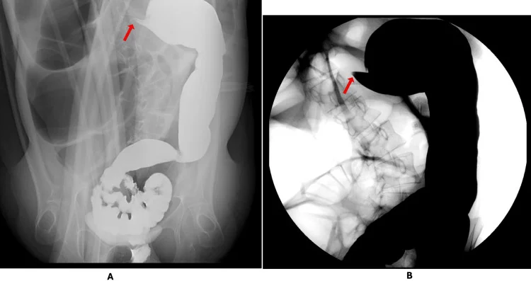
A soluble-contrast (for example Gastrografin) enema has a sensitivity of 96% and a specificity of 98% for confirming large bowel obstruction. [1] It is well suited to distinguishing mechanical obstruction from pseudo-obstruction and to identifying a volvulus. It also shows a "bird-beak" tapering at the point of torsion. [3] It is a valid alternative to CT when CT is not available, and particularly in low-resource settings. It should not be used when perforation is suspected. [1]
Figure 1Water-soluble contrast enema in large bowel obstruction
Long description
Two views of a Gastrografin (water-soluble) contrast enema. In view A and view B red arrows point to the level of obstruction, where the contrast column tapers to a point like a bird's beak. A contrast enema separates a mechanical obstruction, with a fixed narrowing, from a pseudo-obstruction, where contrast passes freely to dilated colon.
In stable patients, colonoscopy allows direct visualisation of the obstructing lesion, biopsy and, when appropriate, stent placement. Its availability is limited in the emergency setting and the quality of the colonic preparation is poor, so it is usually reserved for planned endoscopic treatment. [1]
- Leukocyte count: a raised white cell count supports inflammation, ischaemia or perforation.
- Lactate: any elevation in an obstructed, distended abdomen is worrying.
- Caecal diameter on plain film or CT: a diameter above 9 cm is suspicious in acute colonic distension, and 12 cm is the commonly used level at which perforation risk becomes unacceptable. [4] (See the diagnostic-criteria section.)
Large bowel obstruction remains a clinical diagnosis supported by imaging; there is no single laboratory threshold. Severity is stratified by:
- Haemodynamic status: stable versus hypotensive or septic; unstable patients need damage-control surgery and vigorous resuscitation.
- Presence of ischaemia or perforation: fever, peritonism, free air, pneumatosis, portal venous gas, rising lactate, metabolic acidosis and leukocytosis.
- Caecal diameter in closed-loop and obstructive distension: above 12 cm mandates a much lower threshold to operate; serial imaging every 12 to 24 hours is appropriate in evolving distension. [4]
- Site and cause: left-sided malignant obstruction is where the endoscopic and stenting literature is strongest; right-sided malignant obstruction is treated surgically.
- Fitness (age, ASA grade, co-morbidity such as diabetes and liver disease, immunosuppression) determines whether resection with primary anastomosis or a staged Hartmann procedure is safer.
Volvulus has its own diagnostic clues on imaging. A "coffee-bean" sign on plain abdominal radiography is seen in 60 to 80% of sigmoid volvulus, while X-ray is diagnostic in only 25 to 40% of caecal volvulus. CT has 100% sensitivity and greater than 90% specificity for sigmoid volvulus. A "bird-beak" appearance on contrast study and a mesenteric "whirl" on CT are characteristic. [3]
- Resuscitation first. Assess airway, breathing and circulation. Give intravenous isotonic crystalloid, replace electrolytes (in particular potassium and magnesium), and place a urinary catheter to monitor urine output if the patient is unstable. [1]
- Nil by mouth. No oral intake.
- Decompression. Place a nasogastric tube (and, for colonic decompression, a flatus/rectal tube) to relieve distension, control vomiting and reduce aspiration risk. [3]
- Analgesia and antiemetics as required.
- Antibiotics. Give broad-spectrum intravenous antibiotics covering Gram-negative enteric organisms and anaerobes whenever there is suspected strangulation, perforation, ischaemia, peritonitis, or when the patient is systemically unwell; treatment of complicated intra-abdominal infection is time-critical. The choice and dose should follow local microbiology guidance. [1]
- Thromboprophylaxis and early senior surgical review. Every patient with large bowel obstruction needs surgical assessment; most need admission.
Suspected large bowel obstruction in an adult
- Suspected large bowel obstructionHistory, examination, bloods (CBC, U and E, lactate), plain films; nil by mouth and resuscitate
- Question: Peritonism, fever, free air, rising lactate or sepsis?
Yes
- Urgent laparotomy or damage-control surgeryResuscitate, broad-spectrum antibiotics, resect non-viable bowel; do not detorse ischaemic volvulus before resection
No
- Confirm and characterise: CT with IV contrast (or water-soluble contrast enema if CT unavailable)Locate transition point; identify cause; measure caecal diameter
- Question: Cause and location?
Sigmoid volvulus, stable, viable mucosa
- Rigid or flexible sigmoidoscopy: detorsion and decompressionRectal tube in place; arrange sigmoid colectomy during the same admission
Caecal volvulus
- Emergency surgery after resuscitationRight hemicolectomy/ileocolic resection with primary anastomosis if viable, or resection with diversion if unstable or ischaemic
Malignant left-sided obstruction
- Fit for surgery: emergency resection with primary anastomosis (younger, ASA 1-2) or Hartmann if high riskOr decompressing stoma as a bridge; senior colorectal surgeon
- Unfit or palliative, or as bridge to elective surgery: colonic self-expanding metal stent after shared decisionOnly if no perforation; interval to surgery not more than 2 weeks
Benign stricture or faecal impaction
- Treat cause: decompression, manual or endoscopic disimpaction, stricture dilation, surgery for failureAvoid stenting in benign obstruction unless specific expertise
- Reassess: no resolution or clinical deterioration -> escalate surgical plan
Sigmoid volvulus classically presents in older, constipated, institutionalised patients with neuropsychiatric disease. [3][4] In a stable patient without peritonitis or signs of ischaemia, the first-line management is endoscopic detorsion with rigid or flexible sigmoidoscopy, with the bowel assessed for viability (the twisted mucosa can show a "pinwheel" appearance). [3] Endoscopic detorsion is successful in 60 to 95% of cases. [3]
If detorsion succeeds, a rectal tube is left in place for ongoing decompression, usually for at least 48 hours, and the patient is resuscitated and prepared for definitive surgery. Because recurrence after decompression alone is high (reported at 45 to 90% after successful detorsion, and up to 50 to 70% in other series), sigmoid colectomy should be offered during the same admission. [3][4] Recurrence without prior resection carries the greatest mortality risk. [4]
Non-resectional fixation procedures (sigmoidopexy, mesosigmoidopexy) are associated with unacceptable recurrence and should not be used outside specialist settings. [3][6]
Unlike sigmoid volvulus, caecal volvulus is a surgical disease. Endoscopic reduction is successful in only 5 to 30% of cases, can perforate the thin, distended wall, and delays definitive care, so it is not recommended. [3][4] After resuscitation all such patients should be assessed for emergency surgery.
- Non-viable, perforated or necrotic bowel: resection, with primary anastomosis if the patient is stable and well perfused, or end ileostomy with or without a mucous fistula or a staged discontinuity procedure in the unstable or high-risk patient. [3][4][6]
- Viable bowel: ileocolic (right hemi) colectomy with primary anastomosis is the preferred definitive option, with or without a covering loop ileostomy. Detorsion with caecopexy is a lower-risk alternative when the patient is unfit for resection, but recurrence is higher and caecostomy tube placement has a high complication and mortality rate and should be abandoned. [3][6]
- Published mortality after resection for caecal volvulus ranges from 0 to 32%, and postoperative complications occur in a substantial proportion. [6]
For left-sided malignant obstruction (splenic flexure to rectum), WSES 2017 recommends self-expanding metallic stents (SEMS) as a bridge to elective surgery where expertise exists, because short-term outcomes are better and the stoma rate is significantly lower than with emergency surgery; resection with primary anastomosis is preferred to Hartmann's procedure for uncomplicated obstruction in the absence of other risk factors and where the surgeon's and patient's characteristics permit. [1] Right colectomy is the treatment of choice for right-sided malignant obstruction. [1][2]
ESGE 2020 recommends: stenting is reserved for patients with clinical symptoms and radiological signs of malignant large bowel obstruction, without signs of perforation; prophylactic stent placement is not recommended; CT with contrast must precede stenting; self-expanding stents are discussed with the patient within shared decision-making as an alternative to emergency resection in potentially curable left-sided disease; colonic stenting is the preferred treatment for palliation of malignant obstruction; proximal colonic (right-sided) stenting is a weaker, low-evidence option for bridge to surgery or palliation. [5] A staged approach with stenting should aim for surgery within approximately 2 weeks. [2][5]
Performing the stent: stents are placed with the patient sedated, under fluoroscopic and endoscopic guidance. Uncovered stents have better patency and migration profiles than covered stents in most series; covered stents migrate more often. Complications (perforation, bleeding, migration, occlusion) occur in a minority, but the perforation risk is much higher in benign than in malignant disease (up to 22.4% in a large US series of benign obstruction), so stenting should not be used in benign obstruction without specific expertise. [2] A plain abdominal radiograph after stenting checks position and excludes perforation. [2]
Emergency resection (when stenting is not appropriate, not available, or has failed):
- Right-sided malignant obstruction: right colectomy with primary anastomosis; staged procedures (stent or tube plus delayed resection) are uncommon and reserved for high-risk patients. [2]
- Left-sided malignant obstruction: Hartmann's procedure (resection with end colostomy) or resection with primary anastomosis. Primary anastomosis is generally preferred for uncomplicated obstruction in fit patients, particularly under 70 years of age with few comorbidities; Hartmann's is preferred for high-risk patients, frail patients, or anastomoses judged at risk. [1][2]
- Decompressing (loop) colostomy is reserved for unresectable tumours when a stent is not feasible, or for patients too frail for a major resection; it decompresses the colon with minimal trauma and allows resuscitation and staging. [1]
- Subtotal or total colectomy has an absolute indication when obstruction has produced caecal ischaemia, tears or perforation, or when synchronous proximal cancers are present; otherwise segmental resection is preferred because total colectomy gives poorer bowel function without improving mortality. [1]
The published short-term morbidity and mortality of operating on high-risk, frail patients early remain high (roughly 40% morbidity and 7% mortality), which is part of the rationale for stenting as a bridge where it is safe. [2]
Definitive resection after a stent should not be delayed beyond 2 weeks. [2][5] For cancer, oncological follow-up with complete colon evaluation to exclude synchronous lesions is needed because 3 to 11% of patients have a second, proximal neoplasm. [2] For volvulus, recurrence after detorsion justifies resection in the same admission; late recurrence in megacolon or dysmotility may need subtotal colectomy. [3]
Refer urgently to general and colorectal surgery for every patient with large bowel obstruction; refer to gastroenterology for stent placement and to oncology for malignant disease. Patients with volvulus can progress from viable bowel to gangrene within hours, so early surgical involvement is essential. [3][4]
- Ischaemia and necrosis of the bowel wall proximal to the obstruction, above all the caecum in closed-loop obstruction.
- Perforation, classically caecal, with progression to faecal peritonitis and septic shock; mortality rises sharply.
- Dehydration, electrolyte disturbance and renal impairment from vomiting, third spacing and poor intake.
- Aspiration pneumonia from vomiting.
- Sepsis from bacterial translocation and intra-abdominal sepsis.
- Anastomotic leak after resection (reported leak rates after sigmoid resection for volvulus of approximately 5 to 12%, consistent with colorectal anastomoses; emergency left colonic resection carries 3.5 to 30% in comparison with 5 to 10% electively). [1][3]
- Stoma-related complications after Hartmann or decompressing stoma.
- Recurrent volvulus (45 to 90% after decompression without resection). [3]
- Long-term oncological sequelae of stenting in potentially curable cancer remain uncertain; evidence on long-term survival is conflicting. [2]
Prognosis depends on the cause, the speed of diagnosis and treatment, and the physiological state of the patient.
- Colonic volvulus: mortality is less than 10% when the bowel is viable at presentation, but higher with ischaemia or perforation; published resection mortality for caecal volvulus is 0 to 32%. [3][6]
- Malignant obstruction: most patients are cured only by resection of the primary tumour; survival tracks stage and the presence of perforation, which carries additional long-term oncological risk. [1]
- Electrolyte and metabolic recovery is usually rapid with resuscitation, but older, frail or immunocompromised patients carry a disproportionate burden.
- Recurrence: volvulus recurs in the majority without resection; obstruction from benign strictures recurs if the stricture is not definitively managed.
- Colorectal cancer screening (faecal occult blood or immunochemical testing, followed by colonoscopy where available) prevents malignant obstruction by finding and removing lesions before they obstruct. [2]
- Constipation management in older and institutionalised patients (adequate fibre and fluids, mobilisation, review of constipating drugs, judicious laxatives) is the single most useful preventive measure against sigmoid volvulus.
- Early recognition of the at-risk patient (recent surgery, opioids, ileus) limits the time the colon distends.
- In patients with Hirschsprung disease and other congenital megacolons, definitive surgical correction of the underlying problem reduces volvulus risk. [4]
Large bowel obstruction in children raises a different differential: Hirschsprung disease, congenital megacolon, intussusception, malrotation, anorectal malformation, cystic fibrosis and meconium ileus. Sigmoid volvulus has been described as a presenting sign of Hirschsprung disease. [4] Any child with large bowel obstruction needs specialist paediatric surgical care; the non-surgical manoeuvres and stenting options discussed in this page are not validated in children.
Pregnancy increases the risk of colonic volvulus (the gravid uterus can displace a mobile caecum) and delays diagnosis because concerns about imaging and attribution to other causes are common. [3] Plain films with abdominal shielding, CT or MRI as required, and early surgical consultation are appropriate. Fetal outcomes follow maternal wellbeing; the obstetric and surgical teams should co-manage. There is no adult dosing or imaging advice here that substitutes for that.
Older, frail, constipated and institutionalised patients carry most of the volvulus burden and most of the postoperative risk. Chronic megacolon, dementia and immobility complicate both diagnosis and recovery. [3] Resuscitation and early definitive resection, where the patient can tolerate it, shorten the hospital stay.
Steroids, chronic disease and malnutrition increase the risk of anastomotic failure; Hartmann's procedure or diversion is more often the safer choice in these patients. [3]
- Plain abdominal radiography is widely available and, in experienced hands, diagnoses sigmoid volvulus in most cases; use CT where it exists and is safe in terms of time and transfer. [1]
- Water-soluble contrast enema is cheap, needs only a radiographer, and both diagnoses and distinguishes mechanical large bowel obstruction from pseudo-obstruction. [1]
- Sigmoid volvulus can be decompressed by a flatus tube inserted under direct vision through a rigid or flexible sigmoidoscope by a trained clinician; a blind attempt risks perforation. In East African and similar low-resource settings, percutaneous endoscopic sigmoidopexy has been used as a low-cost alternative where bowel resection is not immediately feasible, although recurrence data are limited. [3][6]
- Caecal volvulus and perforated obstruction cannot be managed endoscopically; they need laparotomy, and early referral or transfer is essential. Where laparotomy is not available locally, arrange transfer before the bowel becomes necrotic.
- Stents (colonoscopy plus fluoroscopy plus consumables) are usually unavailable; decompressing stoma or resection are the practical alternatives. A flatus tube may allow temporary decompression of a high-grade obstruction if that is all that is available, but most patients will need a stoma or resection. [1][6]
- Antibiotics, crystalloid and basic monitoring (urine output, lactate where available) cost little and matter most. The priority is recognition, resuscitation and source control, not sophisticated imaging.
Acute abdomen; acute colonic pseudo-obstruction (Ogilvie syndrome); bowel obstruction (small); colorectal cancer; Crohn disease; constipation in adults; diverticular disease; dysphagia; enhanced recovery after surgery; Hirschsprung disease; ischaemic colitis and bowel infarction; low-resource abdominal emergencies; peritonitis; sepsis recognition and initial management.
- Large bowel obstruction is a distal obstruction: constipation, obstipation, distension and late faeculent vomiting, with less intense pain than small bowel obstruction. [1]
- Colorectal cancer is the most common cause in adults; diverticular stricture and sigmoid and caecal volvulus, faecal impaction, Crohn and ischaemic strictures, extrinsic compression and benign tumours are the other major causes. [1][3]
- With a competent ileocaecal valve the proximal colon is a closed loop; the caecum, with the largest diameter, distends first and perforates first. A caecal diameter above 12 cm is the usual operative threshold. [4]
- Volvulus is a closed-loop obstruction in itself; it is the third most common cause of large bowel obstruction worldwide and very common in the volvulus belt. [3][4]
- Plain X-ray shows peripheral colonic dilation with absent rectal gas; CT with contrast confirms the diagnosis, defines the level and cause, and detects ischaemia and perforation. A water-soluble contrast enema is the alternative where CT is unavailable, provided perforation is not suspected. [1]
- Sigmoid volvulus in a stable, non-peritonitic patient is treated first by endoscopic detorsion, which succeeds in 60 to 95% of cases; resection is offered in the same admission because recurrence after decompression alone is 45 to 90%. [3]
- Cecal volvulus needs emergency surgery; endoscopic reduction works in only 5 to 30% and risks perforation. [3][4]
- Left-sided malignant obstruction: WSES 2017 prefers resection with primary anastomosis to Hartmann's in uncomplicated disease, and supports SEMS as bridge or palliation; ESGE 2020 reserves stenting for symptomatic malignant obstruction without perforation, mandates shared decision-making, prefers stenting for palliation, and advises surgery within 2 weeks of stenting. [1][2][5]
- Keep the ileocaecal valve competent out of this argument; decompression into the small bowel changes the clinical picture. [4]
- Red flags (fever, peritonism, rising lactate, free air) convert large bowel obstruction management to urgent surgery. [1]
Answer each one in your head first, then open the answer.
A 78-year-old nursing-home resident with chronic constipation presents after 3 days of abdominal distension, obstipation and colicky left lower quadrant pain. Plain film shows a coffee-bean sign. What is the diagnosis and first management step if the patient is stable and the abdomen is not peritonitic?
Show answerHide answer
Sigmoid volvulus. The first step is resuscitation and endoscopic detorsion with rigid or flexible sigmoidoscopy to assess mucosal viability; if detorsion succeeds, place a rectal tube and arrange sigmoid colectomy during the same admission, because recurrence after decompression alone is 45 to 90%. [3]
Why does a large bowel obstruction with a competent ileocaecal valve put the caecum at the highest risk of perforation, and at what caecal diameter does that risk become unacceptable?
Show answerHide answer
The competent valve creates a closed loop, so gas and fluid accumulate proximally with no decompression into the small bowel. By the Law of Laplace the wall tension needed to distend a tube is inversely proportional to its radius, and the caecum has the largest diameter, so it distends at the lowest pressure. A caecal diameter above 12 cm (with some authors citing 9 to 12 cm) is the level at which perforation risk becomes unacceptable, particularly if distension is acute. [4]
A patient with computed-tomography-confirmed obstructing sigmoid cancer, no perforation and a performance status that allows surgery, asks about stenting. What do WSES 2017 and ESGE 2020 advise, and within what time frame should surgery follow?
Show answerHide answer
Both guidelines support self-expanding metal stenting as a bridge to surgery after shared decision-making in potentially curable left-sided disease, with better short-term outcomes and lower stoma rates than emergency surgery; stenting should be reserved for symptomatic radiological obstruction without perforation, and ESGE 2020 prefers stenting for palliation. Surgery should follow within approximately 2 weeks. [1][2][5]
How do you distinguish large bowel obstruction from small bowel obstruction on history, examination and first imaging?
Show answerHide answer
Large bowel obstruction tends to have a slower onset with distension as the dominant sign, late and often faeculent vomiting, and less severe colicky pain than small bowel obstruction, whose classic features (early bilious then faeculent vomiting, severe central colicky pain, visible peristalsis) usually appear within hours. On plain film, large bowel obstruction shows peripheral colonic dilation with haustral folds and absent rectal gas; computed tomography is decisive in both. [1][4]
A 52-year-old woman has acute right-sided abdominal pain, vomiting and a plain film suggestive of a small bowel obstruction; CT shows a caecal volvulus. What is the correct management?
Show answerHide answer
Cecal volvulus is a surgical disease: resuscitate, give antibiotics if compromised, and take the patient to theatre. Endoscopic reduction succeeds in only 5 to 30% of cases and risks perforation. If the bowel is non-viable or perforated, resect with anastomosis or diversion depending on stability; if viable, ileocolic resection with primary anastomosis is preferred. [3][4]
List the listed causes of large bowel obstruction and state which is the commonest.
- Pisano M, Zorcolo L, Merli C, Cimbanassi S, Poiasina E, Ceresoli M, et al. 2017 WSES guidelines on colon and rectal cancer emergencies: obstruction and perforation. World Journal of Emergency Surgery 2018;13:36.. Accessed .
- Pavlidis ET, Galanis IN, Pavlidis TE. Management of obstructed colorectal carcinoma in an emergency setting: An update. World Journal of Gastrointestinal Oncology 2024;16(3):598-613.. Accessed .
- Savitch SL, Harbaugh CM. Colonic Volvulus. Clinics in Colon and Rectal Surgery 2023;37(6):398-403.. Accessed .
- Underhill J, Munding E, Hayden D. Acute Colonic Pseudo-obstruction and Volvulus: Pathophysiology, Evaluation, and Treatment. Clinics in Colon and Rectal Surgery 2021;34(4):242-250.. Accessed .
- van Hooft JE, Veld JV, Arnold D, Beets-Tan RGH, Everett S, Gotz M, et al. Self-expandable metal stents for obstructing colonic and extracolonic cancer: European Society of Gastrointestinal Endoscopy (ESGE) Guideline - Update 2020. Endoscopy 2020;52(5):389-407.. Accessed .
- Gingold D, Murrell Z. Management of colonic volvulus. Clinics in Colon and Rectal Surgery 2012;25(4):236-244.. Accessed .
Figure credits
- Figure 1: Luu N. Pham, Jericho Ghanem and Troy Kerner, Cureus 2026, CC BY 4.0, CC BY 4.0. Converted to WebP and resized; no other changes..
Educational content for medical students and doctors. It is not a substitute for clinical judgement or local guidelines.