Crohn disease
Transmural IBD from mouth to anus: episodic pain, diarrhoea, strictures and fistulas, and stepwise then advanced therapy.
- Crohn disease is a chronic, relapsing, often progressive inflammatory bowel disease (IBD). It can inflame any part of the gut from the mouth to the anus, most often the terminal ileum and colon, and the inflammation runs through the whole wall of the bowel (transmural), not just the lining.
- It comes in patches ("skip lesions") rather than a continuous sheet, and repeats in flares and remissions. Complications are its signature: strictures (narrowing), fistulas (abnormal tracts between bowel and skin, bladder, vagina or other bowel), abscesses and malabsorption.
- Cardinal symptoms are abdominal pain (often right lower quadrant and worse after eating), chronic diarrhoea, weight loss, fatigue, fever and, in children, poor growth. Perianal disease (fissures, fistulas, abscesses, skin tags) is common and may appear before the bowel diagnosis.
- Diagnosis combines history, examination, bloods, stool infection and calprotectin, ileocolonoscopy with biopsies, and small-bowel imaging (MR or CT enterography; capsule endoscopy only when no stricture). No single test is pathognomonic.
- Treatment has shifted from repeated steroids to early, objective, treat-to-target care: stop smoking and optimise nutrition; then budesonide or steroid induction and steroid-sparing drugs (thiopurines, methotrexate, anti-TNF, vedolizumab, ustekinumab, IL-23 inhibitors, upadacitinib). Surgery is common and treats complications but is not a cure; recurrence is expected.
By the end of this page you should be able to:
- Describe the distribution, depth and behaviour of Crohn disease, and use the Montreal classification correctly.
- Explain why Crohn disease produces its symptoms, complications and extra-intestinal problems from its pathology.
- Plan investigations for suspected or relapsing disease, including what each test measures and when not to use capsule endoscopy.
- Choose and sequence treatment from mild ileal disease through moderate–severe and fistulising disease, with typical doses.
- Recognise red flags, malignancy risk and the preventive care that travels with Crohn disease, including pregnancy.
- Explain why outcomes in low-resource settings depend on a few key tests and early referral.
DefinitionCrohn disease
Crohn disease is a chronic, relapsing, immune-mediated inflammatory bowel disease in which discontinuous, transmural inflammation can occur anywhere along the gastrointestinal tract from the mouth to the anus. It most often involves the terminal ileum and colon, produces skip lesions, and leads to complications of stricture, penetration and malabsorption. "Crohn disease" and "regional enteritis" describe the same condition.
The two great inflammatory bowel diseases are Crohn disease and ulcerative colitis. The comparison between them is developed in the ulcerative colitis topic; the contrasts that matter most here are that Crohn disease is transmural, patchy and potentially pan-enteric, whereas ulcerative colitis is superficial and continuous from the rectum.
The Montreal classification is used in trials and (in adapted form) in clinic to describe age at diagnosis, maximum extent and behaviour; it is the standard way to convert a diffuse clinical picture into a defined phenotype [3].
| Element | Category | Meaning |
|---|---|---|
| Age at diagnosis | A1 / A2 / A3 | at most 16 years / 17 to 40 years / older than 40 years [3] |
| Location | L1 / L2 / L3 / L4 | ileal / colonic / ileocolonic / upper gastrointestinal, the last as a modifier [3] |
| Behaviour | B1 / B2 / B3 / p | non-stricturing, non-penetrating / stricturing / penetrating / perianal modifier [3] |
Mechanistically, NOD2 (CARD15) variants are linked to ileal rather than colonic disease (odds ratio for ileal versus colonic disease 2.5, 95% confidence interval 2.0 to 3.2) and confer a high risk when both chromosomes carry a variant (relative risk about 17) [3]. NOD2 findings are rare or absent in many Asian, Arab and African populations, so the genotype–phenotype story is not universal [3].
Crohn disease is rising worldwide. Onset is usually in young adulthood but has two peaks: a larger one in the teens to 30s and a smaller one in the 50s [2]. Incidence and prevalence differ more than tenfold between regions: overall 0.1 to 58 cases per 100 000 person-years, with 0 to 20.2 in North America and 0.3 to 12.7 in Europe [2]. Reports of disease location also differ: one series describes terminal ileum plus colon in 50%, colon only in 20% and small bowel only in 30% of patients [4], whereas the ACG 2025 guideline notes ileal, ileocolonic and colonic disease in roughly one third each [1]. Prevalence is highest in Europe and North America; urbanisation, a western diet and industrialisation are implicated [2].
Historically Crohn disease was a disease of white, industrialised populations, and it remains more common in white and Ashkenazi Jewish populations, though incidence is now rising fastest in newly industrialised countries in Asia, Africa and South America [2] [4]. Men and women are affected about equally overall, with a female predominance in Western cohorts and a male predominance in some Asian cohorts [2]. Familial aggregation is strong, and NOD2 variants confer a relative risk of about 17 when both chromosomes are affected [3].
Smoking is the one firmly established modifiable risk factor: it roughly doubles the risk of developing Crohn disease, is associated with more perianal and stricturing disease, accelerates need for immunosuppression and surgery, and worsens postoperative recurrence, while patients who stop smoking have fewer flares and less need for steroids and immunomodulators [1] [2]. Alcohol in moderation, NSAIDs, appendicectomy, urban childhood, oral contraceptives and early-life antibiotics have been variously linked; farm and pet exposure, breastfeeding and high fibre diets are protective in many cohorts [2].
Crohn disease arises from the interaction of genetic susceptibility, an abnormal mucosal immune response to the normal gut flora, and environmental factors, on a background of impaired epithelial barrier function [2].
- Genetics. Over 200 susceptibility loci are recognised, with more than 71 Crohn-specific loci. NOD2 (intracellular bacterial sensor), ATG16L1 and IRGM (autophagy), IL23R and TNFSF15 are the classic examples; their variants are predominantly Caucasian, while in Asian cohorts TNFSF15 is the predominant risk locus [2] [3] [1]. Susceptibility variants do not diagnose the disease; none is common or specific enough.
- Barrier and innate immunity. Paneth-cell and tight-junction (claudin) abnormalities increase permeability; macrophage and neutrophil handling of microbes is impaired; the unfolded-protein/ER-stress response matters [2].
- Adaptive immunity. Excessive T helper 1 and T helper 17 responses driven by IL-12, IL-18 and IL-23 produce TNF, IFN-gamma and IL-17; regulatory T-cell function (IL-10) is impaired [2].
- Microbiome. Dysbiosis with reduced Firmicutes and Bacteroidetes and expansion of pro-inflammatory taxa precedes inflammation in at-risk first-degree relatives; diet (high sugar, processed foods, low fibre) and emulsifiers are implicated [2].
- Environment. Smoking is the main modifiable factor; other associations (NSAIDs, urban living, antibiotics in infancy) are plausible but not proven causal [2] [1].
Clinically, the most important translation is that disease location and depth are stable but behaviour progresses: most patients present with inflammatory (non-stricturing, non-penetrating) disease, yet about 50% develop strictures, fistulas or abscesses within 20 years, faster if disease is ileal, ileocolonic or proximal rather than colonic [1].
The bowel wall is thickened and rubbery with a narrowed lumen; the mesentery is shortened and thickened and fat may wrap the involved segment ("creeping fat"). Ulcers are deep and linear, the mucosal surface between them forms islands, and together these give a cobblestone appearance. Skip areas of normal mucosa lie between involved segments. Internal and external fistulas, abscesses and strictures are the consequences [2].
Inflammation is transmural and lymphoid aggregates track through the wall. The ulceration is deep, often pyloric-gland metaplasia develops, and epithelioid (non-caseating) granulomas are found in a minority of biopsies (up to about one third of patients, and far fewer in small biopsies) [4] [1].
Crohn disease announces itself with abdominal pain, chronic diarrhoea and systemic upset. The pain is often right lower quadrant and worse after eating; the diarrhoea is commonly one to several stools a day and may be bloody when the colon is involved. Weight loss, anorexia, fatigue and low-grade fever are frequent. About one third of patients have perianal disease at some point, which can precede the bowel diagnosis, and up to 10% present first with perianal complications or extraintestinal manifestations [2] [4].
In children, growth failure can be the presenting problem, sometimes before bowel symptoms are recognised [1].
Nutritional deficiency is a disease consequence rather than a coincidence: vitamin B12 deficiency develops with terminal ileal disease or resection (risk rises with ileal resection of more than 20 cm; unclear for 20 to 30 cm), iron deficiency is very common, and fat and fat-soluble vitamin malabsorption follow severe or extensive disease. Gallstones and renal stones, small intestinal bacterial overgrowth, bile-salt diarrhoea after ileal resection, and protein-calorie malnutrition all belong to the disease course [8] [4].
Examination documents nutritional state, pallor, dehydration and (in children) height and growth. Abdominal findings include right lower quadrant tenderness or a palpable mass, distension and audible bowel sounds; careful perianal inspection looks for fissures, fistulas, abscesses and large skin tags. Extraintestinal signs (joints, skin, eyes, mucosa) are part of the routine examination.
Extra-intestinal manifestations occur in up to 43% of patients and may appear before any bowel symptom [2]. They can improve with treatment of the gut, or run their own course:
- Mouth: aphthous stomatitis.
- Musculoskeletal: peripheral arthritis (large joints, tracking with activity) and axial disease (ankylosing spondylitis, sacroiliitis), which is activity-independent.
- Skin: erythema nodosum and pyoderma gangrenosum; psoriasis and hidradenitis also cluster.
- Eyes: uveitis, episcleritis and scleritis.
- Hepatobiliary: primary sclerosing cholangitis (less common in Crohn disease than in ulcerative colitis), gallstones.
- Metabolic/skeletal/renal: osteoporosis and osteopenia, nephrolithiasis, cholelithiasis, and thromboembolism (about threefold higher than baseline) [1].
Ileitis and colitis are common; the history usually separates them. Infections (including tuberculosis, Yersinia and Campylobacter), appendicitis, coeliac disease and lactose intolerance must be excluded first.
| Alternative | Feature that separates it |
|---|---|
| Ulcerative colitis | continuous colonic disease, rectal involvement, mucosal/submucosal depth, no skip, rare fistulas/strictures |
| Intestinal tuberculosis | ileocaecal location, caseating granulomas, mantoux/IGRA positivity, anti-TB response |
| Yersinia enterocolitica infection | acute onset, ileitis, positive serology/stool culture, self-limited |
| Behcet disease | oral/genital ulcers, ileocaecal ulcers, pathergy, HLA-B51 |
| Ischaemic colitis | vascular risk, acute pain, segmental at watershed, abrupt onset and recovery |
| NSAID enteropathy | drug history, diaphragm-like strictures, erosions |
| Coeliac disease | villous atrophy in duodenum, anti-tTG antibodies, response to gluten-free diet |
| Lymphoma / carcinoma | mass, age, lack of chronic mucosal inflammation pattern, biopsy |
| Radiation enteritis | prior radiotherapy, fixed strictures in radiation field |
No test is diagnostic; they measure activity and complications:
- Full blood count: anaemia and thrombocytosis are the commonest changes [1].
- CRP and ESR: CRP is an acute-phase reactant with a short half-life of 19 hours, so it falls quickly and is useful for monitoring response; ESR may be useful in an individual patient but is less discriminatory [1]. Up to 40% of patients with mild inflammation have normal CRP and ESR [1].
- Albumin and micronutrients: hypoalbuminaemia and vitamin deficiencies (B12, iron, folate, zinc, vitamin D) are common with extensive small bowel disease; iron deficiency anaemia occurs in up to 76% of patients with IBD [8].
- Stool: fecal calprotectin is a sensitive marker of neutrophil-driven intestinal inflammation; a value above 50 to 100 micrograms per gram distinguishes inflammatory from non-inflammatory colonic disease, and 50 micrograms per gram has a sensitivity of 88% and specificity of 72% in distinguishing IBD from functional bowel disease [1]. Always test stool for bacterial pathogens and Clostridioides difficile (including toxigenic strains) in active disease, because presentation mimics a flare and IBD patients carry a higher burden of C. difficile [1].
- Serology (ASCA, pANCA) can hint but is neither specific nor sensitive; it is not used to establish the diagnosis [1].
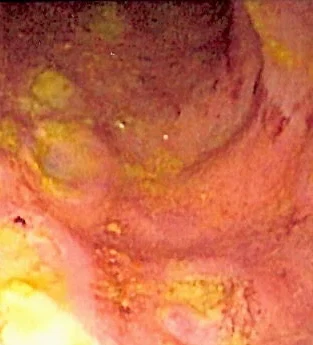
Figure 1Colonoscopy in Crohn colitis: deep ulcers
Long description
Endoscopic view of the sigmoid colon showing several deep ulcers with yellow fibrinous exudate between segments of red, inflamed mucosa.

Ileocolonoscopy with biopsies of involved and apparently uninvolved mucosa is the core investigation: it establishes distribution and severity, mucosal nodularity, aphthoid and serpiginous ulcers producing a cobblestone pattern, skip areas, strictures and rectal sparing, and granulomas when present (minority) [1] [2]. Photograph the ileum at diagnosis; document extent for future cancer surveillance. Upper endoscopy is added when upper symptoms or disease is suspected.
Video capsule endoscopy is a useful adjunct for suspected small-bowel disease that is invisible to the ileoscope, but it is contraindicated in obstruction: patients with obstructive symptoms need cross-sectional imaging and/or a patency capsule first (retention risk 0 to 5.4% in suspected CD, higher in known disease) [1].
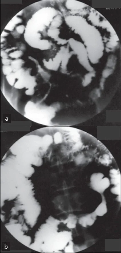
Figure 2Small bowel barium study in Crohn disease: skip lesions and string sign
Long description
Two views of a small bowel barium study. Normal-calibre barium-filled loops alternate with narrowed diseased segments; in view a a long, thin channel of barium through a stricture shows the string sign, and a fistula between loops is also described in the source.
Small-bowel imaging belongs in the initial work-up. MRE is comparable to CTE in sensitivity (reported up to 90%), avoids radiation and is preferred under 35 years or when serial scans will be needed; CTE is widely available and accurate. Intestinal ultrasound is an increasingly used, point-of-care, radiation-free alternative. Pelvic MRI or endoscopic ultrasound characterises perianal fistulas and abscesses; CT of abdomen and pelvis is used when an intra-abdominal abscess is suspected [1] [2].
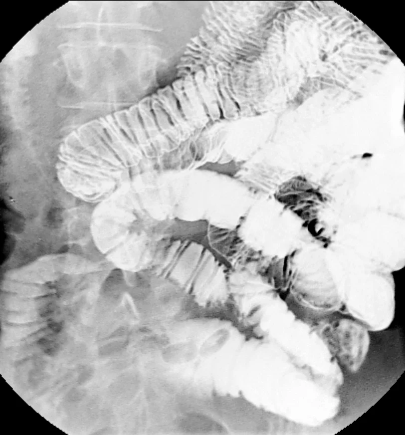
The old barium small-bowel follow-through has largely been replaced; classic findings included the "string sign" of luminal narrowing with proximal dilatation in stricturing disease. Neither the string sign nor any single imaging finding is specific [1].
Figure 3Small bowel barium study in Crohn disease
Long description
Barium enteroclysis image of the small bowel. A fixed narrowing of ileal loops with irregular, thickened mucosal folds is the typical small bowel stricture of Crohn disease; unaffected loops lie between affected segments (skip lesions).

Biopsies show focal, transmural, granulomatous inflammation; granulomas support but are not required for the diagnosis. Pyloric-gland metaplasia and architectural distortion reflect chronic injury [4] [1].
Diagnosis is clinical, integrating history, examination, laboratory, endoscopy, imaging and histology, because no single feature is pathognomonic [1].
Severity is scored for trials and sometimes for practice. The Crohn Disease Activity Index (CDAI) remains the trial gold standard (remission under 150; response a fall of more than 70 or 100 depending on trial; moderate to severe disease between 220 and 450; severe above 450), although it is cumbersome and subjective [4]. The Harvey-Bradshaw Index and the Simple Endoscopic Score for Crohn Disease (SES-CD) are practical alternatives; the Lémann index quantifies bowel damage. For eligibility studies, the Montreal phenotype suffices.
Red flags that mandate urgent reassessment: obstruction (colic, distension, vomiting), abscess/sepsis (fever, tachycardia, raised CRP), perianal disease with abscess, and any abrupt change in symptoms in a patient with small-bowel disease (possible cancer).
Treat to target. Symptoms correlate poorly with inflammation, so therapy is titrated against objective markers (CRP, fecal calprotectin) and endoscopic healing, not symptoms alone. A rapid step-up approach supported by close biomarker monitoring is recommended for many patients, while a top-down approach (early combination of biologic and immunomodulator) is considered for poor prognostic or complicated disease; newer ACG 2025 and AGA 2025 guidance places greater emphasis on initial advanced therapy for moderate to severe disease [1] [6]. Poor prognostic features include young age at diagnosis, extensive or ileal/ileocolonic involvement, perianal or severe rectal disease, extraintestinal manifestations at diagnosis, penetrating or stricturing behaviour, high CRP or calprotectin and low haemoglobin or albumin [1].
The ACG 2025 and ECCO 2024 guidelines agree on this shift from step-up to treat-to-target; ECCO notes there is currently insufficient evidence to direct how advanced therapies should be positioned in a therapeutic algorithm for luminal Crohn disease, so sequencing remains a judgment based on phenotype, comorbidity and cost [6].
Approach to a patient with suspected or newly diagnosed Crohn disease
- Suspected CDHistory, exam, baseline bloods and stool, ileocolonoscopy with biopsies, small-bowel imaging
- Confirm diagnosisExclude infection, TB (if considering biologics), and mimics; define extent, severity and behaviour
- Question: Severity and phenotype?
Mild ileal or ileocaecal
- Budesonide 9 mg daily for 8 to 16 weeksFor ileum and proximal colon only
- Step up if no response or relapseTreat to target with biomarkers
Moderate to severe
- InductionSystemic corticosteroids; start steroid-sparing plan early
- MaintenanceThiopurines 2.0-2.5 mg/kg/day, or methotrexate 15 mg weekly; biologic if high risk or steroid-dependent
Fistulising or perianal
- Antibiotics (metronidazole 500 mg twice daily and/or ciprofloxacin 250 mg twice daily) plus drainage
- Anti-TNF and/or ustekinumab; seton placement; surgery as neededAvoid steroids for fistula
Complications (stricture, abscess)
- Image first; drainage of abscess; medical optimisationEndoscopic dilation or surgery for fibrotic strictures
- Resect/strictureplasty for obstructive stricture
For mild to moderate ileal or ileocaecal disease, budesonide 9 mg daily by coated capsule is first-line induction for 8 to 16 weeks, with about a 50% remission rate at 8 weeks; a mesalamine course is not appropriate [5] [1]. Sulfasalazine may be considered for mild colonic disease but does not maintain remission [4]. Antibiotics have no proven role in luminal disease but are used for perianal disease and abscess.
For systemic disease, conventional corticosteroids (prednisone or prednisolone, commonly 40 mg daily up to 1 mg per kilogram per day) induce remission effectively within 8 to 12 weeks; they do not maintain remission and carry serious long-term risks, so a maintenance plan must be started when they are prescribed [5] [1].
Immunomodulators (thiopurines, methotrexate) are the backbone of maintenance for moderate to severe or steroid-dependent disease [4].
- Azathioprine 2.0 to 2.5 mg per kilogram per day (2 to 3 mg per kilogram per day in some reviews; step up from 1.0 mg per kilogram, increasing at 1- to 2-week intervals, especially with wild-type TPMT/NUDT15 genotypes); 6-mercaptopurine 1.0 to 1.5 mg per kilogram per day [5] [4].
- Methotrexate 25 mg intramuscularly or subcutaneously weekly for induction, then 15 mg weekly for maintenance; oral dosing (10 to 20 mg weekly) has only minimal benefit and is not recommended, so parenteral (intramuscular or subcutaneous) dosing is preferred. Monitor liver function and give folic acid; contraindicated in pregnancy [5] [4].
- TPMT is checked before thiopurines where available; they are associated with an increased risk of lymphoma, particularly in young men and the elderly; liver and marrow monitoring is mandatory. Combination anti-TNF plus thiopurine improves outcomes in biologic-naive moderate to severe disease (the SONIC trial), and lowers immunogenicity; thiopurines can be stopped later with more favourable relapse rates than stopping with monotherapy [4] [6].
Biologic and small-molecule therapy:
- Anti-TNF: infliximab 5 mg per kilogram intravenously at weeks 0, 2 and 6, then every 8 weeks (escalate to 10 mg per kilogram if loss of response); adalimumab 160 mg subcutaneously at week 0, 80 mg at week 2, then 40 mg every other week; certolizumab pegol 400 mg subcutaneously at weeks 0, 2 and 4 then every 4 weeks. All need tuberculosis, hepatitis B (and usually hepatitis C and HIV) screening and updated vaccination before the first dose; anti-TNF can be used when appropriate, combined with an immunomodulator or with therapeutic drug monitoring [5] [11].
- Vedolizumab (anti-alpha4beta7 integrin) is effective in luminal disease; it is given as an intravenous or subcutaneous induction and maintenance schedule per label [2].
- Ustekinumab (anti-p40 of IL-12/23) is effective in TNF-naive and TNF-refractory disease (including perianal), induction by weight (260 mg at 55 kg or less, 390 mg for 55 to 85 kg, 520 mg above 85 kg intravenously) and maintenance 90 mg subcutaneously every 8 weeks per product label (every 8 to 12 weeks in some real-world regimens) [2] [4] [6] [9] [10].
- IL-23 inhibitors (risankizumab, mirikizumab, guselkumab) are the preferred choice after TNF failure; risankizumab induction is 600 mg intravenously at weeks 0, 4 and 8, then 360 mg subcutaneously every 8 weeks [2] [6] [10].
- Upadacitinib (JAK-1 inhibitor) 45 mg orally once daily for 12 weeks induction, then 15 mg once daily maintenance; fast acting and oral, but avoided in pregnancy [2] [7].
Mesalamine is no longer recommended for Crohn disease: aminosalicylates are inferior to steroids and biologics, only sulfasalazine shows modest induction efficacy in colonic disease, and neither mesalamine nor budesonide maintains remission beyond 6 months [4] [1].
About a quarter of patients develop perianal fistulising disease, which may be the presenting feature [1]. Management combines antibiotics (metronidazole 500 mg twice daily and/or ciprofloxacin 250 mg twice daily), drainage of abscesses (with setons), and an effective advanced therapy (infliximab, adalimumab, ustekinumab, upadacitinib and vedolizumab are all recommended for fistulising disease) [1] [5] [2]. Corticosteroids are not beneficial for fistulas and may delay healing.
Small intra-abdominal abscesses (20 to 45 mm) can be treated with antibiotics with or without percutaneous drainage followed by early anti-TNF, avoiding surgery in about 60% at one year, provided there are no fistulas and nutrition is good [6].
Surgery is not curative but essential for complications and for disease not controlled medically. Up to half of patients need a resection within 10 years of diagnosis [4], and about 35% need a second resection within 10 years of the first [1]. Indications include strictures causing obstruction, fistulas and abscesses not controlled medically, perforation, dysplasia or cancer, and refractory or steroid-dependent disease. Options include limited resection, strictureplasty (preserves bowel, preferred when disease is diffuse), abscess drainage and, in selected uncomplicated terminal ileal disease, primary ileocaecal resection (the LIRIC approach) [2] [6]. Laparoscopic resection shortens recovery.
Postoperatively, endoscopic recurrence within 1 year is reported from over 70% to approaching 90%, depending on the series [1] [5]. Risk factors for recurrence include active smoking, shorter disease duration, more than one resection and penetrating disease [1]. Check at 6 to 12 months with ileocolonoscopy (Rutgeerts score); start an effective drug (anti-TNF, or vedolizumab or ustekinumab) shortly after resection in high-risk patients (smokers, penetrating disease, prior resections), while observing low-risk patients [1] [6].
Exclusive enteral nutrition (EEN) (polymeric feed as sole nutrition) is first-line induction in children and adolescents, with remission rates comparable to steroids; in adults the evidence is more limited and EEN is used mainly as an adjunct or short course [2] [6]. Diet (low-emulsifier/Crohn Disease Exclusion Diet) may have adjunctive benefit; in active disease ensure calories, correct micronutrients and, in severe disease, parenteral nutrition when needed.
Most IBD drugs except methotrexate and JAK inhibitors are considered safe in pregnancy and breastfeeding; active disease in pregnancy worsens outcomes, so aim for remission before conception and keep maintenance therapy on [2]. Methotrexate and upadacitinib must be stopped before conception; live vaccines are avoided in pregnant patients on biologics and newborn vaccination is adjusted if anti-TNF was used in the third trimester [11].
Preventive care is part of the diagnosis. Before biologic therapy, screen for latent tuberculosis and hepatitis B (and usually hepatitis C and HIV), and allow vaccination schedules, including live vaccines where possible, to be completed first; isoniazid for latent tuberculosis is started at least 1 month before the biologic [11]. Live attenuated vaccines are contraindicated during immunosuppression, and all patients need the usual inactivated vaccines (influenza annually, pneumococcal, hepatitis A and B, tetanus, HPV) [11].
Deficiency correction: vitamin B12 for ileal resection of more than 20 cm, iron and vitamin D repletion, and calcium and bone monitoring, particularly on steroids.
Patients with Crohn colitis involving at least one third of the colon, or at least 8 years of disease, are at increased colorectal cancer risk (about 2 to 3 times baseline, higher with PSC, younger age at diagnosis, family history of colorectal cancer and severe ongoing inflammation) [4] [1]. The first surveillance colonoscopy follows 8 years of disease, then every 1 to 3 years; with primary sclerosing cholangitis, colonoscopy starts at diagnosis and is annual [4]. Small-bowel adenocarcinoma risk is raised (at least 18-fold) but absolute risk is low and routine surveillance of the small bowel is not recommended; investigate a stable patient with an abrupt change in symptoms [1].
- Stricture and obstruction (inflammatory or fibrotic), including ileal narrowing.
- Fistulas: enterocutaneous, enteroenteric, enterovesical, rectovaginal; abscesses and phlegmon.
- Perforation and toxic colitis; intra-abdominal sepsis.
- Malabsorption, nutritional deficiency and growth failure; osteoporosis and stones.
- Small bowel and colorectal neoplasia in long-standing colonic disease.
- Postoperative recurrence.
Only about one in five patients has a truly indolent course; most relapse or progress [1]. Roughly half progress to a complication within 20 years (about half within 10 years by some estimates [2]), with surgery in up to half within 10 years of diagnosis [4] and symptomatic recurrence in about half within 5 years of surgery [1]; after a first resection a second resection occurs in about 35% within 10 years, and closer to 30% in more recent series [1]. Mortality is slightly increased (standardised mortality ratio about 1.4), mostly from gastrointestinal disease and cancer and respiratory disease in smokers, though the relative mortality is driven largely by the pre-biologic era [1]. Outcomes have improved substantially with early effective therapy and combination regimens [1] [2].
Children carry the same disease but presenting with growth failure and pubertal delay requires early nutritional and immunosuppressive therapy (EEN first-line); the paediatric strategy is handled by specialist teams. Older adults need infection and cancer vigilance; immunomodulators cross the blood-brain barrier poorly and biologics are chosen by comorbidity and frailty.
Where resources are limited:
- A careful history and examination plus a stool calprotectin or CRP screen and a colonoscopy with ileal intubation and biopsies establish most diagnoses; abdominal ultrasound is cheaper than CT/MRI.
- Budesonide is cheap and effective for mild ileal disease; metronidazole, azathioprine and even prednisolone are inexpensive; anti-TNF and IL-23 agents may be unavailable or unaffordable.
- Where imaging is limited, clinical assessment of obstruction and abscess (tenderness, mass, fever) guides urgent surgery.
- Refer early for abscess, obstruction, stricture and paediatric disease; tobacco cessation advice is free and among the most effective interventions.
- Crohn disease is pan-enteric, discontinuous and transmural; ulcerative colitis is contiguous and superficial.
- It most often hits the terminal ileum and right colon, with a teens-to-30s peak and a second peak in the 50s; incidence is rising worldwide and is shifting into newly industrialised countries.
- Smoking is the one well established modifiable risk factor and worsens every outcome.
- NOD2 and autophagy genes explain part of the genetic susceptibility, particularly ileal disease, but are not universal across ethnicities.
- Presentation is pain (often right lower quadrant, postprandial), chronic diarrhoea, weight loss and fatigue; perianal disease and growth failure can come first; only a minority have a truly indolent course.
- Fistulas and strictures are the defining complications: fistulas in about 10 to 26% in population cohorts, abscesses in many, and surgery in roughly half within 10 years.
- Investigations: bloods for anaemia and inflammation, stool pathogens and C. difficile, fecal calprotectin (cutoff above 50 to 100 micrograms per gram), ileocolonoscopy with biopsies, and MR/CT enterography; capsule only without a stricture.
- Endoscopic healing, not just symptoms, is the target; treat to target with CRP and calprotectin.
- Mesalamine is no longer recommended; budesonide (9 mg daily) for mild ileal disease; steroids induce but do not maintain.
- Thiopurines (azathioprine 2.0 to 2.5 mg per kilogram daily; 6-mercaptopurine 1.0 to 1.5 mg per kilogram daily) and methotrexate (25 mg weekly intramuscularly for induction, 15 mg for maintenance) maintain remission.
- Anti-TNF (infliximab 5 mg per kilogram intravenously at weeks 0, 2 and 6 then every 8 weeks; adalimumab 160 mg at week 0, 80 mg at week 2 then 40 mg every other week), vedolizumab, ustekinumab (260, 390 or 520 mg intravenously by weight then 90 mg subcutaneously every 8 weeks), IL-23 inhibitors (risankizumab 600 mg intravenously at weeks 0, 4 and 8 then 360 mg subcutaneously every 8 weeks) and upadacitinib (45 mg daily for 12 weeks then 15 mg daily) are the effective advanced options.
- Antibiotics (metronidazole 500 mg twice daily and/or ciprofloxacin 250 mg twice daily) are for perianal disease and abscess, not for colitis.
- Screen before biologics: latent tuberculosis and hepatitis B (and usually hepatitis C and HIV); vaccinate and supplement (B12 for ileal resection over 20 cm, iron, vitamin D).
- Colorectal cancer surveillance from 8 years of disease (annual from diagnosis if primary sclerosing cholangitis); primary sclerosing cholangitis is less common than in ulcerative colitis.
- Most IBD drugs except methotrexate and JAK inhibitors are safe in pregnancy; surgery treats but does not cure, and recurrence is expected.
Answer each one in your head first, then open the answer.
A 22-year-old is newly diagnosed with ileal Crohn disease after weeks of right lower quadrant pain after meals, 4 kg weight loss and 3-4 loose stools daily. What is the most appropriate initial induction strategy for mild to moderate ileal disease, and what should happen next?
Show answerHide answer
Budesonide 9 mg daily by coated capsule for 8 to 16 weeks, followed by objective reassessment and a plan for maintenance, not repeated steroids alone. If remission is achieved, step up or down according to biomarkers (CRP, fecal calprotectin above 50 to 100 micrograms per gram indicates active inflammation) and endoscopic findings, with a steroid-sparing immunomodulator or biologic for steroid-dependent or high-risk disease.
Which statement best distinguishes Crohn disease from ulcerative colitis?
Show answerHide answer
Crohn disease is transmural, discontinuous and can affect any part of the gut with strictures and fistulas; ulcerative colitis is superficial, continuous from the rectum and rarely penetrates or strictures. Features such as perianal fistulas, rectal sparing and skip lesions point to Crohn disease.
A patient with Crohn disease has ileal resection of 40 cm. What nutritional problems and monitoring should be anticipated?
Show answerHide answer
Vitamin B12 deficiency (supplement when ileal resection exceeds 20 cm), iron deficiency, fat and fat-soluble vitamin malabsorption, and bile-salt diarrhoea. Check B12, iron studies, folate and vitamin D periodically, and replace as needed; diarrhoea may respond to bile acid sequestrants under specialist review.
Why is infliximab given with an immunomodulator in biologic-naive moderate to severe disease, and what does this combination require beforehand?
Show answerHide answer
Combination therapy (infliximab plus azathioprine) produced higher clinical and endoscopic remission at 26 and 54 weeks than either alone in the SONIC trial, partly because azathioprine reduces anti-infliximab antibodies and raises drug levels. It requires exclusion of untreated latent tuberculosis and hepatitis B (and usually hepatitis C and HIV), updated vaccination and careful counselling about infection and small lymphoma risks, and liver/marrow monitoring. Stopping the immunomodulator later is more favourable than stopping anti-TNF monotherapy.
A patient with ileal Crohn disease, two prior resections, a smoking habit and perianal fistulas presents after a flare. Explain your proposed hierarchy of risk and treatment.
Show answerHide answer
This is high-risk (smoking, penetrating/perianal phenotype, multiple resections). Stop smoking first (free, high yield), drain any abscess, give antibiotics and place setons if needed, and use an anti-TNF or other effective biologic, checking tuberculosis and hepatitis B first and giving a treat-to-target plan with biomarkers and 6-to-12-month ileocolonoscopy. Early surgery is reserved for obstruction or failure, and anti-TNF monotherapy after the initial combination phase is an option once remission is secure.
How should colorectal cancer surveillance be planned in a patient with Crohn colitis?
Show answerHide answer
The first surveillance colonoscopy follows 8 years of disease when more than 30% of the colon is involved (or earlier if primary sclerosing cholangitis, in which case it is annual from diagnosis), then every 1 to 3 years according to extent, duration, activity, family history and other risk modifiers; obtain quadrant biopsies every 10 cm and targeted biopsies of abnormal areas.
What screening is mandatory before starting a biologic drug, and what is the minimum practical interval?
Show answerHide answer
Screen for latent tuberculosis and hepatitis B (and usually hepatitis C and HIV), check vaccinations and start isoniazid for latent tuberculosis at least 1 month before the biologic (some regimens allow anti-TNF after 1 to 2 months of treatment), and give HBV prophylaxis with entecavir or tenofovir if active HBV is found, delaying the biologic until HBV DNA is negative. Live vaccines are given before, not during, immunosuppression.
Ulcerative colitis and inflammatory bowel disease overview; primary sclerosing cholangitis; malabsorption and maldigestion; coeliac disease; colorectal cancer; osteoporosis; cholelithiasis and biliary colic; nephrolithiasis; extraintestinal manifestations of IBD; tobacco smoking cessation; vaccination and preventive care in IBD; perianal fistula and abscess.
- Lichtenstein GR, Loftus EV Jr, Afzali A, Long MD, Barnes EL, Isaacs KL, Ha CY. ACG Clinical Guideline: Management of Crohn's Disease in Adults. The American Journal of Gastroenterology 2025;120(6):1225-1264. doi:10.14309/ajg.0000000000003465. Accessed .
- An YK, Gilmore R, Radford-Smith G, Begun J. Advances in Crohn's Disease: A Comprehensive Review of Pathophysiology, Diagnosis, and Emerging Therapeutics. JGH Open 2026;10(4):e70394. doi:10.1002/jgh3.70394. Accessed .
- Satsangi J, Silverberg MS, Vermeire S, Colombel JF. The Montreal classification of inflammatory bowel disease: controversies, consensus, and implications. Gut 2006;55(6):749-753. doi:10.1136/gut.2005.082909. Accessed .
- Cheifetz AS. Management of Active Crohn Disease. JAMA 2013;309(20):2150-2158. doi:10.1001/jama.2013.4466. Accessed .
- Feldman PA, Wolfson D, Barkin JS. Medical Management of Crohn's Disease. Clinics in Colon and Rectal Surgery 2007;20(4):269-281. doi:10.1055/s-2007-991026. Accessed .
- Latorre MA, Rennie M, Rosiou K, Selinger CP. Perspective: Making Treatment Decisions for Crohn's Disease in 2025—Key Considerations. Journal of Clinical Medicine 2025;14(19):6879. doi:10.3390/jcm14196879. Accessed .
- AbbVie. RINVOQ (upadacitinib) extended-release tablets, prescribing information. DailyMed (US National Library of Medicine) label, accessed 2026-10-28. Accessed .
- Lichtenstein GR. Micronutrient Deficiencies in Inflammatory Bowel Disease. Gastroenterology and Hepatology 2021;17(12 Suppl 7):10-11.. Accessed .
- Lusetti F, D'Amico F, Allocca M, Furfaro F, Zilli A, Fiorino G, Parigi TL, Radice S, Peyrin-Biroulet L, Danese S. Positioning risankizumab in the treatment algorithm of moderate-to-severe Crohn's disease. Immunotherapy 2024;16(9):581-595. doi:10.2217/imt-2023-0219. Accessed .
- Janssen Biotech, Inc. STELARA (ustekinumab) injection, solution: prescribing information. DailyMed (US National Library of Medicine) label, accessed 2026-10-28. Accessed .
- Chebli JMF et al. A guide to preparation of patients with inflammatory bowel diseases for anti-TNF-alpha therapy. Medical Science Monitor 2014;20:487-498. doi:10.12659/MSM.889747. Accessed .
Figure credits
- Figure 1: Samir at English Wikipedia, via Wikimedia Commons, CC BY-SA 3.0, CC BY-SA 3.0. Cropped on the left and bottom edges to remove on-screen text; converted to WebP..
- Figure 2: Al-Mofarreh MA, Al Mofleh IA, Al-Teimi IN and Al-Jebreen AM, Saudi Journal of Gastroenterology 2009, CC BY, CC BY. Converted to WebP; no other changes..
- Figure 3: Hellerhoff, via Wikimedia Commons, CC BY-SA 3.0, CC BY-SA 3.0.
Educational content for medical students and doctors. It is not a substitute for clinical judgement or local guidelines.