Ulcerative colitis and inflammatory bowel disease overview
Inflammatory bowel disease: how ulcerative colitis presents, is diagnosed, scored, treated, and followed; Crohn disease compared on the features that matter.
- Inflammatory bowel disease (IBD) is chronic, relapsing inflammation of the digestive tract. Its two main forms are ulcerative colitis (UC), which starts in the rectum and spreads without gaps along the colon, and Crohn disease, which can strike anywhere from mouth to anus and penetrates the full bowel wall.
- UC inflames only the mucosa and submucosa, continuously from the rectum upward. The cardinal symptoms are bloody diarrhoea, urgency to open the bowels, tenesmus (a feeling of unfinished emptying) and lower abdominal cramps. Severity, extent and surface healing, not just symptoms, decide treatment.
- Most disease is mild to moderate and controlled with 5-aminosalicylates, which are given by mouth, as enemas or as suppositories according to where the disease is. Flares need corticosteroids, then steroid-sparing therapy; moderate to severe disease increasingly needs advanced therapies (anti-TNF, anti-integrin, anti-IL-12/23 and anti-IL-23, S1P modulators, JAK inhibitors). Acute severe colitis is a hospital emergency.
- Surgery is curative for UC: removing the colon and joining the small bowel to the anal canal (ileal pouch-anal anastomosis) restores continence, but pouchitis and other pouch complications must be anticipated. Extensive disease raises the long-term risk of colorectal cancer, so surveillance colonoscopy is part of routine care.
- A single patient can have colitis of any extent, with extra-intestinal problems (joints, skin, eyes, bile ducts) that are treated alongside the bowel, and a course that runs from one flare to remission. The ACG 2025 guideline and the AGA 2023 biomarker guideline direct modern monitoring and drug choice.
By the end of this page you should be able to:
- Explain what is shared by all IBD and how ulcerative colitis differs from Crohn disease.
- Classify the extent of UC by the Montreal system and judge severity with the Truelove and Witts criteria, the Mayo score and acute severe criteria.
- Work through the investigation of a patient with suspected or relapsing UC, including when not to perform a full colonoscopy.
- Prescribe the drug classes used in UC with their usual doses, and plan the escalation from proctitis to acute severe disease.
- Outline the surgical indications and complications after colectomy with pouch formation.
- Plan colorectal cancer surveillance and the preventive care (vaccinations, screening, pregnancy advice) that travels with a diagnosis of UC.
DefinitionUlcerative colitis
Ulcerative colitis is a chronic, relapsing inflammatory disease limited to the colon. It begins in the rectum and extends proximally in a continuous sheet; it involves the mucosa and submucosa, never the full thickness of the wall, and does not skip. Microscopically one expects crypt architectural distortion, basal plasmacytosis and crypt abscesses.
DefinitionInflammatory bowel disease (IBD)
IBD is the umbrella term for UC and Crohn disease, two disorders that share chronic intestinal inflammation, a tendency to relapse and remit, systemic extra-intestinal manifestations and an increased risk of colorectal neoplasia. They differ in distribution, depth and behaviour.
UC is a worldwide disease and appears to be rising in incidence, including in parts of Asia where it was rare two decades ago. Incidence and prevalence figures vary by region, by case-finding method and over time; pooled series from high-income countries report annual incidence in the tens per 100,000 person-years and prevalence up to a few hundred per 100,000, while lower-incidence regions report far fewer. East Asian cohorts often describe a slight male predominance, whereas pooled Western cohorts show no overall sex difference, with older men carrying higher risk. Age at peak onset is young adulthood. [7]
IBD is understood as an exaggerated and poorly regulated immune response to the normal gut flora in a genetically susceptible person, triggered or shaped by environmental factors. No single cause is known, and no single gene accounts for the disease.
- Genetics. IBD aggregates in families. Genome-wide studies identify 260 susceptibility loci for IBD, many shared between UC and Crohn disease (NOD2 is the classic Crohn-related locus; none is specific to UC alone). Family history is the strongest known predictor, but most affected people have no affected relative. [7] [10]
- The microbiome. Reduced diversity of gut bacteria, loss of butyrate-producing species and an expansion of pro-inflammatory organisms are described in active disease. Environmental factors such as diet and antibiotics appear to matter, but whether these changes cause, follow or merely accompany inflammation remains uncertain. [7]
- Environment. Urban living, a Westernised diet, appendicectomy status and antibiotic exposure (usually early in life) have been associated with UC; breastfeeding is considered protective in many cohorts. The hygiene hypothesis, that reduced early infection predisposes to immune dysregulation, is often invoked but not proven. [3]
- Smoking paradox. Epidemiological studies have repeatedly found that current smoking is associated with a lower incidence of UC and, sometimes, milder disease, while former smokers carry a higher risk than never-smokers. The opposite holds for Crohn disease, where smoking worsens outcomes. Because smoking harms every other organ, with higher rates of extra-intestinal malignancy and vascular disease and a worse therapy response in IBD overall, smoking cessation is recommended to every patient. Any "benefit" to the colon should never be used to justify tobacco use. [7]
In UC, the final common pathway is a self-sustaining mucosal inflammatory cascade.
- Barrier loss. Defects in the mucus layer and epithelial tight junctions allow luminal contents to engage the innate immune system.
- Innate activation. Macrophages and dendritic cells produce tumour necrosis factor-alpha (TNF-alpha), interleukin (IL)-1, IL-6 and IL-23; neutrophils migrate and form crypt abscesses.
- Adaptive reinforcement. T-helper 2 and T-helper 17 responses amplify the reaction, with atypical natural killer T cells and IL-13 contributing to epithelial damage in UC (an IL-13-rich profile is more characteristic of UC than of Crohn disease).
- Tissue result. Mucosal erosion, ulceration, bleeding and oozing produce visible blood in the stool (haematochezia) and protein loss. Continued inflammation damages the muscularis mucosae; in acute severe disease the myenteric reflex can fail and the colon dilates.
Because the deeper muscular wall and serosa are spared, penetrating complications (fistulae, strictures) belong to Crohn disease, not to typical UC.
| Feature | Ulcerative colitis | Crohn disease |
|---|---|---|
| Site | Colon only, continuous from the rectum | Anywhere mouth to anus; often terminal ileum plus colon, with small-bowel-only disease possible |
| Depth of inflammation | Mucosa and submucosa | Transmural, through the full wall |
| Pattern | Continuous, no skip lesions; rectum almost always involved | Patchy, with skip areas and "creeping" fat around involved segments |
| Ulcer morphology | Diffuse, confluent; friable mucosa and crypt abscesses | Aphthae, deep linear ulcers, a cobblestone surface |
| Perianal disease | Usually absent | Common: fissures, fistulae, abscesses, skin tags |
| Fistulae and strictures | Not typical | Typical later complications |
| Presenting symptoms | Bloody diarrhoea, urgency, tenesmus, mucus | Abdominal pain, diarrhoea that may not be bloody, weight loss, malabsorption |
| Extraintestinal manifestations | Arthritis, erythema nodosum, pyoderma gangrenosum, uveitis, ankylosing spondylitis, primary sclerosing cholangitis | The same spectrum, with erythema nodosum and peripheral arthritis especially common |
| Surgery | Colectomy is effectively curative, with pouch reconstruction possible | Segmental resection, but recurrence is common and curative surgery is not |
| Serology | p-ANCA is often positive in UC | ASCA (anti-Saccharomyces cerevisiae antibodies) is more characteristic of Crohn disease; p-ANCA can also be positive |
| Smoking | Stopping may worsen control; current smoking often milder disease (see aetiology) | Smoking clearly worsens course |
| Malignancy risk | Colorectal cancer rises with extent and duration | Small-bowel and colonic cancer risk where disease is colonic |
Extent is defined at endoscopy and by histology, and it shapes both treatment and cancer risk.
| Stage | Name | Anatomy |
|---|---|---|
| E1 | Ulcerative proctitis | Inflammation limited to the rectum, distal to the rectosigmoid junction, within 18 cm of the anal verge [1] |
| E2 | Left-sided (distal) colitis | Disease extends to the splenic flexure but not beyond it |
| E3 | Extensive colitis (pancolitis) | Disease extends proximal to the splenic flexure, sometimes the whole colon |
- Proctitis (E1) often needs only topical therapy and rarely leads to cancer.
- Left-sided colitis (E2) usually responds to combination oral and topical 5-aminosalicylates.
- Extensive colitis (E3) carries the clearest excess risk of colorectal cancer and more often needs advanced therapy. Among people who present with proctitis, a sizeable minority progress to extensive disease over the decades; progression figures up to half after ten years or more have been cited from clinic cohorts. [4]
History. UC announces itself with bowel frequency and bleeding. The statements to listen for are simple: "I keep having to go", "blood and mucus", "I cannot make it to the toilet" (urgency and incontinence), "a feeling that I still need to go" (tenesmus), "crampy pain low down that is relieved by passing stool", "waking at night to empty the bowels", "waking tired because of disturbed nights", "my joints ache", "a sore, red eye", "sores on my legs". Constipation with a reduced stool frequency is unusual, and pain that worsens rather than improves after defaecation raises another diagnosis.
- Bloody diarrhoea is the hallmark; it may be the only symptom in proctitis, where stool volumes are small and tenesmus dominates.
- Mucus and pus are common and reflect mucosal exudation.
- Urgency and faecal incontinence come from rectal inflammation reducing capacity; they are among the most disabling symptoms.
- Pain is usually crampy, hypogastric or suprapubic, and eased by passing stool.
- Nocturnal symptoms indicate active disease rather than irritable bowel syndrome.
- Weight loss and fever mark more severe or extensive disease.
- Extraintestinal manifestations (EIMs) occur in a substantial minority of patients (higher in Western series, where reported ranges reach 21 to 40 per cent); they may appear before any bowel symptom and must be asked about directly. [3] [7]
- Joints: peripheral arthritis (large joints, tracking with disease activity) and ankylosing spondylitis (axial, independent of bowel activity).
- Skin: erythema nodosum (tender red nodules on the shins, activity-dependent) and pyoderma gangrenosum (painful ulcers on the shins or elsewhere, which may not track disease activity and can be triggered by minor trauma, the pathergy phenomenon).
- Eyes: uveitis, episcleritis and conjunctivitis.
- Hepatobiliary: primary sclerosing cholangitis (PSC), discussed below.
- Other: deep venous thrombosis (pharmacological prophylaxis is therefore standard in hospital), osteoporosis and anaemia.
Primary sclerosing cholangitis is the extra-intestinal manifestation with the gravest implications. It produces cholestatic liver biochemistry, may be silent early on, and is associated with a raised risk of both bile-duct and colorectal cancer. Screening uses liver function tests and, when indicated, magnetic resonance cholangiopancreatography. Patients with UC plus PSC should have annual colonoscopic surveillance from diagnosis. [6]
Examination. Physical examination may be mild even in active proctitis. Look for anaemia, tachycardia and fever in acute severe disease; lower-abdominal tenderness or guarding; a "doughy" abdominal wall suggesting colonic dilatation; extraintestinal signs such as mouth ulcers, arthritis, erythema nodosum, pyoderma or uveitis; and malnutrition in chronic severe disease. Rectal examination finds tenderness, mucus and blood; proctoscopy, where available, shows a granular, friable, oozing mucosa. Biopsy and culture distinguish UC from infection.
The main imitators of UC are infectious colitis (including Clostridioides difficile, which must be excluded at every presentation and flare), ischaemic colitis, Crohn colitis, microscopic colitis, diverticular colitis and radiation proctitis. Atypical endoscopic patterns (patchy disease, rectal sparing, isolated caecal inflammation) can be seen in treated UC and do not by themselves convert the diagnosis to Crohn disease. [3]
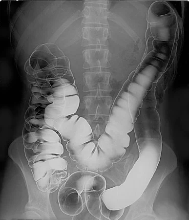
Figure 1Normal barium enema: haustral markings
Long description
Double-contrast barium enema of a normal colon, with regular haustra from caecum to rectum. It serves as the comparison for the lead-pipe colon of long-standing ulcerative colitis.

A new diagnosis starts with confirmation that this is UC and not infection or Crohn disease, then mapping extent and severity.
- Stool studies first. Test for C. difficile (and other pathogens) at presentation, through a multistep approach. Stool culture, ova and parasites, and inflammatory markers such as faecal calprotectin belong in the initial panel. [1] [3]
- Blood tests. Full blood count (anaemia, thrombocytosis, leukocytosis), CRP, ESR, albumin, renal function and liver function; hepatitis B surface antigen, surface antibody and core antibody before any immunosuppression; iron, vitamin D and other nutrients where relevant.
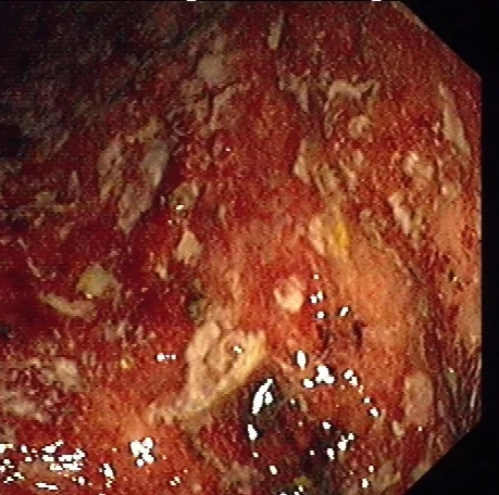
- Colonoscopy with intubation of the terminal ileum and biopsies is the mainstay: diffuse continuous mucosal inflammation from the rectum, with loss of vascular pattern, granularity, friability, erosions and ulcers. Take at least two biopsies from inflamed and uninflamed areas; interpret with a gastrointestinal pathologist. Histology shows crypt architectural distortion, basal plasmacytosis and crypt abscesses (neutrophils in crypt lumens), and allows the exclusion of CMV, TB, amebiasis and cancer.
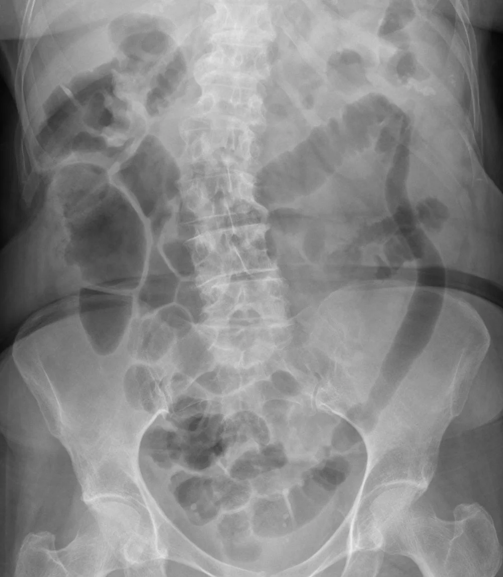
- Abdominal radiograph in suspected acute severe disease to look for colonic dilatation, perforation or obstruction. Colonic dilatation greater than 5.5 to 6 cm, with systemic toxicity, defines toxic megacolon. Computed tomography has superior detail when perforation or abscess is suspected. [3] [4]
- Avoid full colonoscopy in severe disease. In acute severe colitis, perform flexible sigmoidoscopy only; full colonoscopy worsens disease activity and raises perforation risk. Flexible sigmoidoscopy also assesses endoscopic severity and obtains biopsies for CMV. [3] [4]
- Barium enema was once taught for the "lead-pipe" colon with loss of haustra; it is now rarely used and has been replaced by colonoscopy and cross-sectional imaging.
- p-ANCA and ASCA are serological markers occasionally used as supportive clues; p-ANCA is often positive in UC and ASCA in Crohn disease, but neither is diagnostic.
- Intestinal ultrasound, where available, assesses wall thickness, vascularity and extent non-invasively and can reduce colonoscopy use.
Figure 2Lead-pipe colon in ulcerative colitis
Long description
Plain abdominal radiograph in known ulcerative colitis. The descending and sigmoid colon appear as a smooth, featureless gas-filled tube, the 'bicycle tube' or lead-pipe appearance, because chronic inflammation has flattened the haustral folds and shortened the bowel.

Figure 3Colonoscopy in active ulcerative colitis
Long description
Endoscopic image of the colon in ulcerative colitis. The mucosa is diffusely red and granular with shallow ulcers and patches of pale exudate over a continuous stretch of bowel.

Severity is staged from the patient's description and the objective inflammatory burden.
| Domain | Threshold |
|---|---|
| Stool frequency | 6 or more bloody stools a day |
| Plus at least one of | Temperature above 37.8 °C (original wording: 37.8 °C or more on at least 2 of 4 days, or a mean evening temperature above 37.5 °C; condensed criteria often use above 37.5 °C) |
| Pulse above 90 beats per minute | |
| Haemoglobin under 10.5 g/dL (usual modern cut-off, allowing for transfusion) or 75% or less of normal by the original Truelove and Witts criterion | |
| ESR above 30 mm/h |
The temperature and haemoglobin criteria are quoted differently by different adaptations of Truelove and Witts: the original wording is a temperature of 37.8 °C or more on at least 2 of 4 days (or a mean evening temperature above 37.5 °C) with haemoglobin 75% or less, while modern summaries usually state a temperature above 37.8 °C and a haemoglobin under 10.5 g/dL. [4]
Mild disease is defined as 4 or fewer bowel motions a day with only small amounts of visible blood, no fever, no tachycardia, anaemia not severe and an ESR not increased more than 30 mm in 1 hour; severe disease is 6 or more bloody motions a day plus at least one of the systemic features listed; moderate disease falls between the two. Remission is 1 to 2 stools a day without blood, no fever or tachycardia, haemoglobin and ESR returning toward normal and the patient gaining weight. [4]
The Mayo score (sometimes Disease Activity Index) is the workhorse for grading activity in practice and in trials. The stool frequency and bleeding components (each 0 to 3), plus the physician's global assessment and the endoscopy component (0 to 3), are standard; the partial Mayo score uses stool frequency plus rectal bleeding plus physician's global assessment (0 to 9). The total runs from 0 to 12 and the partial score from 0 to 9, with higher values representing more active disease. [4]
| Score | Appearance |
|---|---|
| 0 | Normal or inactive (healed) mucosa |
| 1 | Mild (erythema, decreased vascular pattern, mild friability) |
| 2 | Moderate (marked erythema, absent vascular pattern, friability, erosions) |
| 3 | Severe (spontaneous bleeding, ulceration) |
The ACG 2025 guideline adds faecal calprotectin, endoscopy (MES or UCEIS) and CRP to the classification: faecal calprotectin 150 to 200 micrograms per gram or below is used to separate remission from active disease in the older ACG table, and a CRP persistently above 45 mg/L together with 3 to 8 bowel motions a day despite 3 or more days of high-dose intravenous corticosteroids marks a high risk of needing colectomy. [1] [4]
The AGA 2023 biomarker guideline recommends using faecal calprotectin below 150 micrograms per gram, or normal faecal lactoferrin or normal CRP, to rule out active inflammation during remission and to avoid routine endoscopy, with monitoring every 6 to 12 months. Concentrations above 150 micrograms per gram during symptomatic remission should prompt endoscopic assessment rather than reactive dose escalation. [2]
A stepwise approach to suspected or relapsing ulcerative colitis
- First presentation or suspected relapseHistory, examination, stool culture plus C. difficile testing, blood tests
- Question: Concurrent infection?
Yes
- Treat specifically (for example oral vancomycin or fidaxomicin for C. difficile)
No
- Proceed with UC management
- Judge severity and extentMontreal E1/E2/E3, Truelove and Witts criteria, Mayo score, faecal calprotectin and CRP
- Question: Severity?
Mild to moderate E1/E2
- Proctitis E1: rectal mesalamine 1 g dailyEnema or suppository depending on extent
- Left-sided E2: rectal mesalamine enema at least 1 g daily plus oral mesalamine at least 2.0 g daily
- Extensive E3 plus mild disease: oral mesalamine at least 2.0 g daily
- Reassess response within 8 weeks; step up if no remission
Moderate to severe or 5-ASA failure
- Oral budesonide MMX 9 mg/d for moderately active disease; systemic corticosteroids if severely active
- Then steroid-sparing advanced therapy: vedolizumab, anti-TNF, anti-IL-23/IL-12/23, S1P modulator or upadacitinib/tofacitinibChoice guided by EIMs, comorbidity and patient preference
- Combination thiopurine with infliximab for induction improves outcomes
Acute severe colitis
- Admit: hydrocortisone 100 mg 3 to 4 times daily, or methylprednisolone 60 mg/dNothing by mouth, intravenous fluids, VTE prophylaxis, stop NSAIDs, narcotics and anticholinergics
- Flexible sigmoidoscopy within 24 to 72 hours for severity and CMVAssess for toxic megacolon
- At day 3: if more than 8 stools a day, or 3 to 8 stools plus CRP above 45 mg/L, give infliximab or cyclosporine
- If no response to rescue, or perforation, megacolon, massive bleeding: colectomy
- Maintenance: continue effective therapy; reassess at each visit with symptoms, faecal calprotectin and periodic endoscopy
Induction.
- Proctitis (E1): rectal 5-aminosalicylate, mesalamine 1 g daily by suppository or foam enema, is first line; it is preferred over rectal corticosteroids. [1]
- Proctitis not responding to topical 5-ASA: tacrolimus or beclomethasone suppositories, or topical corticosteroids, are conditional options. [1]
- Left-sided (E2): rectal mesalamine enema at least 1 g daily preferred over rectal steroids, with or (for refractory or partial-response disease) combined with oral mesalamine at least 2.0 g daily. [1]
- Extensive or left-sided not responding to 5-ASA, or oral treatment preferred for mild disease: oral mesalamine at least 2.0 g daily; a large meta-analytic picture from the ACG 2025 review quotes "at least 2 to 4.8 g daily oral mesalamine and/or at least 1 g daily rectal mesalamine" as the appropriate dose range. For mildly active disease of any extent, 2.0 to 2.4 g is as good as 4.8 g, so avoid overdosing. [1]
- Budesonide MMX 9 mg daily for mildly to moderately active disease not controlled by 5-ASA, especially in moderately active colitis; it is non-systemic and has less adrenal suppression, but it is not for maintenance. [1]
- Oral systemic corticosteroids for disease that fails 5-ASA and budesonide, following the disease severity and extent; they are for induction only. [1] Corticosteroids are not acceptable maintenance therapy.
- 5-ASA should not be changed to another formulation of the same class after an adequate dose has failed; move to a different class. Combining 5-ASA with a biologic once advanced therapy is started does not add benefit. [1]
- Reassess within 8 weeks of induction. [1]
Maintenance. Continue the agent that achieved remission. Rectal mesalamine 1 g daily maintains proctitis; oral mesalamine at least 1.5 g daily maintains left-sided or extensive mild disease. Topical and systemic corticosteroids and budesonide MMX are not maintenance therapies. [1]
The ACG 2025 guideline offers a ranked menu; individual choice depends on mechanism, comorbidity and patient preference, and first advanced therapy tends to work better than later ones.
- Infliximab (with a thiopurine, azathioprine, for induction), and adalimumab or golimumab, as anti-TNF agents.
- Vedolizumab (gut-selective anti-integrin), which is superior to adalimumab for induction and maintenance of remission in head-to-head data.
- Ustekinumab (anti-IL-12/23p40).
- Guselkumab, mirikizumab and risankizumab (anti-IL-23p19).
- S1P receptor modulators ozanimod and etrasimod.
- JAK inhibitors tofacitinib and upadacitinib.
- Thiopurines (azathioprine or 6-mercaptopurine) for maintenance, particularly post-corticosteroid remission, but not for induction; methotrexate is not recommended for maintenance of UC (unlike Crohn disease). [1]
Useful practical points from the ACG 2025 recommendations:
- Combination therapy: when infliximab is used for induction, combine with a thiopurine. Infliximab is the preferred anti-TNF in UC. [1]
- Positioning: drug levels and anti-drug antibodies help with suspected loss of response to an anti-TNF; primary non-responders (no benefit despite adequate drug levels) usually need a different mechanistic class rather than a second anti-TNF. Biosimilars of anti-TNFs and ustekinumab are acceptable substitutes, and switches should be made without delay. [1]
- Isolated proctitis that is moderately to severely active should still receive the advanced therapies with proven efficacy, not only topical agents. [1]
- High-risk infections or frailty may favour vedolizumab or an anti-IL-23 strategy over more systemically immunosuppressive options; extraintestinal manifestations guide choice (anti-TNF or ustekinumab for arthropathy, skin or eye disease). [1]
- Thiopurines and methotrexate are not recommended for induction. [1]
Doses commonly used in adults are summarised below for orientation; confirm every dose, route and monitoring requirement against the current product label and local formulary. Class-level maintenance and induction schedules are drug-specific and are not reproduced here beyond the 5-aminosalicylate, budesonide and corticosteroid doses supported by the ACG 2025 guideline. [1]
- Thiopurines and methotrexate: see local protocol and label.
- Infliximab: intravenous induction followed by maintenance every 8 weeks, with higher or shortened intervals in refractory disease.
- Adalimumab and golimumab: subcutaneous induction and maintenance per label.
- Vedolizumab: intravenous (then subcutaneous) induction and maintenance per label.
- Ustekinumab: intravenous induction then subcutaneous maintenance.
- Tofacitinib: induction at high dose then maintenance at lower dose, subject to regulatory restrictions in many jurisdictions.
- Upadacitinib: induction then maintenance dosing per label.
- Ozanimod and etrasimod: oral daily.
- Mirikizumab, guselkumab, and risankizumab per label.
- S1P modulators and JAK inhibitors carry cardiovascular, infection and malignancy warnings that must be discussed; baseline lipids, liver function and infection screening apply.
Acute severe ulcerative colitis (ASUC) is a medical emergency present in up to one quarter of UC patients over their lifetime, and is the setting in which up to 40 per cent ultimately require colectomy. [4]
- Admission, resuscitation and assessment. Intravenous hydrocortisone 100 mg 3 to 4 times daily or intravenous methylprednisolone 60 mg daily; nothing by mouth, intravenous fluids, correction of electrolytes and anaemia, pharmacological venous thromboembolism prophylaxis; avoid NSAIDs, opioids, anticholinergic and antidiarrhoeal drugs. [1]
- Stool frequency, rectal bleeding, examination, vital signs and serial CRP monitor response. [1]
- C. difficile should be re-tested, and flexible sigmoidoscopy performed within 72 hours, preferably 24 hours, to grade inflammation and biopsy for CMV. [1]
- Assess for toxic megacolon clinically and radiologically. [1]
- Medical rescue at day 3. If stools exceed 8 per day, or stool frequency is 3 to 8 per day with a CRP above 45 mg/L, give infliximab at standard induction dosing (with possible dose intensification where serum albumin is below 2.5 g/dL) or cyclosporine (standard low-dose intravenous regimen, with lipid levels and renal function monitored). These criteria predict colectomy in roughly 85 per cent of patients, though modern rescue therapy has lowered the observed rate. [1] [5]
- Surgical escalation when there is no response, suspected toxicity, toxic megacolon, perforation, massive haemorrhage or refractoriness to rescue; surgery should not be deferred on account of prior infliximab or cyclosporine exposure. [1]
- Maintenance after rescue: continue infliximab for maintenance where infliximab achieved remission; use a thiopurine after cyclosporine rescue. [1]
- Toxic megacolon management: nil by mouth and nasogastric decompression, intravenous fluids and electrolyte repletion, broad-spectrum antibiotics guided by culture, intravenous corticosteroids (or continuation of the therapy that precipitated nothing new), stopping anticholinergics, opioids, antimuscarinic and NSAID drugs, frequent clinical and radiographic monitoring of colonic diameter, and immediate surgical consultation; colectomy is indicated for perforation, uncontrolled bleeding, clinical deterioration over 24 to 48 hours, or failure to improve. [3] [4] [1]
Indications for colectomy include toxic megacolon, perforation, massive bleeding, dysplasia, cancer, and medically refractory disease.
- Elective total proctocolectomy with ileal pouch-anal anastomosis (IPAA, the J-pouch, very occasionally S-pouch or W-pouch) is now the standard restorative operation; it removes the diseased mucosa while preserving continence through a spinal (neorectal) reservoir emptied by the anal sphincter. Laparoscopic and robotic approaches are increasingly standard. [8]
- Complications of the pouch: pouchitis (the commonest, presenting with increased stool frequency, urgency, bleeding, fever and malaise; treated with metronidazole or ciprofloxacin, typically a 2-week course, with chronic antibiotic-dependent or refractory courses treated as in the AGA pouchitis guideline), cuffitis (inflammation of the retained rectal mucosal cuff), Crohn-like disease of the pouch (non-caseating granulomas, fistulae, afferent limb disease, rising anti-glycan markers), strictures, pelvic sepsis and anastomotic leak after surgery, and sexual dysfunction or reduced fertility in some patients. [8]
- Fertility and delivery: IPAA does not exclude pregnancy; infertility rates rise modestly after pelvic surgery, so counselling and, where indicated, in vitro fertilisation should be discussed early. Vaginal delivery is generally acceptable, though some centres prefer caesarean section after pouch surgery.
- Proctocolectomy without pouch (Brooke or continent ileostomy) remains a good option where the anal sphincter is incompetent or cancer requires ultra-low resection.
- Goals of therapy are symptom control (urgency foremost) and mucosal healing (Mayo endoscopic subscore 0 or 1; histological healing is increasingly tracked). [4]
- Treat to target. The STRIDE framework targets clinical and endoscopic remission; biomarker normalisation supports but does not replace endoscopy for major decisions.
- Step down carefully: de-escalate only after sustained remission (at least a year in many cases) and never stop a thiopurine abruptly in high-risk patients. Restart therapy for a documented flare rather than relying on symptom-based perceptions alone, since symptoms and inflammation dissociate.
- Reassessment: every 3 to 6 months in active disease, then 6 to 12 monthly; colonoscopy when escalation is considered, and annually when surveillance indications exist.
- Acute: massive haemorrhage, toxic megacolon, perforation.
- Chronic: stricture (unusual in pure UC; think Crohn or cancer), anaemia from blood loss, osteoporosis, venous thromboembolism in flares.
- Colorectal cancer (below) and cholangiocarcinoma in PSC.
Most patients have a relapsing and remitting course: roughly one patient in five has no further attack after the first episode; most relapse within the first year; and a minority develops chronic continuous activity. Corticosteroid dependence and refractory disease mark a poor trajectory. Colectomy rates fall with modern therapy, but figures near 10 per cent at 10 years in population cohorts are commonly cited. Extent tends to evolve, not usually to regress.
Chronic colonic inflammation drives the inflammation-dysplasia-carcinoma sequence, and the risk rises with extent and duration.
- Risk becomes appreciable from about 7 years after diagnosis in extensive disease. The observed annual incidence is of the order of 0.3 per cent, and the cumulative risk has historically been about 1.6 per cent at 10 years, 8.3 per cent at 20 years and 18.4 per cent at 30 years; more recent cohorts report lower figures (about 2.5 per cent at 20 years, 7.6 per cent at 30 years and 10.8 per cent at 40 years). Extensive colitis carries roughly a 15-fold excess, left-sided disease about 3-fold, and proctitis about population risk. A family history of colorectal cancer or PSC raises risk further. [6]
- Screening colonoscopy at 8 to 10 years after diagnosis for all patients with left-sided or extensive colitis (colonic, not isolated proctitis), and immediately on the diagnosis of PSC. [6] [7]
- Surveillance intervals every 1 to 2 years thereafter, with the precise interval risk-stratified. [6]
- Low risk: quiescent disease, mucosal healing and at least two negative examinations may stretch to 5 years.
- Intermediate risk: mild persistent inflammation, or family history without high-risk features: every 2 to 3 years.
- High risk: severe active inflammation, extensive pseudopolyposis, family history in a first-degree relative under 50 years, failure of compliance: annually.
- PSC: annually from diagnosis. [6] [7]
- Chromoendoscopy (pancolonic dye spraying with targeted biopsies of mucosal irregularities) detects dysplasia two to three times better than conventional white light endoscopy; where it is unavailable, white light with extensive random biopsies (four quadrants every 10 cm, at least 33 biopsies) remains the fall-back, though it samples under 1 per cent of mucosa. [6]
- Histology of dysplasia: high-grade or multifocal low-grade dysplasia, and non-adenoma-like dysplasia-associated lesions or masses, prompt total (procto)colectomy; unifocal low-grade dysplasia is managed with resection plus intensified surveillance when colectomy is unacceptable. [6]
- Chemoprevention. Sustained mucosal healing with medical therapy, including thiopurines and anti-TNF agents in observational data, appears to reduce neoplasia risk. 5-ASA may have a preventive effect at doses above 1.2 g daily; the evidence is observational and inconsistent. Ursodeoxycholic acid below 25 mg per kg daily is reasonable in PSC-UC, whereas higher doses may be harmful. [6]
- Tuberculosis screen before biologics, JAK inhibitors and other immunosuppression; treat latent disease first. Repeat periodically in risk groups or endemic settings. [7]
- Hepatitis B: screen at diagnosis; vaccinate the non-immune (three-dose schedule at 0, 1 and 6 months, or two-dose Heplisav-B over 1 month); check anti-HBs 1 to 3 months later (target 10 mIU per mL or more) and consider double-dose or accelerated schedules and boosters where response is poor. Hepatitis A likewise. [3] [7]
- Vaccinations: follow the standard age-appropriate schedule, ideally before immunosuppression; all inactivated vaccines are safe. Recommended: inactivated influenza annually, pneumococcal (one of PCV20/21, or PCV15 followed by PPSV23), hepatitis A and B, tetanus-diphtheria-pertussis, human papillomavirus (9 to 45 years, matching the product licence; routine use is targeted at 9 to 26 years, with administration at 27 to 45 years a shared decision), inactivated recombinant herpes zoster vaccine (over 50, and in selected immunosuppressed patients over 19), meningococcal, COVID-19, and (over 60 to 75, especially if immunosuppressed or older) a single RSV dose. Live vaccines (measles-mumps-rubella, varicella, live influenza, yellow fever, oral typhoid) are contraindicated on immunosuppression and may be given only after 3 months off therapy and at least 6 weeks before restarting. Household contacts should keep live vaccines current. [7]
- Bone health: dual-energy X-ray absorptiometry at diagnosis in those with conventional risk factors; repeat every 5 years if normal, or sooner; calcium 1000 to 1500 mg daily, vitamin D, weight-bearing exercise and osteopenia or osteoporosis treatment as indicated; corticosteroid use is the commonest acquired risk. [7]
- Nutrition and mental health: Mediterranean diet for most; correct iron (usually intravenous if severe or malabsorbed), B12 (especially after ileal resection or pouch), folate, vitamin D, calcium and magnesium deficiency; screen for depression and anxiety; regular exercise. [7]
- Diabetes and cardiovascular risk: glucocorticoids and, occasionally, JAK inhibitors need risk assessment in patients with cardiac disease; control blood pressure, lipids and smoking. [7]
Most women with UC achieve good pregnancy outcomes, and the single most important determinant is disease activity at conception: active disease raises the risks of miscarriage, preterm delivery, low birth weight and stillbirth (though still rare), whereas quiescent disease tracks close to population risks. A relapse during pregnancy occurs in roughly one third of patients, more commonly in those who were active at conception.
- Aim for deep remission (symptom-free with mucosal healing) for a sustained period before attempting conception, and coordinate with gastroenterology, obstetrics and, where needed, paediatrics. [9]
- Continue effective maintenance therapy through pregnancy. 5-aminosalicylates, thiopurines (azathioprine or 6-mercaptopurine), anti-TNF agents and vedolizumab are generally continued; when a biologic is used, timing of the final pre-delivery dose is individualised to limit neonatal exposure while keeping the mother in remission. Infliximab is often delayed in the third trimester and adalimumab may be continued closer to term. [9]
- Stop methotrexate at least 3 months before conception (it is teratogenic); JAK inhibitors and S1P modulators are generally avoided in pregnancy because of fetal safety signals from animal data, and the choice of each advanced agent should follow the latest consensus. Corticosteroids are used for acute flares only, at the lowest effective dose. [9]
- Delivery: vaginal delivery is standard; caesarean section is favoured where there is active perianal disease (more relevant to Crohn disease), an ileal pouch, or obstetric indications. Avoid routine peri-partum immunosuppression changes.
- Post-partum and infant care: breastfeeding is generally safe on 5-ASA, thiopurines and anti-TNF agents; biologics in breast milk are largely IgG fragments degraded in the infant gut. In infants whose mothers (or fathers) were exposed to biologics in pregnancy, live vaccines such as rotavirus should be deferred for a time after birth, and the infant vaccinated once drug levels have cleared; inactivated vaccines proceed on schedule. [9]
- VTE: pregnancy and the post-partum period add to the thromboembolic risk of active UC; thromboprophylaxis is standard when disease is active or in late pregnancy and the puerperium. [1]
Where colonoscopy, calprotectin or all advanced drugs are unavailable, the ACG and Taiwan documents still support good care:
- Diagnosis rests on careful history, sigmoidoscopy where available, and exclusion of dysentery, amebiasis and C. difficile; flexible sigmoidoscopy reproduces most of what matters for UC assessment.
- 5-ASA, corticosteroids and thiopurines are inexpensive, widely listed on essential medicines lists, and produce remission in the majority of mild to moderate disease; treat what is available and use the highest tolerated oral dose.
- Subcutaneous or biosimilar anti-TNF is the most accessible advanced option; vedolizumab and anti-IL-23 classes may be unobtainable.
- Stool calprotectin can be replaced by symptom scores and CRP, which still predict relapse; endoscopy for assessment within 8 weeks of induction may not be possible, so treat clinically with a lower threshold for escalation.
- Toxic megacolon remains a clinical and plain-radiograph diagnosis (colonic diameter above 5.5 cm): nil by mouth, intravenous fluids, broad-spectrum antibiotics (including metronidazole where appropriate), intravenous corticosteroids and urgent surgical review must happen without waiting for a CT.
- Cancer surveillance every 1 to 2 years cannot be delivered everywhere; prioritise patients with extensive disease beyond 10 years, PSC, long-standing pseudopolyp and family history, and use high-definition white light with as many targeted biopsies as resources allow.
- Encourage smoking cessation in all patients while acknowledging the paradox, promote vaccination (influenza and, if affordable, pneumococcal and hepatitis B), and screen for and treat TB before any immunosuppression in endemic regions. [3] [7]
- IBD shares inflammation, relapse and extraintestinal manifestations; UC is the purely colonic, superficial, continuous form, Crohn disease is transmural and may affect any part of the gut.
- Extent (Montreal E1 to E3), severity (Truelove and Witts, Mayo, acute severe criteria) and mucosal healing, not just symptoms, drive treatment decisions.
- First-line therapy for mild to moderate disease is 5-aminosalicylate, placed where it is needed: suppository or foam for proctitis, enema plus oral tablets for left-sided disease, oral tablets alone for extensive mild disease; escalation is to budesonide MMX, systemic corticosteroids and then to advanced therapies.
- Acute severe UC is a hospital emergency: intravenous corticosteroids, daily monitoring, flexible sigmoidoscopy, rescue (infliximab or cyclosporine) at day 3 using the Oxford criteria, and timely colectomy where needed.
- Surgery is curative in UC; colectomy with IPAA restores continence but pouchitis and cuffitis are common and must be anticipated.
- Colorectal cancer risk rises with extent and duration; surveillance colonoscopy starts at 8 to 10 years and continues every 1 to 2 years, supported by chromoendoscopy.
- Screening for TB and hepatitis B, vaccinations, bone health, nutrition and mental health travel with the diagnosis for life.
- Pregnancy outcome is best when conception happens in remission; most maintenance therapy can be continued and only live vaccines are delayed in the infant.
- Low-resource settings can still achieve high remission rates with inexpensive 5-ASA, corticosteroids and thiopurines, aided by simple clinical scoring.
Answer each one in your head first, then open the answer.
A 28-year-old woman has 10 bloody stools a day, fever 38.2 °C, pulse 100 and haemoglobin 9.9 g/dL. What is the classification and what are the first management steps?
Show answerHide answer
Acute severe ulcerative colitis by Truelove and Witts criteria (6 or more bloody stools a day plus at least one systemic feature). She should be admitted for intravenous hydrocortisone 100 mg 3 to 4 times daily (or methylprednisolone 60 mg), intravenous fluids, electrolyte correction, pharmacological VTE prophylaxis, stopping NSAIDs, narcotics and anticholinergics, monitoring of stool frequency and CRP, flexible sigmoidoscopy within 24 to 72 hours for severity and CMV, and assessment for toxic megacolon. Rescue with infliximab or cyclosporine is indicated at day 3 if the Oxford criteria are met. [1] [4] [5]
How do you choose the initial drug and route for a patient with newly diagnosed ulcerative colitis?
Show answerHide answer
By extent. Proctitis: rectal mesalamine 1 g daily. Left-sided disease: rectal mesalamine enema at least 1 g daily, plus oral mesalamine at least 2.0 g daily when needed. Extensive or mild disease: oral mesalamine at least 2.0 g daily. Mesalamine doses top around 4.8 g daily and, for mildly active disease, low doses (2.0 to 2.4 g) are as effective as 4.8 g. Reassess within 8 weeks and step up to budesonide MMX 9 mg daily or systemic corticosteroids and then advanced therapy if remission is not achieved. [1]
Your patient has ulcerative colitis with a confirmed Mayo endoscopic subscore of 3, raised CRP and 14 stools a day. Why is a plain abdominal film and sigmoidoscopy enough, rather than ileocolonoscopy?
Show answerHide answer
Acute severe disease can be worsened and perforated by full colonoscopy. Plain radiography screens for toxic megacolon (colonic diameter greater than 5.5 to 6 cm) and sigmoidoscopy grades inflammation and excludes CMV without excessive risk; cross-sectional imaging is reserved for suspected perforation or abscess. [3] [4] [1]
A 45-year-old man in symptomatic remission has a faecal calprotectin of 210 micrograms per gram. What should you do next, according to the AGA 2023 biomarker guideline?
Show answerHide answer
Do not escalate treatment blindly. Concentrations above 150 micrograms per gram during remission call for endoscopic assessment of disease activity rather than reactive empiric adjustment, with routine biomarker monitoring every 6 to 12 months thereafter. [2]
When should colonoscopic surveillance start in ulcerative colitis, and how often should it be repeated?
Show answerHide answer
Screening colonoscopy at 8 to 10 years after diagnosis for left-sided or extensive colitis, immediately on diagnosis of PSC. Surveillance every 1 to 2 years thereafter, risk-stratified: annually for high risk or PSC, every 2 to 3 years for intermediate risk, up to 5 years for low risk after two negative examinations. Chromoendoscopy improves dysplasia detection. High-grade or multifocal low-grade dysplasia, or non-adenoma-like dysplasia-associated lesions, warrant total (procto)colectomy. [6] [7]
What must be done before starting a biologic or JAK inhibitor in UC?
Show answerHide answer
Screen for latent tuberculosis, hepatitis B, hepatitis C and, where relevant, HIV and varicella; treat latent TB before immunosuppression. Check pregnancy status where relevant, vaccinate all age-appropriate inactivated vaccines first, assess bone health and nutritional status, and agree a monitoring plan. Repeat TB screening periodically in endemic settings. [7] [1]
A patient with quiescent UC plans pregnancy. Outline the principles.
Show answerHide answer
Aim for deep remission for 3 to 6 months before conception; most maintenance therapy, including 5-ASA, thiopurines, anti-TNF and vedolizumab, can be continued with an individualised delivery dose plan; stop methotrexate at least 3 months beforehand and generally avoid JAK inhibitors and S1P modulators. Vaginal delivery is standard, and live vaccines in the infant are deferred if biologics were used late in pregnancy. [9]
Crohn disease is the other main form of inflammatory bowel disease and is covered in its own topic; other neighbours are colorectal cancer and dysplasia surveillance, primary sclerosing cholangitis, coeliac disease and microscopic colitis, venous thromboembolism, osteoporosis, vaccination and health maintenance, pregnancy and inflammatory bowel disease, and nutrition in inflammatory bowel disease.
- Rubin DT, Ananthakrishnan AN, Siegel CA, Barnes EL, Long MD. ACG Clinical Guideline Update: Ulcerative Colitis in Adults. The American Journal of Gastroenterology 2025;120:1187-1224.. Accessed .
- Singh S, Ananthakrishnan AN, Nguyen NH, et al. AGA Clinical Practice Guideline on the Role of Biomarkers for the Management of Ulcerative Colitis. Gastroenterology 2023;164(3):344-372.. Accessed .
- Yen HH, Wu JF, Wang HY, et al. Management of ulcerative colitis in Taiwan: consensus guideline of the Taiwan Society of Inflammatory Bowel Disease updated in 2023. Intestinal Research 2024;22(3):213-249.. Accessed .
- Pabla BS, Schwartz DA. Assessing Severity of Disease in Patients with Ulcerative Colitis. Gastroenterology Clinics of North America 2020;49(4):671-688.. Accessed .
- Travis SP, Farrant JM, Ricketts C, et al. Predicting outcome in severe ulcerative colitis. Gut 1996;38(6):905-910.. Accessed .
- Bae SI, Kim YS. Colon Cancer Screening and Surveillance in Inflammatory Bowel Disease. Clinical Endoscopy 2014;47(6):509-515.. Accessed .
- Jaoude CRB, Bitar ER, Mourad FH, Gnanapandithan K, Farraye FA, Hashash JG. A guide to healthcare maintenance for the patient with inflammatory bowel disease. Annals of Gastroenterology 2026;39(5):509-522.. Accessed .
- Barnes EL, Agrawal M, Syal G, et al. Management of pouchitis and inflammatory pouch disorders. Gastroenterology 2024;166(1):59-85.. Accessed .
- Mahadevan U, et al. Global Consensus Statement on the Management of Pregnancy in Inflammatory Bowel Disease. American College of Gastroenterology, 2025.. Accessed .
- Porter RJ, Kalla R, Ho GT. Ulcerative colitis: recent advances in the understanding of disease pathogenesis. F1000Research 2020;9:F1000 Faculty Rev-294.. Accessed .
Figure credits
- Figure 1: The original uploader was Glitzy queen00 at English Wikipedia., via Wikimedia Commons, Public domain, Public domain.
- Figure 2: Hellerhoff, via Wikimedia Commons, CC BY-SA 3.0, CC BY-SA 3.0.
- Figure 3: Joachim Guntau = J. Guntau, via Wikimedia Commons, CC BY-SA 2.0 de, CC BY-SA 2.0 de. Cropped on the left edge to remove on-screen text; converted to WebP..
Educational content for medical students and doctors. It is not a substitute for clinical judgement or local guidelines.