Gastro-oesophageal reflux disease and Barrett oesophagus
What GERD and Barrett oesophagus are, how they present and are diagnosed, and how they are managed from lifestyle measures and PPIs to surgery.
- Gastro-oesophageal reflux disease (GERD) is the condition in which reflux of gastric contents into the oesophagus causes symptoms or complications. Heartburn and regurgitation are the classic symptoms; cough, hoarseness, asthma and chest pain are recognised extra-oesophageal ones.
- The main mechanisms are transient lower oesophageal sphincter relaxations, made more likely by a hiatus hernia, obesity, pregnancy, delayed gastric emptying, scleroderma and several drugs.
- Most patients can be diagnosed by history plus an 8-week trial of a proton pump inhibitor (PPI). Alarm features (dysphagia, weight loss, bleeding, anaemia, vomiting) or an older age at first symptoms should prompt endoscopy.
- Treatment steps up through lifestyle measures, antacids or alginate, H2 receptor antagonists, once-daily then twice-daily PPIs, and (where available) potassium-competitive acid blockers such as vonoprazan. Endoscopic and surgical options are reserved for confirmed refractory disease.
- Complications include erosive oesophagitis, peptic stricture, Barrett oesophagus and oesophageal adenocarcinoma. Barrett surveillance and endoscopic eradication are guided by the grade of dysplasia.
By the end of this page you should be able to:
- Explain the pathophysiology of reflux from transient lower oesophageal sphincter relaxations to symptoms and tissue injury.
- Recognise typical, atypical and alarm presentations, and say when endoscopy is needed.
- Outline the stepwise diagnosis, from empiric PPI trial to endoscopy, pH or pH-impedance monitoring and manometry.
- Prescribe the drug classes used for GERD with their typical doses, and weigh the adverse-effect data on long-term PPIs honestly.
- Outline the surgical and endoscopic options for refractory GERD, including the special case of obesity.
- Describe Barrett oesophagus, who to screen, how to biopsy and the surveillance intervals and endoscopic eradication strategies by dysplasia grade.
- Outline management in infants and children and in low-resource settings.
DefinitionGastro-oesophageal reflux disease (GERD)
GERD is the condition in which reflux of gastric contents into the oesophagus results in symptoms and/or complications. It is objectively defined by mucosal injury seen at endoscopy and/or abnormal oesophageal acid exposure on a reflux monitoring study. [1]
GERD is best understood as a spectrum. At one end is non-erosive reflux disease (NERD), with reflux symptoms but no visible oesophageal injury. In the middle is erosive oesophagitis, with visible inflammation. At the other end are complications: stricture, Barrett oesophagus and adenocarcinoma. The same person can move along this spectrum over time, and the presence of one stage does not reliably predict symptoms.
GERD is one of the most common conditions seen in gastroenterology and becomes more common with age and with increasing body weight. [1]
- Sex. Overall prevalence is similar in men and women; men are more likely to develop Barrett oesophagus and oesophageal adenocarcinoma.
- Age. Symptoms are common in the second and third decades and persist; complications accumulate with time.
- Obesity. Central adiposity is a strong and dose-dependent risk factor for GERD and for Barrett oesophagus.
- Region. GERD is reported more often in Western and in "westernised" populations, partly from diet and partly from diagnostic and reporting differences.
The fundamental problem is a weak anti-reflux barrier or excessive exposure to refluxate. The factors below act on that barrier.
A TLESR is a brief, inappropriate relaxation of the lower oesophageal sphincter that is not triggered by a swallow. It is the dominant mechanism of reflux in otherwise healthy people and in NERD, and it is especially common after meals, with gastric distension, and on lying down. [1]
A sliding hiatus hernia impairs the mechanical support the diaphragm gives to the lower oesophageal sphincter and increases the size of the acid pocket above the junction. Large hernias make reflux more frequent and more severe and are a reason to consider surgery. [1]
Gastroparesis and gastric stasis increase gastric distension and the number of TLESRs. It is an important and often overlooked contributor. [1]
Central obesity raises intra-abdominal pressure and reduces lower oesophageal sphincter tone. Pregnancy, especially in the third trimester, combines hormonal relaxation of smooth muscle with mechanical displacement of the stomach. [1]
Scleroderma (systemic sclerosis) produces aperistalsis and an incompetent lower oesophageal sphincter, leading to severe, often silent reflux and, eventually, Barrett changes and stricture. [1]
Drugs that reduce lower oesophageal sphincter pressure or relax smooth muscle include:
- calcium-channel blockers
- nitrates
- anticholinergics and other antimuscarinic drugs
- beta-adrenergic agonists
- theophylline and related methylxanthines
- diazepam
- opioids
When a patient's symptoms began or worsened with one of these, review the indication. [1]
Three events determine whether reflux causes harm:
- The anti-reflux barrier fails. The lower oesophageal sphincter and the crural diaphragm form a high-pressure zone. TLESRs, a low sphincter pressure, or a hiatus hernia weaken it.
- The refluxate is injurious. Gastric acid, and sometimes bile and pancreatic juice, reaches the mucosa. Acid is the principal injurious agent; pepsin and bile salts aggravate it.
- Clearance fails. Peristalsis and saliva normally wash the acid away and neutralise it. Ineffective motility or reduced salivation (as in Sjogren syndrome, anticholinergic use, or radiotherapy) prolongs acid contact.
Symptom generation. Classic heartburn is produced by acid stimulation of chemosensitive nerve endings in the oesophageal mucosa. The degree of acid exposure correlates only loosely with symptoms: some patients with severe erosive disease are asymptomatic, while some with normal acid exposure have debilitating heartburn.
Extra-oesophageal symptoms arise from micro-aspiration of refluxate into the airway or from vagal reflexes. Chronic cough, laryngitis, hoarseness, asthma exacerbations, dental enamel erosion and non-cardiac chest pain are all recognised. Objective reflux is found in only a minority of patients with isolated cough or asthma, so not every patient with these symptoms has GERD as the cause. [1]
- Heartburn: a burning rising from behind the sternum toward the throat, often after meals, when bending or lying, and when full.
- Regurgitation: effortless return of sour or bitter fluid or food into the mouth or throat, especially at night.
- Nocturnal symptoms: waking at night with cough, choking or sour taste.
- Dysphagia: common in older patients with long-standing reflux; should raise the possibility of stricture or a motility disorder.
- Chest pain: retrosternal, sometimes indistinguishable from angina; always consider cardiac causes first in the right patient.
Physical examination is usually normal. Look for:
- dental erosion
- hoarseness or a chronic sore throat
- asthma on examination (wheeze)
- signs of an underlying motility disorder, such as sclerodactyly in scleroderma
- alarm features: weight loss, anaemia, melaena or haematemesis
Red flagAlarm features that warrant prompt endoscopy
Dysphagia, odynophagia, unintentional weight loss, gastrointestinal bleeding or anaemia, vomiting, a palpable mass, new symptoms at age 50 years or older, or any finding suggesting malignancy. Patients with multiple risk factors for Barrett oesophagus also warrant endoscopy. [1]
Use the history, alarm features and investigations to distinguish:
| Condition | How it differs |
|---|---|
| Eosinophilic oesophagitis | Young atopic patient, dysphagia and food impaction, endoscopy trachealisation and furrows, eosinophils on biopsy. Does not respond to PPI as consistently. |
| Peptic ulcer disease | Epigastric pain related to meals; melena or haematemesis possible; H. pylori or NSAID use. |
| Biliary colic / gallstones | Right upper quadrant pain after fatty meals, may radiate to scapula; normal endoscopy. |
| Cardiac chest pain (angina) | Exertional, relieved by rest or GTN, may have ECG changes; rule out in the right patient. |
| Achalasia and other motility disorders | Dysphagia to solids and liquids early, regurgitation of undigested food, bird-beak on barium, absent peristalsis on manometry. |
| Functional dyspepsia | Post-prandial fullness and early satiety predominate; Rome criteria apply; a true GERD component must be excluded. |
| Functional heartburn | Reflux symptoms with a normal endoscopy and normal acid exposure on pH testing; hypersensitive oesophagus. |
- Most patients with typical symptoms and no alarm features need no investigation beyond an 8-week empiric PPI trial and a review of the response.
- An upper endoscopy is first-line for alarm features, for symptoms starting at age 50 years or older, for treatment failure, and for patients with multiple risk factors for Barrett oesophagus. [1]
- Endoscopy finds erosive oesophagitis, Barrett segments and strictures, and allows biopsy. It can be normal in NERD.
Figure 1Oesophagus-Striktur bei Reflux kurzstreckig - 66w - RF - 001
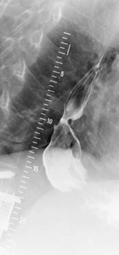
Long description
Barium-contrast fluoroscopic X-ray whose file page describes a short stricture of the lower esophagus in long-standing gastroesophageal reflux disease.

- Upper endoscopy (EGD). Anatomy and mucosa. Grade erosive injury by the Los Angeles classification. Obtain biopsies for Barrett, eosinophilic oesophagitis or H. pylori when indicated, and for any lesion.
- Ambulatory pH monitoring (wireless capsule or catheter). Abnormal oesophageal acid exposure quantified as total acid exposure time and a composite DeMeester score. Monitor off PPI for 7 days when the diagnosis is uncertain, or prior to surgery or endoscopic therapy, to document reflux. [1]
- pH-impedance monitoring. Adds weakly acidic and non-acid reflux, proximal extent of reflux and symptom correlation. Useful for persistent symptoms on PPI and for extra-oesophageal presentations.
- Symptom correlation indices. Symptom index of 50 percent or more, or symptom association probability above 95 percent, support a link between reflux and symptoms, although both have limitations. [1]
- High-resolution manometry (HRM). Should be performed before any anti-reflux procedure to exclude achalasia and absent contractility. Also useful in refractory GERD when endoscopy and pH testing are unrevealing. [1]
The Los Angeles classification of erosive oesophagitis grades mucosal injury from A (small mucosal breaks) to D (circumferential large mucosal breaks). [1] Los Angeles grades C and D represent more severe disease and, with large hiatus hernias or persistent troublesome symptoms, are indications for maintenance therapy or intervention. [1]
A clinical definition of GERD is symptom-based reflux that either improves with acid suppression or is proven abnormal on reflux monitoring. Objective confirmation (mucosal injury or abnormal acid exposure) is required for research and before invasive treatment.
See the dedicated Barrett oesophagus section below for who to screen, how to biopsy and surveillance intervals.
A stepwise approach to GERD
- Typical symptoms (heartburn, regurgitation) with no alarm featuresEmpiric PPI once daily 30-60 minutes before breakfast for 8 weeks; optimise lifestyle
- Question: Symptom response?
Complete or partial response
- Step down: lowest effective PPI dose, on-demand or intermittent therapyReassess; switch to H2 blocker at night if nocturnal symptoms only
No or only partial response
- Verify diagnosis: exclude alarm features and consider endoscopyOptimise PPI timing and dose (twice daily) before declaring failure
- Question: Endoscopy findings?
Los Angeles C or D, Barrett, stricture
- Treat accordingly; maintain PPI; consider surgery
Normal or Los Angeles A or B
- Ambulatory pH or pH-impedance testing off PPI; HRM if refractory or pre-surgicalCorrelate symptoms with reflux; exclude eosinophilic oesophagitis
- Objective documentation before any anti-reflux surgery or endoscopic therapy
The evidence behind lifestyle advice is mixed, so be honest about what works:
- Weight loss in overweight or obese patients improves reflux symptoms in a dose-dependent way: a BMI reduction of about 3.5 improved frequent symptoms in about 40 percent of women in one cohort. [1]
- Smoking cessation improved symptoms in about 44 percent of successful quitters versus 18 percent of continuing smokers in a cohort study. [1]
- Avoidance of late meals and lying down after eating, elevating the head of the bed and, in some patients, left-side-down positioning reduce nocturnal reflux. Randomised trials show benefit for head-of-bed elevation and wedge pillows. [1]
- Avoidance of "trigger foods" (coffee, tea, soda, chocolate, high-fat or spicy foods, alcohol, peppermint, citrus) is often advised but rests on low-quality evidence; their effect is mainly to reduce lower oesophageal sphincter pressure or to irritate. [1]
- Alcohol reduction is sensible, though evidence is less consistent than for weight and smoking.
- Baclofen reduces TLESRs and can be considered in patients with documented continued reflux despite optimal PPIs (5 to 20 mg three times daily). [1]
United States
In the United States, the ACG 2022 guideline recommends weight loss in overweight or obese patients (strong), and suggests avoidance of tobacco, trigger foods and head-of-bed elevation for night symptoms (conditional, low evidence). [1]
- Confirm the diagnosis before escalating beyond PPIs when the clinical picture is atypical.
- Match the drug to the severity: antacids and alginate for intermittent or mild symptoms; H2 receptor antagonists for milder or nocturnal symptoms; PPIs for erosive oesophagitis, complications and maintenance. [1]
- PPIs bind active proton pumps, so they work best when taken 30 to 60 minutes before the first meal of the day; twice-daily dosing should be taken before breakfast and before dinner. [1]
- Severe disease (Los Angeles grades C and D) needs maintenance PPI therapy or intervention, not just a short course. [1]
| Class | Typical dose | Role and evidence |
|---|---|---|
| Antacids (aluminium, calcium or magnesium salts) | 10-30 mL (1-2 tablespoonfuls) or 2-4 tablets after meals and at bedtime, on demand | Rapid, short-lived symptom relief; little healing benefit for erosive oesophagitis. [1][3][4] |
| Alginates (e.g. sodium alginate with antacid, "Gaviscon") | Product label dose after meals and at bedtime (combined products commonly 10-20 mL) | Forms a floating raft over gastric contents; reduces post-prandial reflux and symptoms; useful as add-on therapy. No universal standard alginate dose is established; follow the product label. [3] |
| H2 receptor antagonists | Famotidine 20-40 mg twice daily; nizatidine 300 mg twice daily; cimetidine up to 800 mg twice daily (1600 mg/day) | Less effective than PPIs for healing (about 50 percent vs 84 percent for erosive oesophagitis at standard doses); tolerance (tachyphylaxis) limits long-term utility; ranitidine withdrawn from some markets. [3] |
| Proton pump inhibitors (PPIs) | Omeprazole 20 mg; esomeprazole 20-40 mg; lansoprazole 15-30 mg; pantoprazole 40 mg; rabeprazole 20 mg; dexlansoprazole 30-60 mg (once daily; double the dose or give twice daily for severe disease) | Most effective class; heals 75-95 percent of erosive oesophagitis by 8 weeks. Standard once daily for non-erosive and mild disease; twice daily or double dose for severe, complicated or refractory disease. [1][3] |
| Potassium-competitive acid blockers (P-CABs) | Vonoprazan 20 mg once daily for healing erosive oesophagitis; 10 or 20 mg once daily for maintenance | Non-inferior to PPIs for healing, with advantages in advanced (Los Angeles C or D) and PPI-resistant erosive oesophagitis. [3] |
- Sucralfate 1 g four times daily (an aluminium salt that coats the mucosa) is first-line only in pregnancy; routine use in GERD is not recommended, because evidence is limited. [1][3]
- Metoclopramide can be used cautiously for gastroparesis, but is not recommended for GERD itself because of limited efficacy and adverse central nervous system effects with long-term or high-dose use. [1]
- Baclofen 5 to 20 mg three times daily reduces TLESRs in documented reflux (see Lifestyle). [1]
About two-thirds of pregnant women report heartburn. Start with lifestyle measures. If those fail, antacids (aluminium, calcium or magnesium containing), alginates and sucralfate are first-line. All H2 blockers are FDA pregnancy category B and all PPIs are category B except omeprazole (category C). [1]
United States
In the United States, the ACG 2022 guideline carries over the older FDA pregnancy categories; local formularies and current labeling should be checked. [1]
The safety of long-term PPIs is one of the most contested topics in gastroenterology, and an honest summary matters:
- The ACG 2022 GERD guideline advises telling patients that high-quality studies have found PPIs do not significantly increase the risk of pneumonia, fracture, cardiovascular disease, dementia, chronic kidney disease, cancer or all-cause mortality, except for a small increase in enteric infections. PPIs do not cause a significant increase; the well-established benefits for GERD generally outweigh the theoretical risks. [1]
- The strongest available evidence comes from a randomised trial (3 years) in which pantoprazole 40 mg daily produced no significant difference from placebo for any tested outcome except enteric infection (1.4 percent versus 1.0 percent; odds ratio 1.33, 95 percent confidence interval 1.01-1.75). [1]
- PPIs should still be used at the lowest effective dose and de-prescribed when the indication has resolved. Enteric infection (including C. difficile) is the one outcome with credible, consistent signal.
- Long-term PPI use can also cause hypomagnesaemia; routine monitoring of serum magnesium is not recommended except in symptomatic patients or those on interacting drugs. [1]
- Routine monitoring of vitamin B12 or creatinine is not recommended in GERD patients on PPIs with no other risk factors. [1]
Definition. Symptoms of GERD that persist despite optimised medical therapy, usually 8 or more weeks of twice-daily PPI taken correctly.
Workup (stepwise):
- Optimise the PPI. Verify adherence and correct timing; in one study only about 12 percent improved when omeprazole was dosed before breakfast and dinner with explicit instructions. Doubling the dose also helps some. [1]
- Endoscopy. Exclude erosive oesophagitis, Barrett oesophagus, stricture, infection and eosinophilic oesophagitis.
- Ambulatory reflux monitoring. Off PPI when the original diagnosis is uncertain; pH-impedance on PPI when diagnosis was previously proven. Look for acid, weakly acidic or non-acid reflux, symptom association, and reflux hypersensitivity. [1]
- High-resolution manometry and gastric emptying if reflux testing is unrevealing, to look for motility disorders, achalasia, rumination or delayed gastric emptying.
- Anxiety, depression and functional heartburn account for a substantial share of patients who turn out to have normal acid exposure and no erosive disease. [1]
Regurgitation as the main symptom that persists on PPI, with objectively abnormal reflux, is the best predictor of benefit from anti-reflux surgery or a transoral incisionless fundoplication. [1]
Surgery and endoscopy are reserved for patients with objectively documented GERD: erosive disease, Barrett oesophagus, large hiatus hernia or proven abnormal reflux, with troublesome symptoms. They are not a shortcut around the workup.
- Laparoscopic Nissen fundoplication is the gold standard among anti-reflux procedures. It is widely regarded as the long-term treatment of choice when performed by an experienced surgeon, especially in Los Angeles grades C or D oesophagitis, large hiatus hernias and persistent, troublesome symptoms. [1]
- Pre-operative high-resolution manometry is recommended to exclude achalasia and absent contractility (a contraindication to fundoplication). Tailoring the operation to peristalsis (complete vs partial fundoplication) is not supported by evidence. [1]
- Medical therapy response predicts surgical success. The best outcomes (about two-thirds success) are in patients with typical symptoms who previously responded to PPIs; pooled data show laparoscopic Nissen fundoplication (66.7 percent) significantly outperformed active medical (28.0 percent) and placebo medical therapy (11.5 percent) at one year in rigorously confirmed refractory GERD (P less than 0.01). [1]
- Magnetic sphincter augmentation (MSA, a ring of magnets around the lower oesophageal sphincter) is suggested as an alternative to fundoplication in patients with regurgitation failing medical therapy, with objective documentation of abnormal reflux. [1]
- Transoral incisionless fundoplication (TIF) creates a partial fundoplication endoscopically and is suggested for troublesome regurgitation or heartburn in patients who do not want surgery and who do not have Los Angeles C or D disease or a hiatus hernia above 2 cm. [1]
- Reproducible durability caveat: a substantial proportion of patients need reflux medication again after fundoplication (up to about 43 percent in cohort studies), although surgical patients report higher satisfaction in the short to medium term. [1]
Obesity complicates and strains a standard fundoplication. The ACG 2022 guideline suggests consideration of Roux-en-Y gastric bypass (RYGB) as an option to treat GERD in obese patients who are candidates and willing to accept its risks and lifestyle demands. [1] The small gastric pouch produces little acid and the long Roux limb prevents bile reflux. Fundoplication has poorer expected outcomes in this group. [1]
There is little evidence that emergency surgery is superior to a prompt planned operation; surgery can be timed according to the patient's condition, for example after a recent stroke. [1]
Figure 2Barrett oesophagus at endoscopy
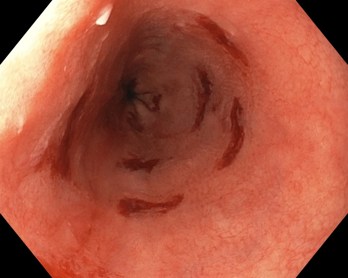
Long description
Endoscopic view of the lower oesophagus lined by salmon-pink columnar mucosa, with fresh biopsy sites around the lumen. The salmon-coloured segment extends up from the gastro-oesophageal junction; biopsies are taken in four quadrants at intervals along it to look for intestinal metaplasia and dysplasia.

Figure 3Oesophagus-Ca AEG I
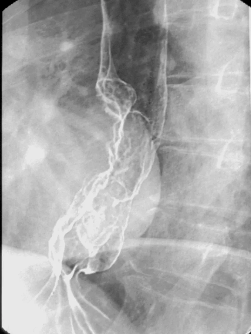
Long description
Barium swallow (Breischluckuntersuchung) described on its file page as an adenocarcinoma of the gastroesophageal junction, type 1, with the main mass above the cardia.

- Erosive oesophagitis. Los Angeles A to D; higher grades often need maintenance PPI and predict recurrence on stopping treatment.
- Peptic stricture. From chronic ulceration and scarring; presents with progressive dysphagia; treated by dilation plus strong acid suppression.
- Barrett oesophagus. Survival-critical precursor to adenocarcinoma (see below).
- Oesophageal adenocarcinoma. The feared end of the metaplasia-dysplasia-carcinoma sequence.
- Extra-oesophageal disease. Chronic cough, laryngitis, hoarseness, asthma exacerbations, dental enamel erosion, non-cardiac chest pain and aspiration; objective reflux is found in only a minority, so evidence of causation is required before attributing these to GERD. [1]
Barrett oesophagus is a change of the distal oesophageal epithelium to an intestinal (columnar) type with specialised intestinal metaplasia (goblet cells), usually arising through chronic reflux injury. [2] It is a precursor to oesophageal adenocarcinoma. Risk factors include male sex, White race, age above 50 years, central obesity, tobacco smoking, a family history in a first-degree relative and long-standing GERD. [2]
The annual incidence of progression to cancer is estimated at roughly 0.2 to 0.5 percent per year for non-dysplastic Barrett and about 0.7 percent per year for low-grade dysplasia. [2] Most patients never progress to cancer, but a minority does, which is why structured surveillance and risk stratification matter.
A single screening endoscopy is suggested for patients with chronic GERD symptoms and three or more additional risk factors (male sex, age above 50 years, White race, tobacco smoking, obesity, family history of Barrett or adenocarcinoma). Screening women with chronic GERD symptoms alone is unlikely to be cost-effective, though screening a woman with multiple risk factors may be appropriate after discussion. [2]
Screening is only useful where follow-up surveillance and (if needed) endoscopic treatment are available locally.
- The distal oesophagus is inspected carefully under high-definition white light and virtual chromoendoscopy.
- Prague classification records the circumferential and maximal extent of the Barrett segment.
- Seattle protocol: visually targeted biopsies of any lesion, then four-quadrant biopsies at intervals of no more than 2 cm from the lower oesophageal sphincter to the squamocolumnar junction. [2]
- Two pathologists should confirm grades of dysplasia, especially indefinite for dysplasia and low-grade dysplasia. [2]
- If endoscopy and biopsies refute Barrett, record a clear negative result; repeat findings and technique vary.
Ten good-practice steps of the endoscopic examination include adequate cleaning and insufflation, use of a distal cap, systematic review, virtual chromoendoscopy, Prague and Paris classifications, Seattle biopsies and retroflexion of the gastric cardia. [2]
| Baseline finding | Suggested interval (ACG 2022) |
|---|---|
| Non-dysplastic Barrett, segment under 3 cm | Every 5 years |
| Non-dysplastic Barrett, segment 3 cm or more | Every 3 years |
| Indefinite for dysplasia (confirmed by a second pathologist) | Repeat endoscopy within 6 months after optimising PPI to twice daily; thereafter follow the result |
| Low-grade dysplasia (confirmed by a second pathologist, opting for surveillance) | 6 months, then 12 months, then annually |
| High-grade dysplasia / intramucosal carcinoma | See endoscopic eradication below |
Segment length itself predicts progression (longer segments, higher risk), so assign intervals by length as above. [2]
- Non-dysplastic or indefinite for dysplasia: PPI therapy (often twice daily) and surveillance at the intervals above. [2]
- Low-grade dysplasia: endoscopic eradication therapy is preferred over surveillance for most, but surveillance is a reasonable option for patients with risk of treatment. Endoscopic eradication therapy removes (radiofrequency ablation or endoscopic resection) the Barrett segment. [2]
- High-grade dysplasia and intramucosal carcinoma: complete endoscopic eradication is the standard.
- Endoscopic mucosal resection (EMR) (cap-assisted or multiband) gives tissue diagnosis for any visible lesion before ablation and is the preferred resection technique; endoscopic submucosal dissection is an alternative where needed. [2]
- Radiofrequency ablation (RFA) is the most widely studied ablative modality and is the preferred ablative method for flat dysplastic tissue. [2]
- After complete eradication of intestinal metaplasia, surveillance is tailored to the worst pretreatment histology: low-grade dysplasia at 1 year, 3 years, then every 2 years; high-grade dysplasia or intramucosal carcinoma at 3, 6 and 12 months, then annually. [2]
- Antireflux control (PPI or surgery where indicated) is part of post-treatment care because new or recurrent metaplasia and dysplasia remain possible. [2]
Gastro-oesophageal reflux is physiological in infants. Pathological GERD in children can present with feeding refusal, vomiting, poor weight gain, irritability, apnoea or cough and aspiration. Investigations are reserved for complications or a doubtful diagnosis. Treatment begins with lifestyle measures (feeding changes, thickened feeds, positioning) and weight gain; medical therapy follows age-appropriate dosing from guideline and label; surgery is reserved for complications or failure. A joint Italian consensus (SIP/SIGENP 2026) provides detailed guidance on paediatric management; regional paediatric guidelines take precedence over adult algorithms. [1]
See Medical therapy - Pregnancy above.
Older patients often present late with strictures or Barrett changes. In scleroderma and other motility disease, reflux is frequently severe and silent; screen with low threshold and treat aggressively. [1]
- Diagnosis can be mostly clinical. A careful history of heartburn and regurgitation, the response to an affordable 4- to 8-week trial of a PPI (8 weeks in ACG 2022; 4 to 8 weeks accepted in the Seoul Consensus 2025 for non-erosive or mild erosive disease), and the exclusion of alarm features are sufficient for most patients. [1][4]
- Endoscopy is not always available; when it is, prioritise alarm features, age above 50 at onset, treatment failure and suspected Barrett.
- Ambulatory pH testing and manometry are often unavailable. Invasive anti-reflux procedures should not be offered without objective documentation where it can be obtained; otherwise keep treatment medical.
- PPIs are on the WHO essential medicines list and are inexpensive; where only one PPI exists use whatever is available at standard doses, taken 30-60 minutes before meals.
- Fundoplication requires a skilled surgical team and pre-operative testing; if unavailable locally, refer to a tertiary centre and treat medically in the meantime.
- H. pylori eradication is cheap and relevant because peptic ulcer disease mimics GERD.
- Screening for Barrett is impractical where endoscopy is scarce; focus resources on patients with multiple risk factors and alarm features.
Peptic ulcer disease and Helicobacter pylori infection; dysphagia; eosinophilic oesophagitis; gastric emptying disorders; oesophageal squamous and adenocarcinoma; weight management and bariatric surgery.
- GERD is reflux of gastric contents causing symptoms or complications; heartburn and regurgitation are classic, cough, hoarseness, asthma and chest pain are extra-oesophageal possibilities.
- Transient lower oesophageal sphincter relaxations, worsened by a hiatus hernia, obesity, pregnancy, delayed gastric emptying, scleroderma and several drugs, are the dominant mechanism.
- Most patients are diagnosed by typical symptoms and an 8-week empiric PPI trial; alarm features mean urgent endoscopy.
- Upper endoscopy grades erosive injury (Los Angeles A to D), finds Barrett and stricture; ambulatory pH or pH-impedance confirms reflux; manometry precedes any anti-reflux procedure.
- Lifestyle measures that genuinely help are weight loss, smoking cessation, elevation of the head of the bed and avoidance of late meals; "trigger food" avoidance has weak evidence.
- Antacids and alginates relieve symptoms; H2 receptor antagonists (famotidine 20-40 mg twice daily) are less effective; PPIs (omeprazole 20 mg and equivalents, timed before breakfast) heal most erosive disease in 8 weeks; vonoprazan 20 mg is an effective alternative.
- Long-term PPIs: high-quality evidence does not show significant increases in pneumonia, fracture, renal, cognitive, cardiovascular or mortality risk except a modest increase in enteric infection, and benefits for GERD outweigh theoretical risks; use the lowest effective dose.
- Refractory GERD demands optimisation, endoscopy, pH or pH-impedance testing and manometry; functional heartburn and rumination are common explanations.
- Anti-reflux surgery (laparoscopic Nissen) and MSA or TIF are reserved for documented disease; obesity argues for Roux-en-Y gastric bypass; objective proof is required.
- Barrett oesophagus is intestinal metaplasia at risk of adenocarcinoma; screen those with chronic reflux plus three or more additional risk factors; biopsy by the Seattle protocol (four quadrants at intervals of 2 cm or less); survey by grade (non-dysplastic: 5 yearly if under 3 cm, 3 yearly if 3 cm or more; low-grade dysplasia: 6, 12 months then annually); treat low-grade and higher with endoscopic eradication (RFA and EMR).
- Infants and children need age-specific assessment; pregnancy changes drug choice; low-resource settings rely on clinical diagnosis and affordable PPIs.
Answer each one in your head first, then open the answer.
A 45-year-old has heartburn and regurgitation for six months, no alarm symptoms, BMI 32, and a normal examination. What is the best first step in management?
Show answerHide answer
Diagnose clinically and treat with an 8-week trial of a PPI taken 30-60 minutes before breakfast, combined with weight loss. No investigation is needed unless symptoms persist or alarm features appear. [1]
Which acid-reducing drug classes are used for GERD, and which is most effective for healing erosive oesophagitis?
Show answerHide answer
Antacids and alginates for symptom relief, H2 receptor antagonists (famotidine 20-40 mg twice daily) as a weaker option, PPIs (omeprazole 20 mg once daily and equivalents) as the most effective, and P-CABs such as vonoprazan 20 mg once daily for healing. PPIs heal 75-95 percent of erosive oesophagitis by 8 weeks. [1][3]
Your patient has persistent heartburn despite 8 weeks of correctly timed omeprazole 20 mg twice daily, with a normal endoscopy and normal acid exposure on pH monitoring. What is the most likely explanation and what should you avoid?
Show answerHide answer
Functional heartburn or a behavioural condition such as rumination or supragastric belching. Avoid escalating to surgery without objective evidence of reflux; optimise therapy and consider neuromodulation, diaphragmatic breathing and referral to a specialist team. [1]
Who should be screened for Barrett oesophagus, and how are biopsies taken?
Show answerHide answer
A single screening endoscopy is suggested for patients with chronic reflux symptoms and three or more additional risk factors (male sex, age above 50 years, White race, obesity, tobacco smoking, family history). Biopsies follow the Seattle protocol: targeted biopsy of visible lesions plus four quadrants at 2 cm intervals along the segment. [2]
A patient with Barrett oesophagus has confirmed low-grade dysplasia. What are the management and surveillance options?
Show answerHide answer
Endoscopic eradication therapy (radiofrequency ablation and endoscopic mucosal resection of visible lesions) is preferred, though surveillance is acceptable in selected patients; if surveillance is chosen, endoscopy at 6 months, 12 months and annually thereafter. If complete eradication is achieved, follow 1 year, then 3 years, then every 2 years. [2]
A 28-year-old man with obesity and a sliding hiatus hernia has documented reflux unresponsive to medical therapy. Which surgical option fits best and why?
Show answerHide answer
In a patient with obesity, Roux-en-Y gastric bypass is the preferred anti-reflux and bariatric option, because fundoplication is prone to poor outcomes with elevated abdominal pressure and the small pouch produces little acid. Standard laparoscopic Nissen fundoplication is otherwise the gold standard in non-obese patients with confirmed disease. [1]
How should PPIs be prescribed to get the best acid suppression?
Show answerHide answer
Take the PPI 30 to 60 minutes before the first meal of the day for once-daily dosing, and before breakfast and dinner for twice-daily dosing, because proton pumps are activated by meals. [1]
- Katz PO, Dunbar KB, Schnoll-Sussman FH, Greer KB, Yadlapati R, Spechler SJ, et al. ACG Clinical Guideline: Guidelines for the Diagnosis and Management of Gastroesophageal Reflux Disease. The American Journal of Gastroenterology 2022;117:27-56.. Accessed .
- Shaheen NJ, Falk GW, Iyer PG, Souza RF, Yadlapati RH, Sauer BG, Wani S, et al. Diagnosis and Management of Barrett's Esophagus: An Updated ACG Guideline. The American Journal of Gastroenterology 2022;117:559-587.. Accessed .
- Huh CW, Lee DH, et al. Seoul Consensus 2025: focused update on acid suppressive therapy in gastroesophageal reflux disease. Journal of Neurogastroenterology and Motility 2025/2026; focused update.. Accessed .
- Haleon US Holdings LLC. Gaviscon liquid antacid product label (sodium alginate-containing formulation). DailyMed product labeling; directions 1-2 tablespoonfuls four times daily.. Accessed .
Figure credits
Educational content for medical students and doctors. It is not a substitute for clinical judgement or local guidelines.