Hiatal hernia
A hernia of stomach through the diaphragmatic hiatus: types, symptoms, Cameron anaemia, imaging, surgery for paraesophageal hernia, and emergencies.
- A hiatal hernia is present when part of the stomach (or other abdominal contents) pushes up through the oesophageal opening in the diaphragm into the chest.
- Type I (sliding) hernias are by far the commonest. The gastro-oesophageal junction (GEJ) itself is displaced upward, and reflux is the usual problem.
- Types II to IV are paraesophageal (rolling) hernias. The GEJ may stay below the diaphragm while the stomach (and sometimes other organs) rolls up beside the oesophagus, with a risk of twisting, obstruction and ischaemia.
- Most sliding hernias are managed exactly as gastro-oesophageal reflux disease (GERD): lifestyle changes, then acid suppression.
- Symptomatic paraesophageal hernias are treated surgically. In elderly or minimally symptomatic patients, careful watchful waiting is increasingly accepted.
- Severe upper abdominal pain with retching but no vomiting and inability to pass a nasogastric tube is Borchardt's triad: think gastric volvulus and treat as an emergency.
By the end of this page you should be able to:
- Describe the normal diaphragmatic hiatus and gastro-oesophageal junction, and where they fail in a hiatal hernia.
- Classify a hiatal hernia into its four types and say which type you are looking at.
- List the presenting features, including when a hernia causes iron-deficiency anaemia through Cameron lesions.
- Choose and interpret the investigations used for diagnosis and pre-operative planning.
- Manage a sliding hernia as reflux, and manage a paraesophageal hernia including when to watch and when to operate.
- Recognise volvulus with Borchardt's triad and outline emergency management.
- Describe the common post-operative problems and how to practise in a low-resource setting.
DefinitionHiatal hernia
A hiatal hernia is the protrusion of the stomach, or (in type IV) other abdominal viscera, through the oesophageal hiatus of the diaphragm into the posterior mediastinum. "Hiatus hernia" and "hiatal hernia" are the same thing; "paraesophageal hernia" refers specifically to types II to IV.
The term tells you the anatomy, not the symptom. A small sliding hernia can be completely silent; a paraesophageal hernia can slowly strangulate with little warning. As one review puts it, the two look similar on a scan, but their risks and treatments are quite different.[1]
The diaphragm is a muscular partition. The oesophageal hiatus is the slit in its crural part through which the oesophagus enters the abdomen. Around that entry sit three structures that matter for reflux and herniation.
- The crural diaphragm. The right and left crura of the diaphragm form a muscular sleeve around the oesophagus at the hiatus. Their "pinch" assists the lower oesophageal sphincter (LOS) in closing, and they act as an external sphincter.[2]
- The lower oesophageal sphincter (LOS). This is not a single ring but a zone of high pressure in the distal oesophagus just above the stomach. In health, part of it lies in the abdomen, where positive intra-abdominal pressure keeps it closed.
- The gastro-oesophageal junction (GEJ or EGJ). The point where oesophageal mucosa meets gastric mucosa (the Z-line). The "angle of His" between the oesophagus and the gastric fundus is an important flap-valve component of the anti-reflux barrier.
In a hiatal hernia, the supporting ligaments (the phrenoesophageal membrane) and the muscular hiatus are lax or widened. Part of the stomach is pulled or pushed up through the opening. Two anti-reflux defences are then lost: the intra-abdominal segment of the sphincter is shortened, and the crural pinch no longer works. That is why sliding hernias are associated with reflux. In a paraesophageal hernia, the greater danger is mechanical: the stomach can rotate or become trapped above a relatively tight hiatus.
Figure 1Digestive system - Paraesophageal hernia -- Smart-Servier
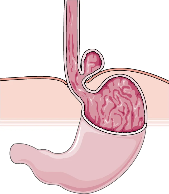
Long description
A medical illustration titled 'Paraesophageal hernia' from SMART-Servier Medical Art showing a paraesophageal hiatal hernia.

Figure 2Hiatus hernia
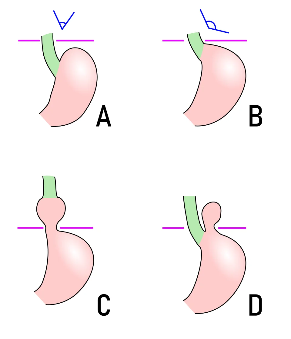
Long description
Schematic diagram of types of hiatus hernia: normal anatomy, pre-stage, sliding hiatus hernia (panel C), and paraesophageal hiatus hernia, with color-coded esophagus, stomach, diaphragm, and HIS angle.

The type depends on where the GEJ is and on what else has herniated. A plain classification of the four types is the single most useful thing to learn, because it drives management.
| Type | What herniates | Where the GEJ is | Frequency | Key point |
|---|---|---|---|---|
| Type I (sliding) | GEJ and gastric cardia/fundus slide up together | Above the hiatus | By far the commonest: more than 90% of cases in one review, and more than 95% in others[1][6] | Often asymptomatic; behaves as reflux disease |
| Type II (pure paraesophageal) | Gastric fundus rolls up beside the oesophagus | Remains below the diaphragm, in normal position | Part of the remaining small fraction; type II PEH is reported as around 5% or less of all hiatal hernias[5][6] | Risk of volvulus despite few reflux symptoms |
| Type III (mixed) | Both fundus and GEJ herniate through the hiatus; fundus lies above the GEJ | Above the hiatus (combined with fundus) | Uncommon, a subset of paraesophageal cases | Combines reflux with mechanical complications |
| Type IV | Stomach plus another organ (small bowel, colon, omentum, spleen, peritoneum) | Above the hiatus | Rare | May present as a large mediastinal mass |
One review's summary is representative: type I is the "sliding" hernia, while types II to IV are paraesophageal hernias whose main importance is their potential for ischaemia, obstruction or volvulus.[1] A current surgical guideline gives the same anatomical definitions.[2]
Hiatal hernia is common. In most studies, how common depends on the method used to look and on the age of the people studied.
- On upper endoscopy, hiatal hernias are seen in about 0.8 to 2.9% of all patients.[3] Hiatal hernia is also one of the most frequent findings at autopsy and chest imaging in older people.
- Sliding hernias are reported with a prevalence ranging from 10% to 60% depending on the series, and they represent the overwhelming majority of cases.[5]
- The prevalence rises with age. Older individuals are affected far more often than people under the age of 50.[5] A surgical guideline specifically notes that prevalence increases alongside increasing age, and that age alone should not exclude operative management.[2]
- A large proportion of patients have no symptoms. In one hiatal-hernia series only about 9% of patients had symptoms.[6] Another guideline notes that most patients undergoing repair actually do have symptoms that were not initially attributed to the hernia.[2]
The key clinical lesson is that finding a hernia is not the same as finding the cause of the patient's symptoms. Always correlate the image with the history.
No single cause explains every hiatal hernia. The current view is that the hiatus widens and the phrenoesophageal membrane stretches or tears, allowing the stomach to move up. Recognised contributing factors include:
- Age. The membrane and crural muscle weaken over time, which explains the steep rise in prevalence with age.[1][5]
- Raised intra-abdominal pressure. Obesity, pregnancy, chronic cough, heavy lifting, constipation and straining all increase pressure and predispose to herniation. "Being overweight and elderly are the key risk factors" in one review's words.[1]
- Obesity. A common comorbidity and a known risk factor for recurrence after repair; obese patients are discussed below and in the bariatric surgery topics.[1][2]
- Prior surgery. A history of oesophageal surgery, partial or total gastrectomy, or iatrogenic injury can predispose to herniation.[1]
- Skeletal conditions associated with bone decalcification and degeneration are also listed as known associations.[1]
- Multiple pregnancies are a recognised risk factor.[1]
Sliding and paraesophageal hernias partly share these factors, but a paraesophageal hernia is more likely to be found in an older person who has had a long-standing, slowly enlarging defect. Female sex is not a defining feature; the strongest associations are age and obesity.
Sliding hernia (type I). The GEJ and gastric cardia move up through the hiatus. The intra-abdominal sphincter segment is lost and the crural pinch fails, so the barrier against reflux fails. Acid reflux and its complications (oesophagitis, Barrett's metaplasia) follow; sliding hernias are associated with more severe degrees of oesophagitis and with Barrett's oesophagus. A small hernia may cause no symptoms at all and may be found incidentally.
Paraesophageal hernia (types II to IV). The stomach, and sometimes other organs, lies in the chest, usually in an upright person above the diaphragm. Problems come from:
- Mechanical competition. A large volume sits in the chest, compressing lung and heart, which can cause breathlessness, cough and postprandial fullness.
- Twisting. The stomach can rotate on its own axis (gastric volvulus) within a tight hiatus, causing obstruction.
- Strangulation. The blood supply to the herniated stomach can be compromised, leading to ischaemia, ulceration and perforation.
- Bleeding from Cameron lesions. The gastric mucosa at the neck of a large hernia is subject to mechanical trauma and pressure ischaemia at the diaphragmatic impression; linear erosions or ulcers (Cameron lesions) form and can bleed slowly. These lesions are seen in about 5% of patients with a known hiatal hernia and are an important cause of occult iron-deficiency anaemia.[3]
Most type I hernias present, if at all, like reflux disease, and many are silent.
- Heartburn and acid regurgitation are the dominant complaints when the patient is symptomatic.
- Dysphagia is common with a large hernia, because a misaligned junction or a pouch of retained food obstructs or compresses the oesophagus.
- Retrosternal or epigastric chest pain, sometimes postprandial and sometimes relieved by antacids.
- Respiratory symptoms: cough, wheeze, hoarseness and recurrent aspiration, especially at night.
- Asymptomatic detection on chest radiograph or CT. Remember that a sliding hernia found incidentally in an asymptomatic person does not by itself need surgery.
The symptoms are more mechanical and postprandial, and reflux is often a minor feature unless a type III (mixed) hernia is present.
- Postprandial fullness, early satiety and inability to finish meals, from the stomach being compressed in the chest.
- Regurgitation of undigested food or liquids, sometimes with relief of chest pressure.
- Dysphagia, particularly for solids, because the junction is angulated.
- Chest pain or pressure after meals, sometimes with shortness of breath.
- Anaemia from Cameron lesions: chronic occult bleeding leads to microcytic, hypochromic iron-deficiency anaemia. All patients with an unexplained iron-deficiency anaemia should be asked about dysphagia and postprandial chest symptoms, and examined carefully for a large hernia.[3]
- Respiratory symptoms from lung compression or aspiration.
- Acute catastrophic presentation: sudden severe epigastric or lower chest pain, retching without vomiting, inability to pass a nasogastric tube (Borchardt's triad), which means gastric volvulus or obstruction.
Among patients undergoing repair of a paraesophageal hernia, studies report presenting anemia in 24 to 57%, dyspnoea in 21 to 67%, and chest pain in 40 to 60%.[2] Many such patients had been labelled asymptomatic or mildly symptomatic.
Most patients have few signs. Large hernias can produce:
- Bowel sounds or a succussion splash audible in the chest on auscultation, or over the lower chest if a large part of the stomach is intrathoracic.
- Microcytic anaemia on routine blood tests.
- Signs of GERD such as dental enamel erosion in severe, long-standing reflux.
- Acute obstruction or volvulus: tender epigastrium, a large air-filled viscus on the chest radiograph, and (in organo-axial volvulus) the inability to pass a nasogastric tube.
For a hiatal hernia, the key distinction is with GERD without hernia, and with complications that mimic it.
| Mimic | What separates it |
|---|---|
| GERD without hernia | No axial herniation on imaging; barium swallow or manometry shows the GEJ below the diaphragm. Treat as reflux alone. |
| Achalasia and motility disorders | Dysphagia to both solids and liquids; dilated oesophagus with a bird's beak; no gastric intrathoracic component. Manometry is diagnostic. |
| Oesophageal carcinoma / peptic stricture | Progressive dysphagia, weight loss, anaemia; endoscopy and biopsy required. |
| Mediastinal mass (type IV confusion) | A type IV hernia can look like a mediastinal mass; CT with contrast shows stomach or bowel containing air and contrast, not a solid tumour. |
| Cardiac chest pain | Exertional, not postprandial; ECG changes; a hernia found on scan may be incidental. |
| Diaphragmatic eventration or other hernia | Different defect in the diaphragm; the hiatus itself may be normal. |
Only tests that will alter management need be done.[1] A complete history and physical examination come first. Investigations map directly to type and to surgical planning.
Often the first clue. A retrocardiac air-fluid level behind the heart, an intrathoracic stomach bubble, or an unusual mediastinal shadow can suggest a large hernia. Air-fluid levels in the chest on an erect film are particularly suggestive of gastric volvulus or obstruction.
Still essential. It gives the size of the herniated stomach, the position of the GEJ, and the shape of the hernia.[1] According to Siegal and colleagues, hiatal hernia can be diagnosed on barium studies if the axial herniation is greater than 2 cm.[1] A video oesophagram adds information on bolus transit, and the study can pick up motility dysfunction, stricture and short oesophagus.[1] Barium swallow is contraindicated in pregnancy and in barium or iodine hypersensitivity, and it carries a radiation dose.
Real-time mucosal inspection. It detects reflux oesophagitis, Barrett's oesophagus, Cameron ulcers and lesions suspicious for malignancy, all of which must be documented before surgery.[1] Air insufflation during the procedure can exaggerate hernia size, and massive hernias (especially with organo-axial rotation) are hard to assess accurately; combine endoscopy with barium swallow for an exact description.[1]
Not routine, but useful. CT shows gastric volvulus, perforation, pneumoperitoneum or pneumomediastinum, and helps in the assessment of large paraesophageal hernias.[1] It is frequently discovered incidentally. CT also stages other pathology when malignancy is a concern.
Esophageal manometry provides valuable information on motility. A separation between the crural diaphragm and lower oesophageal sphincter of 2 cm or more is diagnostic for hiatal hernia.[1] It should be performed before surgery, to rule out achalasia or other motility disorders and to confirm adequate peristalsis before fundoplication; high-resolution manometry is the current tool. Placement of the catheter can be difficult, and the test is completed in fewer than 50% of cases.[1] pH testing is not essential to diagnose the hernia, but quantifies reflux and is the practical "gold standard" to document acid exposure.[1]
For surgical planning, the combination of barium swallow, upper endoscopy and manometry is regarded as essential by most authors.[1]
Approach to a suspected hiatal hernia
- Symptoms or incidental findingReflux, regurgitation, dysphagia, postprandial fullness, chest pain, anaemia, or an air-fluid level on chest X-ray
- Clinical assessment firstHistory for volvulus red flags: sudden severe pain, retching without vomiting, inability to pass NGT
- Question: Red flags of acute volvulus or obstruction?
Yes
- Emergency pathwayResuscitate, attempt NGT decompression, CT if stable, emergency surgery
No
- Confirm and characterise the herniaBarium swallow for size and configuration; endoscopy for mucosa, Barrett's, Cameron ulcers; consider manometry
- Question: Which type?
Type I (sliding)
- Treat as GERDLifestyle, weight loss, PPI; surgery only if reflux persists despite optimal therapy
Types II to IV (paraesophageal)
- Consider surgical repair when symptomaticEspecially obstruction, volvulus, bleeding anaemia, disabling mechanical symptoms
- Truly asymptomatic?Discuss; watchful waiting is reasonable if no reflux or micro-aspiration is documented
Management follows the type:
- Type I (sliding) is treated as reflux disease.
- Types II to IV (paraesophageal) require a surgical decision. Symptomatic cases, particularly with obstructive symptoms or volvulus, need surgery. Asymptomatic cases are discussed and watched.
Because reflux is the problem, start with the measures that apply to GERD.
- Lifestyle modifications are first line. Weight loss; elevate the head of the bed by 8 inches during sleep; avoid meals 2 to 3 hours before bedtime; eliminate trigger foods (chocolate, alcohol, caffeine, spicy foods, citrus, carbonated drinks).[1]
- Proton pump inhibitor (PPI) for symptom relief: an 8-week course is the therapy of choice, with no major efficacy difference between the different PPIs. Use the minimal dose that controls symptoms.[1]
- Twice-daily PPI can be recommended where the response to once daily is inadequate.[1]
- Alternatives are histamine-2 receptor antagonists and antacids, used on demand for moderate symptoms or as an add-on when symptoms persist on a PPI.[1]
- Prokinetic drugs are not recommended, either alone or as add-on treatment.[1]
- Surgery (fundoplication, usually laparoscopic Nissen 360 degrees, or Toupet 270 degrees where there is dysmotility) is considered when a sliding hernia accompanies reflux symptoms that persist despite medical treatment.[1] The SAGES guideline strongly recommends not repairing a type I hernia in the absence of reflux disease and symptoms.[1]
For a large hernia with Cameron lesions, the initial treatment is long-term high-dose PPI plus iron replacement, as in one reported case using pantoprazole 40 mg twice daily with oral or intravenous iron.[3] Persistent anaemia or re-bleeding occurs in about 20% of patients, in whom surgical repair should be considered.[3]
Surgery (laparoscopic fundoplication, often with reduction and crural repair) is the definitive treatment for paraesophageal hernia, and is strongly recommended for symptomatic patients, especially when obstructive symptoms or volvulus are present.[1]
For patients with few or no symptoms, the position is less clear and patient-centred:
- A current surgical guideline makes no evidence-based recommendation for asymptomatic patients (no trials exist), and offers expert opinion instead. It stresses first confirming that the patient is truly asymptomatic, then looking for objective evidence of reflux or micro-aspiration; patients with objective reflux or micro-aspiration may be offered repair if medically fit. Watchful waiting is reasonable where work-up is unrevealing and the patient understands the small risk of acute gastric volvulus.[2]
- Older patients: a study of octogenarians found that elective repair in symptomatic patients should be the preferred approach.[1] Age alone should not exclude surgery where symptoms and fitness support it.[2]
- Young asymptomatic patients: one review cites studies suggesting that even asymptomatic patients younger than 50 should be considered for surgery.[1] Others, with the same evidence, favour watchful waiting; this is genuinely an area where surgeons disagree.
- Fundoplication during repair: a 2024 guideline suggests that patients undergoing type II to IV repair may benefit from adding a fundoplication (conditional recommendation, low-certainty evidence), in view of large reductions in objective reflux with small increases in late dysphagia.[2]
- Bariatric surgery and hiatal hernia can be combined; sleeve gastrectomy and hiatal hernia repair may be performed together, and RYGB may be considered after recurrent hernia failure in carefully selected patients.[2]
Acute obstruction, volvulus, strangulation or perforation requires emergency operation: reduction, assessment of viability, resection of non-viable stomach, crural repair, and often gastropexy. The mortality of acute gastric volvulus rises to as high as 60% in older reports when strangulation occurs; contemporary series with prompt surgery report lower figures.[4]
The goal of surgery is relief of mechanical symptoms and prevention of complications. Post-operative issues to anticipate include:
- Dysphagia early after fundoplication or crural repair, which usually settles; persistent late dysphagia occurs in a small minority.
- Gas-bloat syndrome: inability to belch, with bloating and epigastric pressure; dietary advice and, occasionally, revision help.
- Recurrence of the hernia, particularly in very large defects or where mesh is not used; modern series report varied recurrence, and the role of mesh is still debated.[1][2]
- Persistent anaemia if Cameron lesions were present and the hernia was repaired but bleeding recurred; resolution of anaemia is expected in most after successful repair.
- Reflux recurrence or dysphagia requiring re-evaluation, including testing for a slipped or migrated wrap.
Complications are the reason paraesophageal hernias are not simply watched.
- Incarceration: a non-reducible hernia in the chest.
- Obstruction/volvulus: acute gastric volvulus, with closed-loop obstruction, ischaemia and perforation.
- Borchardt's triad describes acute organo-axial volvulus: sudden severe epigastric or lower-chest pain, retching without vomiting, and inability to pass a nasogastric tube. Reviews cite it in about 70% of cases, but series differ and the full triad is often absent, so its absence does not exclude volvulus.[4]
- Strangulation and ischaemia: gastric ischaemia is more common with the organo-axial type and with greater degrees of rotation; it can progress to gangrene.[4]
- Perforation and mediastinitis, with high mortality.
- Chronic blood loss and iron-deficiency anaemia from Cameron lesions.
- Respiratory compromise from compressive atelectasis or aspiration pneumonia.
- Mass effect: chest pain, dyspnoea and dysphagia, and in type IV a mediastinal mass.
- Pregnancy. Reflux and hiatus hernia are common. Manage sliding hernias medically (lifestyle, sucralfate, antacids, selected H2-blockers or PPIs per obstetric guidance). Surgery, if needed, is usually deferred to the postpartum period except in life-threatening complications.
- Children. Congenital and acquired hernias occur; management follows size and symptoms, and other contributors (congenital diaphragmatic hernia, neurodevelopmental conditions) must be considered.
- Older adults. Common; elective repair is preferred when symptomatic and medically fit. Age alone is not a contraindication.[1][2]
- Obesity. Associated with recurrence; weight loss (including bariatric options) is part of long-term management.[2]
Even where advanced imaging and surgery are unavailable, most of the care can be delivered.
- Clinical recognition is the starting point: postprandial fullness, dysphagia and chest pressure in an older patient, or unexplained iron-deficiency anaemia, suggest a large hernia.
- Chest radiograph (and a simple flat-plate abdomen where appropriate) remains valuable: an air-fluid level behind the heart, a large gastric bubble in the chest, or a mediastinal mass with an air-fluid level points to a hernia or volvulus.
- Barium swallow is cheap and diagnostic where CT or manometry is not available; it gives size and configuration and screens for stricture.
- Endoscopy (where available) excludes Barrett's change and Cameron lesions, and documents malignancy before any operation.
- Medical first line for sliding hernia and Cameron lesions: lifestyle advice, PPI where affordable, iron replacement. H2-blockers and antacids remain usable if PPIs are not available.
- Referral for surgery in symptomatic paraesophageal hernia: even a basic hospital with laparotomy capability can manage an acute volvulus; the mortality rises sharply if operation is delayed.
- Attempt nasogastric decompression early in suspected volvulus; in organo-axial volvulus the tube may not pass, which is itself a diagnostic clue, and the patient should not be left without definitive surgery.
- A hiatal hernia is stomach (or other viscera) pushing through the oesophageal hiatus; "hiatus" and "hiatal" mean the same thing.
- Sliding (type I) hernias are the commonest, produce reflux, and are managed as GERD.
- Paraesophageal (types II to IV) hernias carry mechanical risks: volvulus, obstruction, strangulation, perforation.
- Hiatal hernia prevalence rises sharply with age and with raised intra-abdominal pressure and obesity.
- Many hernias are asymptomatic; never assume a hernia on a scan explains the patient's symptoms.
- Iron-deficiency anaemia from Cameron lesions (∼5% of patients with a known hiatal hernia) should be suspected in every patient with unexplained microcytic anaemia.
- Investigation triad for surgical planning: barium swallow, endoscopy, manometry; hiatus-LOS separation of 2 cm or more on manometry is diagnostic.
- An axial herniation greater than 2 cm on barium swallow is diagnostic; NGT may not pass in organo-axial volvulus.
- Acute volvulus with Borchardt's triad is an emergency with historical mortality up to 60% if strangulation occurs.
- Post-operative dysphagia, gas-bloat and recurrence are the common long-term issues to discuss in consent.
Answer each one in your head first, then open the answer.
What is the anatomical difference between a sliding (type I) and a paraesophageal (type II) hiatal hernia?
Show answerHide answer
In a type I sliding hernia the gastro-oesophageal junction itself slides above the hiatus, taking the gastric cardia with it, so the GEJ is intrathoracic. In a type II (pure paraesophageal) hernia the gastro-oesophageal junction stays in its normal position below the diaphragm; instead, the gastric fundus (and sometimes other organs) herniates up beside the oesophagus. This distinction determines why type I produces reflux and types II to IV produce mechanical complications.
A 72-year-old man has a known large hiatal hernia and newly found microcytic anaemia with ferritin 4 micrograms/L. OGD shows no bleeding source except a few linear erosions at the diaphragmatic impression. What are these lesions and how are they first treated?
Show answerHide answer
They are Cameron lesions: linear erosions on the mucosal folds at the neck of a large hiatal hernia. They are seen in about 5% of patients with a known hiatal hernia and cause occult iron-deficiency anaemia. First-line treatment is long-term high-dose proton pump inhibitor plus iron replacement, for example pantoprazole 40 mg twice daily with oral or intravenous iron. Consider surgical repair if anaemia persists or re-bleeding occurs (about 20%).
A 65-year-old woman presents with sudden severe epigastric pain, repeated retching with no vomit, and the emergency doctor cannot pass a nasogastric tube. Her chest X-ray shows a large air-fluid level in the mediastinum. What is the diagnosis and immediate management?
Show answerHide answer
Acute organo-axial gastric volvulus complicating a paraesophageal hiatal hernia, producing Borchardt's triad (pain, inability to vomit, inability to pass a nasogastric tube), which is often incomplete in practice. Resuscitate, give intravenous fluids, attempt gentle nasogastric decompression, obtain CT if the patient is stable, and proceed to emergency surgery to reduce the volvulus and assess gastric viability. Mortality rises to as high as 60% with strangulation.
An 82-year-old man with a paraesophageal (type III) hiatal hernia has intermittent postprandial chest pressure, mild dysphagia and no reflux. He is medically fit. His daughter asks whether he should have surgery. What does the current evidence support?
Show answerHide answer
The data do not give a clear evidence-based answer for asymptomatic or minimally symptomatic patients. A current surgical guideline relies on expert opinion: confirm that he is truly minimally symptomatic, check for objective reflux or micro-aspiration (endoscopy and pH testing), and weigh the genuine but unpredictable risk of acute gastric volvulus against operative risk. Studies in octogenarians favour elective repair once patients are symptomatic. Because his symptoms are mechanical and recurrent, many surgeons would offer repair, but the decision must be shared.
A 40-year-old woman has typical reflux and barium swallow shows a sliding (type I) hiatal hernia with 2 cm of axial herniation. She is otherwise well. Does she need hernia repair?
Show answerHide answer
No. The SAGES guideline strongly recommends not repairing a type I hernia in the absence of reflux disease, and this patient should be managed as GERD: lifestyle changes and an 8-week course of a proton pump inhibitor, with surgery reserved for reflux that persists despite optimal medical therapy.
Why does manometry help in a patient who is being considered for fundoplication for a large hiatal hernia?
Show answerHide answer
It rules out achalasia and other motility disorders, and confirms that peristalsis is adequate before a fundoplication is created. It also supports the diagnosis: a separation of 2 cm or more between the crural diaphragm and the lower oesophageal sphincter is diagnostic of a hiatal hernia. Catheter placement can be difficult.
Gastro-oesophageal reflux disease (GERD); dysphagia and odynophagia; Barrett's oesophagus; iron-deficiency anaemia; peptic stricture; oesophageal motility disorders; achalasia; bariatric surgery and obesity; gastric volvulus; pneumonia and aspiration; dyspepsia.
- Sfara A, Dumitrascu DL. The management of hiatal hernia: an update on diagnosis and treatment. Medicine and Pharmacy Reports 2019;92(4):321-325. doi:10.15386/mpr-1323. Accessed .
- Daly S, Kumar SS, Collings AT, et al. (SAGES). Guidelines for the Surgical Treatment of Hiatal Hernias. Society of American Gastrointestinal and Endoscopic Surgeons 2024 (online guidelines). Accessed .
- Kimer N, Schmidt PN, Krag A. Cameron lesions: an often overlooked cause of iron deficiency anaemia in patients with large hiatal hernias. BMJ Case Reports 2010;2010:bcr0620103129. doi:10.1136/bcr.06.2010.3129. Accessed .
- Gyawali S, et al. Sleeve gastrectomy for acute gastric volvulus: a salvage surgery. International Journal of Surgery Case Reports. 2026;138:3783-3786. doi:10.1097/RC9.0000000000000848. Accessed .
- Watson TJ, Moritz T. Sliding Hernia (Archived). StatPearls [Internet]. Treasure Island (FL): StatPearls Publishing; 2023 (NCBI Bookshelf NBK459270). Accessed .
- Puri R, Sharma S. Hiatal Hernia. StatPearls [Internet]. Treasure Island (FL): StatPearls Publishing; 2026 (NCBI Bookshelf NBK562200). Accessed .
Figure credits
Educational content for medical students and doctors. It is not a substitute for clinical judgement or local guidelines.