Benign breast lesions
Benign breast disease: classification, fibrocystic change, fibroadenoma, phyllodes, atypical hyperplasia, cancer risk, and triple assessment.
- Most breast lumps and breast symptoms are benign. The whole problem of assessment is to separate the common benign conditions from the uncommon cancer, without over-treating either.
- Benign breast disease splits into nonproliferative lesions (fibrocystic change, cysts, mastalgia), proliferative lesions without atypia, cellular fibroepithelial lesions (fibroadenoma, phyllodes tumour), and atypical hyperplasia, which is where future breast-cancer risk rises.
- The rule of assessment is triple assessment: clinical history and examination, imaging, and a needle biopsy. All three should agree before calling a lesion benign. If they disagree, treat it as suspicious.
- A fibroadenoma in a young woman is usually watched; a phyllodes tumour is graded by the pathologist and usually needs wider excision; a cyst that is simple on ultrasound needs no biopsy.
- Atypical hyperplasia raises long-term breast-cancer risk roughly fourfold, and women with it should be moved into a high-risk pathway with risk assessment, chemoprevention discussion, and tailored surveillance. [3]
By the end of this page you should be able to:
- Classify the common benign breast lesions as nonproliferative, proliferative without atypia, fibroepithelial, or atypical, and say why the classification matters.
- Explain fibrocystic change, breast cysts and mastalgia from normal hormonal breast physiology.
- Describe fibroadenoma and phyllodes tumour, and say when each needs excision and what margin is needed.
- Explain atypical ductal and lobular hyperplasia and the difference between short-term upgrade risk and long-term breast-cancer risk.
- Carry out and document a triple assessment of a breast lump and choose the right imaging for the patient's age.
- Plan management for a cyst, a fibroadenoma, a phyllodes tumour and an atypical lesion.
- Counsel a woman with benign or atypical breast disease about cancer risk and follow-up, including in pregnancy, adolescence and older age.
- Know the red flags that force re-biopsy or referral.
DefinitionBenign breast disease
Any non-cancerous condition of breast tissue: ducts, lobules, stroma or nipple. It is extremely common, and most women will have some degree of it during life. It includes cyclical lumpiness, cysts, fibroadenomas, mastalgia and the whole spectrum of hyperplastic and atypical epithelial changes.
The old name, "fibrocystic disease", is misleading. It suggests a single disease with a predictable course. In reality the breast responds to monthly oestrogen and progesterone in many different ways, and "benign breast disease" is now split into pathologically meaningful groups, because the groups differ in future cancer risk.
- Nonproliferative (no increase in cell number beyond normal): fibrocystic architectural change, cysts, apocrine metaplasia, mild duct ectasia, and most mastalgia. These do not add to breast-cancer risk above baseline.
- Proliferative without atypia (more cells present, but the cells look normal under the microscope): usual ductal hyperplasia, sclerosing adenosis, radial scar / complex sclerosing lesion, and papilloma. These carry a modest rise in future risk; the jump happens only when focal atypia appears.
- Fibroepithelial lesions (stromal overgrowth): fibroadenoma (benign) and phyllodes tumour (benign, borderline or malignant on grading).
- Atypical epithelial proliferations (cells look abnormal under the microscope): atypical ductal hyperplasia (ADH, also called atypical intraductal epithelial proliferation, AIDEP), flat epithelial atypia (FEA), and lobular neoplasia (atypical lobular hyperplasia and classical lobular carcinoma in situ). These are the lesions that matter for long-term risk.
- Other high-risk / borderline lesions: a whole family of "B3" entities diagnosed on biopsy (radial scar with atypia, papilloma with atypia, mucocele-like lesion, adenomyoepithelioma, spindle-cell lesions) that need multidisciplinary management because a focus of cancer may have been missed by the needle.
A biopsy report labels lesions as B1 to B5. B3 is the "of uncertain malignant potential" group. B3 is not one entity but a diagnostic category, and management depends on which entity it is.
| Group | Members | Effect on future breast-cancer risk |
|---|---|---|
| Nonproliferative | Fibrocystic change, cysts, apocrine metaplasia, mastalgia | No added risk over baseline |
| Proliferative without atypia | Usual ductal hyperplasia, sclerosing adenosis, radial scar, papilloma | Modest increase |
| Fibroepithelial | Fibroadenoma, phyllodes tumour | Not increased by the lesion itself; risk is of misclassification |
| Atypical epithelial | ADH / AIDEP, FEA, lobular neoplasia, mixed atypia | Roughly fourfold increase after atypia [3] |
| High-risk / B3 | Papilloma with atypia, radial scar with atypia, cellular FEL, mucocele-like lesion | Governed by the highest-risk component present [3] |
Benign disease is far more common than cancer. Most women who feel a breast lump have a benign cause, and breast symptoms are one of the most common reasons for a primary-care visit. The book splits of this topic (nonproliferative lesions, fibroadenoma, phyllodes tumour, atypical hyperplasia) map onto conditions that are usually diagnosed in adult women, but the ages differ:
- Fibroadenoma clusters in adolescence and the early twenties and is the commonest breast lump in women under 35.
- Cysts and fibrocystic change peak in the thirties to fifties and often settle after the menopause.
- Mastalgia is very common in premenopausal women and is often cyclical.
- Phyllodes tumours usually present later: in a consecutive series the mean age was 40 years for benign, 50 years for borderline and 48 years for malignant tumours. [4]
- Atypia (ADH, FEA, lobular neoplasia) is diagnosed more often in women in their forties and older, and is found more often as age rises. [3]
Breast cancer remains the important comparator. Worldwide, breast cancer caused an estimated 694 000 deaths in 2024 and was the most common cancer in women in 164 of 186 countries; about 2.4 million women were diagnosed in the same year. Approximately 80% of breast cancers occur in women with no specific risk factor other than sex and age. [1] In very high human-development countries, about 1 in 12 women will be diagnosed with breast cancer in their lifetime and about 1 in 71 will die of it. [1] For a woman with benign disease the relevant message is not the headline number but the reassurance that the lesion she has is not cancer, combined with the honest caveat about which lesions do raise future risk.
The normal breast is a branching system of ducts ending in lobules, set in stroma. Each menstrual cycle exposes the epithelium to oestrogen and progesterone; pregnancy and lactation mature the lobules. Benign disease is what happens when that hormonal machinery over-responds or when the stroma overgrows.
Fibrocystic change is not a disease but an exaggerated physiological response: terminal duct lobular units enlarge, cysts form from blocked or dilated ductules, fibrosis follows, and apocrine metaplasia develops in cyst walls. The result is a lumpy, sometimes tender, breast that varies through the cycle. Cysts are round, smooth, tense and mobile; they can appear and disappear quickly and often change size in the days before a period. Mastalgia is the same physiology expressed as pain rather than a lump.
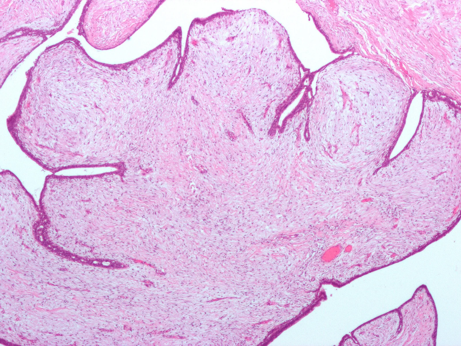
A fibroadenoma is a benign, biphasic tumour in which both ducts and stroma proliferate together, bound by a capsule. It is hormone-sensitive: it grows in pregnancy and in women on the combined oral contraceptive, and it usually shrinks after the menopause. A phyllodes tumour looks similar clinically but is distinguished by stromal overgrowth that folds into leaf-like (phyllodes) clefts; it is classified by microscopic grade (benign, borderline, malignant) on the WHO 2019 criteria. [4]
Atypical hyperplasia (ADH/AIDEP, FEA, lobular neoplasia) is not cancer, but it behaves like a window onto risk. The cells are monomorphic and fill part of a space, and the change can be multifocal. Adjacent cancers are sometimes found on excision, which is the short-term "upgrade" problem; equally important, the risk extends indefinitely into the future and affects either breast, which is why atypia is now treated as a marker of field-wide susceptibility rather than as a local pre-cancer. [3]
Risk factors for developing breast cancer itself (age, family history, inherited mutations such as BRCA1, BRCA2 and PALB2, reproductive history, radiation to the chest wall in youth, obesity, alcohol) are covered in the surgery breast topic and apply to every woman with or without benign disease. Most women who develop breast cancer have no single identifiable cause. [1]
Figure 1Phyllodes tumour
Long description
A benign phyllodes tumour of the breast H&E micrograph at low power magnification showing a myxoid cellular stroma separated by long clefts of compressed epithelial tissue.

- Hormonal stimulation → epithelial proliferation and stromal oedema → premenstrual heaviness, tenderness and lumpiness.
- Duct obstruction or fluid retention → cyst formation → a smooth, tense, often tender lump that may fluctuate in size.
- Stromal proliferation around ducts → fibroadenoma → a firm, rubbery, mobile nodule, or a phyllodes tumour if stroma outgrows the epithelium.
- Epithelial mutation and clonal growth → usual or atypical hyperplasia → an imaging or palpable abnormality with no distinctive feel; only microscopy separates the two.
- Progression in some atypical lesions → ductal carcinoma in situ or invasive cancer; the probability of this over the next five to ten years is the reason atypia changes management.
Cyclical mastalgia follows the same pathway. Tenderness tracks falling progesterone and rising prolactin, and improves after the menopause unless hormone replacement therapy is taken.
Ask about, and document:
- Lump: site, duration, change over the cycle, tenderness, growth, any new or fixed mass.
- Pain: timing (cyclical or not), relation to menses, severity, whether it stops normal activity or sleep.
- Nipple discharge: spontaneous or only on squeezing; from one breast or both; colour (clear, serous, bloody, milky, green) and smell.
- Skin change: dimpling, redness, peau d'orange, nipple retraction or eczema.
- Recent pregnancy or lactation.
- Constitutional symptoms: weight loss, bone pain, breathlessness (these point away from benign disease).
- Risk factors: family history, prior breast disease or radiation, medications (including hormone therapy), and ethnicity, which can modify the chance of carrying a BRCA mutation.
Cyclical mastalgia is a premenstrual ache or heaviness, often bilateral, in the upper outer quadrants, improving once bleeding starts. Non-cyclical pain is focal, persists, and may be burning or pulling; it is often musculoskeletal, dermatological or ductal. Bilateral, diffuse, cyclical pain is almost always benign; unilateral, focal, non-cyclical pain still needs a look.
Red flags for cancer include: a new, hard, irregular, fixed mass; nipple or skin change; spontaneous bloody unilateral discharge; a new axillary node; inflammatory change in a non-lactating breast; any breast symptom in a man; a non-healing breast wound. These do not prove cancer but demand a prompt, structured assessment. [1]
Inspect with the patient sitting and then lying, arms at sides and then raised: look for asymmetry, skin and nipple change, puckering, ulceration, visible veins and inflammation. Palpate with the flat of the fingers in a systematic pattern (a vertical strip is usually easiest), covering the whole breast, the tail into the axilla, and each axilla and supraclavicular fossa. Document the mass position on a clock face with its distance from the nipple in centimetres. Note size, site, shape, consistency, tenderness, mobility and fixity. A useful bedside clue: fibroadenomas are mobile and rubbery, cysts are smooth and tense, and cancer is usually hard, irregular and fixed. If discharge is not visible, ask the patient to express it before you try.
A 32-year-old woman finds a smooth, very mobile lump in her right breast three months after stopping breastfeeding. It does not change with her cycle. Imaging with ultrasound shows a solid 2.2 cm mass with benign features; core biopsy confirms a fibroadenoma. She is asymptomatic and reassured. Six months later she returns because the lump has grown to 3 cm and is now tender. She is referred for excision, not because of cancer risk but because of size and symptoms.
| Condition | Typical features | What separates it |
|---|---|---|
| Fibroadenoma | Young woman; firm, rubbery, smooth, mobile, well-defined | Does not change with cycle; mobile; no skin change |
| Simple cyst | Woman 30 to 50; smooth, tense, mobile; may be tender | May enlarge and shrink; cystic on ultrasound |
| Mastitis / abscess | Lactating woman; erythema, heat, tenderness, fever | Infectious, acute, often recent breastfeeding |
| Fat necrosis | History of trauma or surgery; firm, ill-defined lump | May mimic cancer on imaging; biopsy settles it |
| Fibrocystic change | Lumpy, tender breasts premenstrually; no discrete dominant mass | Diffuse, bilateral, cyclical |
| Intraductal papilloma | Spontaneous unilateral nipple discharge, often bloody | Discharge with otherwise normal imaging common |
| Mammary duct ectasia | Middle age; thick multicoloured discharge from several ducts | Multiple ducts, thick discharge, subareolar |
| Phyllodes tumour | Any age; large or rapidly growing firm mass | Can look like a fibroadenoma; grade only on excision |
| Breast cancer | Any age; hard, irregular, fixed, non-tender; skin or nipple change | Does not change with cycle; grows; malignant biopsy |
No clinical feature alone is reliable. Inflammatory breast cancer can look like mastitis; fat necrosis and phyllodes tumour can look like a fibroadenoma. When doubt remains, biopsy.
The standard way to assess any discrete breast symptom is triple assessment: clinical evaluation, imaging, and pathological (cellular or tissue) diagnosis. Each of the three should agree. Done well, the combination is highly accurate and avoids both missed cancers and unnecessary operations.
History and examination as above, with risk factors and menstrual status documented, and the mass position recorded.
Imaging is chosen by age and presentation, and is usually bilateral. For the patient with symptoms, imaging is the second pillar; it is not a screening test.
- Ultrasound is the best first test in women at younger ages and in pregnancy. It distinguishes solid from cystic, evaluates the axilla, and guides needle biopsy. It is fast, cheap and radiation-free.
- Mammography is the main test with increasing age and is also used to assess focal symptoms. It finds microcalcifications and distortion that ultrasound can miss.
- Digital breast tomosynthesis (3D mammography) and contrast-enhanced mammography are acceptable alternatives where available.
- MRI is reserved for problems the standard tests cannot settle, or for special situations: known BRCA or PALB2 mutation, lobular cancer, suspicion of multifocal or multicentric disease, breast implants, dense breasts, and preoperative planning. MRI should not be used routinely for every new lump. [2]
- Staging imaging for diagnosed cancer: CT of the chest with abdominal imaging and a bone scan should be considered for clinically positive axillary nodes, larger tumours, aggressive biology, or any sign, symptom or blood test suggesting metastasis. PET-CT is an option for high-risk patients or when conventional imaging is inconclusive.
NICE pretreatment recommendations include pretreatment ultrasound evaluation of the axilla for invasive breast cancer, with ultrasound-guided needle sampling if nodes are abnormal, and MRI when the extent of disease is not clear, when mammographic assessment is difficult because of density, or to plan breast-conserving surgery for invasive lobular cancer. [2]
- Fine-needle aspiration (FNA) obtains cells. Useful for cysts and for confirming simple cysts; it is less reliable for distinguishing invasive from in situ disease and for receptor testing.
- Core needle biopsy (CNB) obtains tissue and is the standard for a solid or complex mass. It gives histological type, grade, and receptors. It is usually image-guided, most often ultrasound.
- Open surgical biopsy is now seldom needed; reserved for discordant or non-diagnostic needles, or when a lesion cannot be sampled safely.
- Cyst: a simple cyst diagnosed by classic imaging with clear aspirate can be observed. A complex cyst (internal echoes, septations, solid components) or a bloody or atypical aspirate needs histology. Excise if it recurs, contains an intracystic solid element, or contains atypical cells.
Assessing a new breast symptom in an adult woman
- New breast symptom or lumpDocument size, site, duration; check nodes. Same-day assessment if red flags
- Triple assessmentClinical history and examination, imaging, needle biopsy
- Question: Which imaging first?
Under 35-40 or pregnant
- Ultrasound of both breasts and axillaeDistinguish solid and cystic; guide biopsy
- Mammography if suspicious or high risk
Age 40 and over
- Bilateral mammography plus targeted ultrasoundAdd axillary lymph node assessment
- Question: What does biopsy show?
Cyst fluid clear, benign features
- Reassure and observe or aspirate
Fibroadenoma, papilloma or benign fibrocystic change on core biopsy
- Correlate with clinical and imaging
- Conservative management or excision by symptoms and growth
Atypia or discordant / indeterminate
- MDT review; repeat biopsy or excision
- Risk-based management and follow-up
Malignant
- MDT review and oncology pathway
When all three parts of triple assessment agree on benign disease, the patient can be reassured with confidence. If they disagree, the lesion is managed as suspicious.
B3 lesions are "of uncertain malignant potential". Their management is driven by the upgrade rate for that entity (the percentage in which a further sample or excision shows in situ or invasive cancer), and by the long-term risk of atypia.
| Lesion | Upgrade risk on further sampling | Management in symptomatic practice |
|---|---|---|
| ADH (AIDEP) | 22% (range 5 to 50%) | Vacuum-assisted excision or open excision; then high-risk pathway [3] |
| Lobular neoplasia (ALH, LCIS) | ALH 12%, LCIS 22% (overall LN 0 to 40%) | Same approach; smaller sampled lesions may be surveyed [3] |
| Flat epithelial atypia (FEA) | 5% if pure, 11% if mixed with atypical intraductal proliferation | Excision if mixed or discordant; isolated concordant FEA may be surveyed [3] |
| Papilloma with atypia | 20 to 36% (vs 4 to 9% without atypia) | Open excision of the duct segment [3] |
| Radial scar / complex sclerosing lesion with atypia | 2 to 36% (vs 0 to 10% without atypia) | Excision, especially if large or discordant [3] |
| Cellular fibroepithelial lesion | Risk of phyllodes tumour rather than carcinoma | Excision to distinguish cellular fibroadenoma from phyllodes [3] |
| Mucocele-like lesion as a mass | Higher than screen-detected rate | Excision to exclude mucinous carcinoma [3] |
Two different risks must be kept separate:
- Upgrade (short-term) is the chance that a cancer already exists in the lump and was missed by the needle.
- Long-term risk is the chance of a future cancer in either breast, which is raised roughly fourfold after a diagnosis of atypia. [3]
Fibrocystic change needs no treatment beyond reassurance and a supportive bra. The lumpiness and tenderness are real but harmless. Teach breast awareness and document the mass position so that a future change is noticed.
Breast cysts. A simple cyst on ultrasound in a woman with classic features needs no biopsy and no surgery. Aspirate for pain or diagnostic uncertainty; the fluid should be straw-coloured or clear. Observe recurrences. Excise a cyst that recurs, that has a solid or septated internal component, or that yields bloody or atypical fluid.
Mastalgia is usually benign and self-limiting. The first step is explanation, reassurance and a well-fitting supportive bra. Avoiding caffeine and high-fat intake is commonly advised, although the evidence is weak. Cyclical pain that persists can be treated with topical or oral non-steroidal analgesia. Evening primrose oil is widely used but meta-analyses have not shown a clear benefit. For severe, persistent cyclical pain, short courses of hormonal agents (such as tamoxifen, danazol or bromocriptine, and in some systems goserelin) are effective but have side effects; prescribe them under specialist supervision and according to national guidance, and stop them once symptoms are controlled. A new, focal, non-cyclical pain, or pain with a lump or skin change, should be assessed rather than dismissed as mastalgia.
Usual ductal hyperplasia, sclerosing adenosis, radial scar and papilloma without atypia are generally managed as B3 lesions: they may be removed by vacuum-assisted excision or open surgery when they form a mass or are discordant with imaging, and selected small concordant lesions may be left in place with limited follow-up. Papilloma is excised when it causes spontaneous bloody or single-duct discharge, forms a palpable mass, or contains atypia. Complete removal of the index lesion with radiological-pathological concordance is what justifies conservative follow-up. [3]
Most small, classic, non-growing fibroadenomas in young women can be observed after explanation and imaging, with agreement on clinical and imaging features.
- Diagnose: clinical and imaging agreement; add a core biopsy if the lump is uncertain, growing, or atypical on imaging.
- Manage: observe small, stable, classic lesions; excise if the diagnosis is uncertain, if the lump grows, if it is giant, if it is painful or worrying, or if the patient prefers.
- A surgical excision through a cosmetically placed incision is straightforward.
Fibroadenomas do not transform into cancer and do not need removal for cancer prevention. Juvenile (giant) fibroadenoma in adolescence can distort the developing breast and usually needs excision with attention to cosmesis.
Phyllodes tumours make up fewer than 1% of breast tumours and are graded by the WHO 2019 criteria into benign, borderline and malignant. Core biopsy often cannot distinguish a phyllodes tumour from a cellular fibroadenoma, so a rapidly growing "giant fibroadenoma-like" mass should raise suspicion and be excised. [4]
Management follows the grade:
- Benign phyllodes tumour: complete excision with intact capsule and microscopically negative (ink-negative) margins. A fixed 1 cm gross margin is not required for benign disease. [4]
- Borderline or small malignant tumour: wide local excision with a gross margin of at least 1 cm. [4]
- Large, multifocal, recurrent or chest-wall-invasive tumour: total mastectomy, sometimes with full-thickness chest-wall resection and immediate reconstruction. [4]
- Axillary surgery is not routine, because nodal metastasis is rare; biopsy proven concurrent carcinoma is the exception. [4]
- Adjuvant radiotherapy is used for malignant disease (7 of 15 malignant tumours in the cited series, 46.7%). [4]
- Systemic chemotherapy is reserved for metastatic or unresectable malignant disease; routine adjuvant chemotherapy is not used. Endocrine therapy has no established role, and targeted or immune therapy is not recommended. [4]
- Reconstruction, when needed, follows the same oncological principles as breast cancer.
Atypical hyperplasia (ADH / AIDEP, FEA, lobular neoplasia) is the point at which benign disease becomes a breast-cancer risk problem. In the cited meta-analyses and cohorts, a histological diagnosis of atypia is associated with roughly a fourfold increase in long-term breast-cancer risk, with 5- and 10-year incidence varying by lesion type and sampling technique; atypia is best regarded as a marker of field-wide susceptibility, with subsequent cancers arising many years later in either breast. [3]
Management rests on three pillars:
- Exclude malignancy now. Upgrade to in situ or invasive cancer occurs in roughly 22% of ADH, 22% of classical LCIS, 12% of ALH, and 5 to 11% of FEA on further sampling. Excise or vacuum-assist the lesion and review with a breast pathologist. [3]
- Risk assessment and counselling. Formally quantify lifetime risk (for example with validated tools) and discuss both the reassuring message (most women with atypia never develop cancer) and the need for follow-up.
- Risk-reducing strategy. Patients should ideally be moved into established high-risk pathways that include formal risk assessment, discussion of risk-reducing strategies including chemoprevention, and risk-adapted imaging surveillance. [3]
Chemoprevention is the standard risk-reducing drug strategy for selected women at moderate to high risk, usually tamoxifen in premenopausal women and an aromatase inhibitor in postmenopausal women, prescribed with full discussion of thrombosis, menopausal symptoms and bone effects. [3]
Surveillance after atypia is not standardised worldwide. Some centres return women to routine population screening after complete excision, others use annual mammography for a period, and women with a lifetime risk at or above about 20% are often offered annual (or abbreviated) MRI in addition. [3] Follow your national protocol.
Pregnancy. Fibroadenomas often enlarge in pregnancy and may be multiple; most are observed and excised after delivery unless they are suspicious, giant or ulcerated. Cysts and mastitis are common. Breast surgery in pregnancy is possible when needed and is usually planned with an obstetric and breast team; imaging is ultrasound-first.
Adolescents. The breast changes of puberty can look alarming. Most adolescent masses are fibroadenomas, cysts or normal developing tissue. Excise for diagnostic doubt, growth, or a giant lesion, and protect the developing breast and nipple plate. Medication with hormonal agents is rarely needed.
Older adults. New breast symptoms in older women are taken seriously, even though most turn out benign. Treatment is guided by fitness and frailty rather than age alone; observation with clear follow-up is reasonable for a small, stable, well-characterised benign lesion in a frail patient.
Lactation and the postpartum period. Mastitis, abscess and galactocele dominate. These are usually managed with support, feeding, analgesia and antibiotics; abscess needs drainage. Any inflammatory change that does not respond to treatment should be biopsied to exclude inflammatory breast cancer.
Low-resource settings. The essentials transfer well: breast awareness and prompt referral; examination; ultrasound where available; and core needle (or even FNA) biopsy. A core biopsy requires only a trained operator and basic pathology. Aspiration of a simple cyst, and excision biopsy of a growing mass, are low-cost interventions that prevent both missed cancers and unnecessary mastectomies.
- Missed cancer: the consequence of calling a malignant lump benign. Triple assessment and excision of discordant or B3 lesions are the defence.
- Unnecessary surgery: the consequence of not trusting concordant benign assessment. Core biopsy agreement prevents this.
- Infection or haematoma after aspiration, biopsy or excision.
- Recurrent cysts and ongoing cyclical pain, which can be disabling.
- Phyllodes tumour local recurrence, and, for the malignant grade, haematogenous metastasis (most often to lung and bone). [4]
- Long-term breast-cancer risk after atypia, needing surveillance and, in selected women, chemoprevention. [3]
- Nonproliferative lesions, cysts and mastalgia have an excellent prognosis and often settle after the menopause.
- Fibroadenoma neither turns into cancer nor raises cancer risk. Observation is safe.
- Benign phyllodes tumour has a good prognosis after complete excision; borderline and malignant tumours recur locally more often and metastasise in a minority.
- Atypia changes the long-term outlook: roughly a fourfold rise in breast-cancer risk that persists for years and affects either breast, and it justifies entry into a high-risk pathway. [3]
There is no lifestyle change that prevents benign breast disease, and most of it is not preventable. What is preventable is the harm of late diagnosis.
- Breast awareness: women should know what is normal for their own breasts and report change promptly. This is the single most transferable intervention and works even without mammography. [1]
- Mammography screening in apparently healthy women is usually offered from about 50 to 69 years of age; national programmes set their own start age and interval, and you should follow the policy of the country you work in. [1]
- High-risk women (BRCA1, BRCA2, PALB2 carriers, and women with a high lifetime risk) are offered enhanced surveillance, often including MRI from young adult life. See the familial breast-cancer guidance for eligibility and schedule. [3]
- Risk reduction for atypical disease: consider chemoprevention and risk-reducing surgery only for the small group at genuinely high genetic risk, in a specialist multidisciplinary setting.
- Population-level action: the WHO Global Breast Cancer Initiative aims to reduce global breast-cancer mortality by 2.5% per year and to avert 2.5 million deaths by 2040, through health promotion for early detection, timely diagnosis, and comprehensive management. [1]
Covered above in Management: pregnancy, adolescents, older adults, lactation and low-resource settings.
Some MedGrasp topics to read alongside this one: surgery/breast-disease-and-breast-cancer; obstetrics/lactation-breastfeeding-and-mastitis; gynecology/abnormal-uterine-bleeding for the hormonal context; genetics topics on BRCA1 and BRCA2 once published.
- Most breast lumps and symptoms are benign. The assessor's job is to find the few that are cancer without over-treating the many that are not.
- Classification (nonproliferative, proliferative without atypia, fibroepithelial, atypical, B3) exists because groups differ in cancer risk.
- Triple assessment is the rule: clinical, imaging and pathological diagnosis, all concordant.
- Nonproliferative lesions (fibrocystic change, cysts, mastalgia) are not pre-cancers and do not raise risk above baseline.
- Fibroadenoma in a young woman is observed; it does not turn into cancer.
- A phyllodes tumour needs excision; benign disease needs ink-negative margins only, borderline and malignant need at least 1 cm.
- Atypical hyperplasia raises long-term breast-cancer risk roughly fourfold and justifies entry into a high-risk pathway.
- Distinguish short-term upgrade risk (cancer missed by the needle) from long-term risk (a future cancer in either breast).
- Cysts that are simple on ultrasound need no biopsy; excise if complex, recurrent, or atypical.
- Mastalgia is usually cyclical, benign and self-limiting; red flags still need assessment.
- BRCA and family history modify management and are governed by the familial breast-cancer guidance.
- Low-resource essentials: breast awareness, prompt referral, examination, ultrasound, and core or FNA biopsy.
Answer each one in your head first, then open the answer.
A 22-year-old woman has a 2 cm, firm, rubbery, very mobile breast lump that does not change with her cycle. Ultrasound shows a solid mass with benign features. What is the diagnosis and the management?
Show answerHide answer
Fibroadenoma is the most likely diagnosis in a younger woman with a firm, rubbery, smooth, mobile lump. If clinical and imaging findings agree, there is no need for biopsy; small, classic, stable lesions are observed with reassurance. Excision is indicated if the diagnosis is uncertain, the lump grows, the lump is giant or painful, or the patient prefers removal.
The same lump grows to 4 cm over six months and she now asks for it to be removed. The pathology shows a phyllodes tumour. What principles govern further management?
Show answerHide answer
A cellular fibroepithelial lesion on core biopsy cannot reliably be graded; a rapidly growing mass should be excised. The definitive grade (benign, borderline, malignant by WHO 2019 criteria) is assigned on the excision specimen. Benign phyllodes tumour needs complete excision with ink-negative margins but not a fixed 1 cm margin. Borderline or malignant tumour needs wide excision with at least a 1 cm gross margin, or mastectomy if large. Axillary surgery is not routine. Adjuvant radiotherapy is for malignant disease; chemotherapy is only for metastatic or unresectable disease; endocrine therapy has no established role. [4]
A 48-year-old woman has a core biopsy labelled 'atypical ductal hyperplasia, ADH'. What are the two different risks you must keep in mind, and what is the next step?
Show answerHide answer
Short-term (upgrade): further excision shows in situ or invasive cancer in roughly 22% of cases. Long-term: a roughly fourfold increase in breast-cancer risk in either breast, years later. The next step is careful review by a breast pathologist and excision (vacuum-assisted or open) to exclude malignancy at the index site, followed by formal risk assessment, discussion of chemoprevention, and a risk-adapted surveillance plan within a high-risk pathway.
A 35-year-old woman presents with a smooth, tense breast lump that enlarges before her period and shrinks afterwards. Ultrasound is cystic with no solid component. What do you do?
Show answerHide answer
This is a classic simple cyst. No biopsy is needed: reassure, aspirate if it is painful or for certainty, and observe. Excise (or further investigate) if the aspirate is bloody or atypical, if there is an intracystic solid element on ultrasound, or if the cyst recurs.
A 28-year-old woman has bilateral cyclical breast pain without any lump. Examination and ultrasound are normal. What is the management, and when should you investigate further?
Show answerHide answer
This is cyclical mastalgia, which is benign and usually self-limiting. Explain and reassure, advise a well-fitting supportive bra, and offer simple analgesia. Investigate with imaging and biopsy if the pain is focal and non-cyclical, if a lump, nipple change or skin change appears, or if the pain does not settle and the patient is worried.
A woman with biopsy-proven atypical lobular hyperplasia asks what her follow-up should be. How do you answer honestly?
Show answerHide answer
There is no single worldwide standard. Upgrade to cancer on excision occurs in roughly 12% of ALH, and long-term breast-cancer risk is raised roughly fourfold in either breast, persisting for years. Complete excision with radiological-pathological concordance favours risk-based long-term surveillance; incomplete sampling favours closer early follow-up to exclude residual disease. Women with a lifetime risk at or above about 20% are often offered MRI in addition to mammography. The local high-risk pathway and national guidance set the interval. [3]
- World Health Organization. Breast cancer. WHO fact sheet, 3 July 2026. Accessed .
- National Institute for Health and Care Excellence. Early and locally advanced breast cancer: diagnosis and management. NICE guideline NG101, published 18 July 2018, last updated 14 April 2025. Accessed .
- Rakha EA, Shaaban A, Floris G, et al. Management of high-risk (B3) breast lesions in symptomatic practice: evidence review and practical recommendations for multidisciplinary care. Virchows Archiv 2026;489(4):783-796. doi:10.1007/s00428-026-04672-2. Accessed .
- Kang B, Kim SG, Park TH, et al. Management of Breast Phyllodes Tumors: A Grade-Stratified Surgical and Reconstructive Strategy. Journal of Clinical Medicine 2026;15(18):7226. doi:10.3390/jcm15187226. Accessed .
Figure credits
Educational content for medical students and doctors. It is not a substitute for clinical judgement or local guidelines.