Breast cancer: screening, staging and systemic therapy
Breast cancer: how it is staged and how endocrine, HER2-targeted and chemotherapy treatment are chosen by receptor subtype, plus screening and surveillance.
- Breast cancer becomes dangerous once the abnormal cells have broken through the lining of a duct or lobule. Found while it is still small and confined, it is usually curable. [1]
- In 2024 an estimated 2.4 million women were diagnosed with breast cancer and about 694 000 died from it; it is the most common cancer in women in 164 of 186 countries. [1]
- Roughly 80% of breast cancers develop in women with no modifiable risk factor beyond being female and over 40. [1]
- About 99% of breast cancers occur in women; 0.5-1% occur in men. [1]
- Once the histology and receptors are known, treatment follows the tumour's biology: all oestrogen receptor (ER)-positive cancers receive endocrine therapy, all HER2-positive cancers receive anti-HER2 therapy, and triple-negative cancers rely mainly on chemotherapy. [1][2][3]
By the end of this page you should be able to:
- Define breast cancer and list the invasive histological types and the receptor-defined subtypes, with their natural history.
- Sort the risk factors into the well-established ones and explain the mechanisms behind them.
- Explain how a breast lesion is staged by TNM and by biological risk.
- List the standard tests in triple assessment and state what each result means.
- Plan adjuvant systemic treatment for each receptor subtype, including the role of neoadjuvant therapy, and name the circumstances in which trastuzumab, olaparib, abemaciclib and pembrolizumab are added.
- Describe surveillance after treatment and the special considerations for men, for older adults and during pregnancy.
- State how screening advice differs between WHO, USPSTF, ESMO and NCCN, and why local policy is the one to follow.
DefinitionBreast cancer
Breast cancer is uncontrolled growth of epithelial cells arising from the milk ducts or the milk-producing lobules, which becomes clinically important when cells breach the basement membrane and invade the surrounding breast tissue. Invasion gives access to lymphatic and blood vessels, so the disease can spread to the nodes and then to bone, liver, lung and brain. [1]
The key distinction is between in situ disease, where abnormal cells still sit inside a duct or lobule, and invasive disease, where they have crossed the basement membrane. Ductal carcinoma in situ (DCIS) is the commonest in situ lesion; it is not immediately life-threatening but is treated because it can progress to invasive cancer.
The receptors that define the biological subtypes are:
- Estrogen receptor (ER) and progesterone receptor (PR): markers of dependence on hormonal stimulation for growth.
- Human epidermal growth factor receptor 2 (HER2): overexpression drives proliferation.
- Triple-negative breast cancer: none of ER, PR or HER2 is expressed.
Biological subtyping follows the laboratory profile: luminal A-like (ER-positive, HER2-negative, lower proliferation), luminal B-like (ER-positive, HER2-negative, higher proliferation), HER2-positive and triple-negative. [3] A single tumour can be mixed, and ER, PR and HER2 are re-checked at recurrence because they can change.
- In 2024 an estimated 2.4 million women were diagnosed with breast cancer and about 694 000 died from it. [1]
- Breast cancer is the most common cancer in women in 164 of 186 countries. [1]
- About 99% of breast cancers occur in women and 0.5-1% in men. [1]
- Roughly 80% of breast cancers occur in women with no modifiable risk factor other than being female and older than 40. [1]
- Age-standardised breast cancer mortality in high-income countries fell by about 40% between the 1980s and 2020, through the combination of earlier detection and modern systemic therapy. Mortality remains much higher where presentation is later. [1]
- ER-positive disease is the commonest molecular group; ER-positive cancers make up roughly 53-76% of cases in different populations. [3]
Risk is multifactorial; most women who develop breast cancer have no single identifiable cause.
Non-modifiable
- Female sex and increasing age.
- Family history, particularly breast or ovarian cancer in close relatives.
- High-penetrance inherited mutations, chiefly BRCA1, BRCA2 and PALB2. [1]
- Menstrual and reproductive history: early menarche, late menopause, first pregnancy at older age, nulliparity, and never breastfeeding.
- Previous breast disease with atypia or lobular carcinoma in situ.
- Radiation to the chest wall when young.
Modifiable
- Obesity, especially after the menopause.
- Alcohol.
- Tobacco in some populations.
- Postmenopausal hormone replacement therapy.
- Physical inactivity.
A family history needs careful interpretation rather than age alone: the pattern (early-onset disease, multiple relatives, male breast cancer, ovarian cancer) matters more than a single older relative. BRCA-related risk is quantified and managed by specialist genetic clinics using their criteria.
The reproductive factors share a biological basis: each menstrual cycle exposes breast epithelium to oestrogen and progesterone, and pregnancy and lactation mature the lobules, which lowers risk. [1]
The progression from normal breast to cancer follows five steps:
- In situ change. Abnormal epithelial cells proliferate but remain within the duct or lobule. DCIS is the commonest in situ lesion and is non-invasive but can progress.
- Invasion. Cells break through the basement membrane into the stroma, becoming invasive cancer. Invasive carcinoma of no special type is the commonest.
- Local spread. Through lymphatics, tumour cells reach the regional nodes; clinically positive axillary nodes are a sign of spread.
- Receptor drives. ER/PR expression reflects dependence on oestrogen for growth. HER2 overexpression amplifies growth signals. Triple-negative tumours lack these targets and are driven by other pathways.
- Distant spread. Haematogenous spread reaches bone, liver, lung and brain, giving bone pain, breathlessness, jaundice or neurological symptoms.
Because receptor biology and stage together determine prognosis, modern care treats patients by both anatomy (TNM) and biology.
Breast cancer is usually a symptomatic disease in most of the world; screening detects a smaller, earlier fraction. Symptoms include a breast lump or thickening, change in size, shape or appearance of the breast, skin dimpling, redness, pitting, nipple change, nipple discharge that is spontaneous, unilateral or bloody, and axillary swelling. General symptoms such as weight loss or bone pain occur with late disease. [1]
Red flags that must trigger prompt, structured assessment include a new, hard, irregular, fixed mass; skin or nipple change; spontaneous bloody or clear unilateral discharge; a new axillary node; inflammatory change in a non-lactating breast; and any breast symptom in a man. These do not always mean cancer, but they must not be observed. [1]
Inspection sitting and then lying, arms at the sides and then raised, looks at asymmetry, skin and nipple change, retraction, ulceration and inflammation. Palpation with the flat of the fingers covers the whole breast, the tail and the axillae, and each supraclavicular fossa. The findings are recorded by clock face and distance from the nipple, which is the position a surgeon and radiologist need.
Most palpable lumps in younger women are benign; the chance of malignancy rises with age.
| Condition | Typical features | What separates it from cancer |
|---|---|---|
| Fibroadenoma | Younger woman; firm, rubbery, smooth, very mobile lump | Mobile, well-defined, no skin change |
| Simple cyst | Smooth, tense, mobile lump that fluctuates with the cycle | Cystic on ultrasound; clear aspirate if drained |
| Mastitis or abscess | Lactating woman; erythema, heat, tenderness, fever | Infectious and acute; associated with breastfeeding |
| Fat necrosis | After trauma or surgery; firm, ill-defined lump | May mimic cancer on imaging; biopsy settles it |
| Fibrocystic change | Lumpy, tender breasts premenstrually | Diffuse, bilateral, cyclical; no discrete fixed mass |
| Intraductal papilloma | Unilateral spontaneous bloody or serous discharge | Discharge with normal imaging |
| Mammary duct ectasia | Middle life; thick, multicoloured, multi-duct discharge | Several ducts, subareolar |
| Breast cancer | Any age; hard, irregular, fixed, non-tender; skin or nipple change | Persistent; malignant histology |
| Phyllodes tumour | Rapidly growing, firm mass | Needs excision; resembles fibroadenoma |
Figure 1Ultrasound showing left breast 9 to 10 OC mass with significant shadowing and echogenic foci
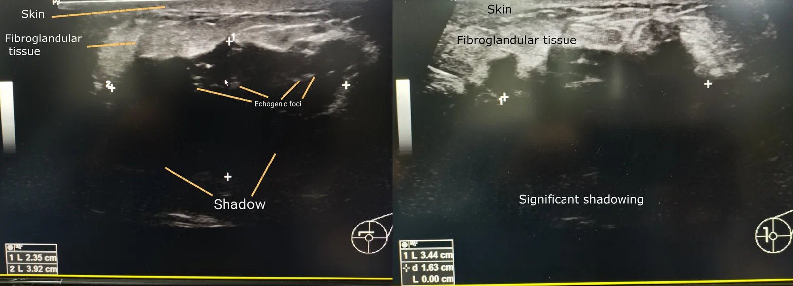
Long description
Left breast ultrasound showing a 9 to 10 o'clock mass measuring 2.4 x 4.0 x 3.4 cm with significant shadowing and echogenic foci; the biopsy was invasive breast carcinoma.

Figure 2Mammogram images of clustered calcifications at CC view and MLO view
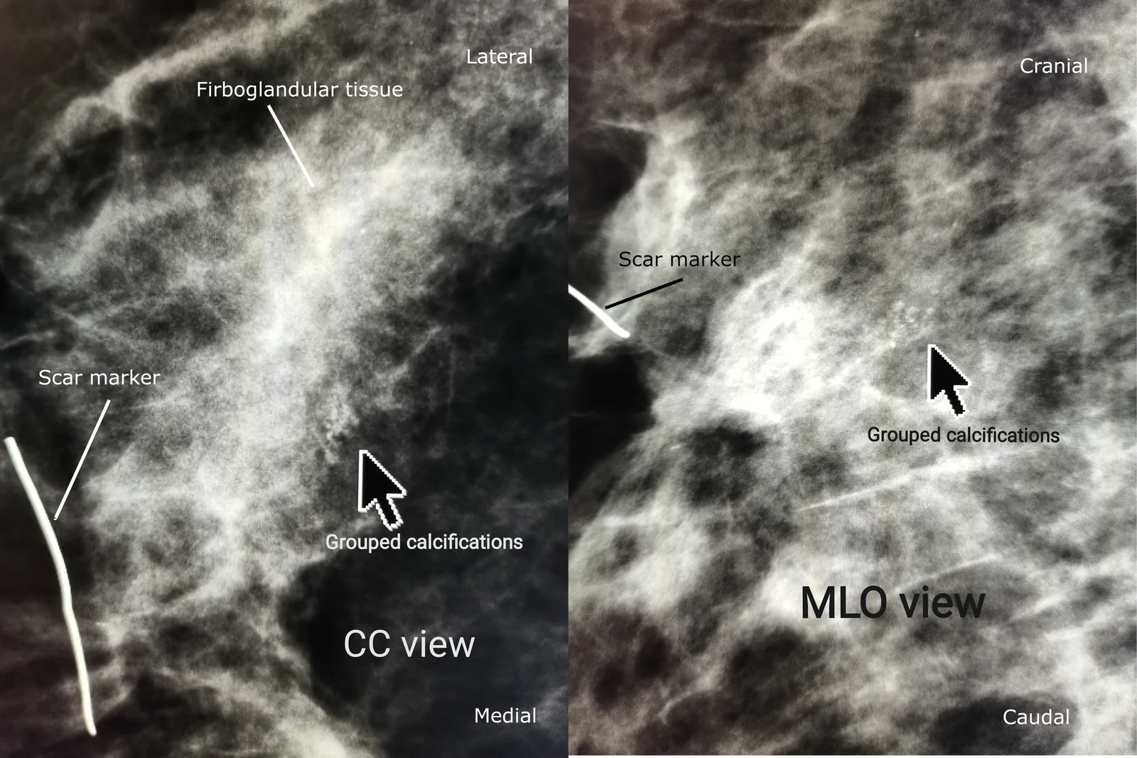
Long description
A Wikimedia Commons mammogram image showing grouped calcifications in the breast at craniocaudal (CC) and mediolateral oblique (MLO) views.

Figure 3Mammography and scintimammography of carcinoma
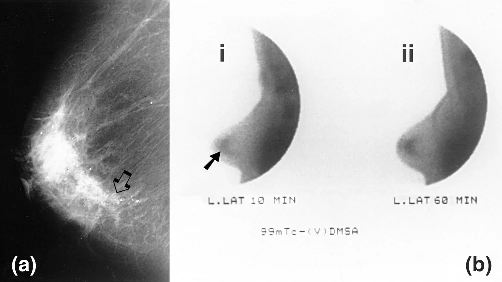
Long description
Medio-lateral mammographic projection of a 4.5-cm ductal carcinoma in situ of the left breast showing microcalcifications behind the nipple, with adjacent scintimammography images.

The standard assessment of any discrete breast symptom is triple assessment: clinical evaluation, imaging and pathological (cellular or tissue) diagnosis, with all three agreeing before a mass is called benign. [2]
Imaging
- Bilateral mammography plus ultrasound of both breasts and regional nodes is the standard for symptomatic disease. [3]
- Ultrasound is the best first test in younger women, in pregnancy and in the assessment of an axillary node.
- Mammography is the main screening tool and also detects microcalcifications that ultrasound can miss.
- Digital breast tomosynthesis and contrast-enhanced mammography are acceptable alternatives where available. [3]
- Breast MRI is reserved for problem solving, multifocal or multicentric suspicion, dense breasts, lobular cancer, BRCA carriers, or preoperative planning. [2][3]
- Minimum blood tests before surgery and systemic therapy: full blood count, liver and renal function tests, alkaline phosphatase and calcium. [3]
Pathology
- Core needle biopsy is the standard for a solid mass and provides type, grade, ER, PR, HER2 and Ki-67.
- ER, PR and HER2 should be assessed together and reported quantitatively at diagnosis. [2]
- HER2 is assessed by immunohistochemistry, with in-situ hybridisation for an equivocal (2+) score. [3]
- Axillary ultrasound with ultrasound-guided needle sampling is used when nodes look abnormal before surgery. [2]
Staging investigations
Staging imaging is not used in every case; it is reserved for higher risk. ESMO recommends computed tomography of the chest, abdominal imaging and bone scan for stage IIb and above, larger tumours (about 5 cm), aggressive biology, or symptoms or laboratory values suggestive of metastasis, and positron emission tomography-computed tomography when conventional imaging is inconclusive or for high-risk patients. [3]
| Test | What it establishes | Key read |
|---|---|---|
| Clinical examination | Lump characteristics and nodes | Site by clock face and distance from nipple |
| Mammography | Microcalcifications, distortion, multifocality | Abnormality reported in standard categories |
| Ultrasound | Solid vs cystic; axillary nodes | Correlates with clinical findings |
| Core needle biopsy | Histological type and grade | ER, PR, HER2 and Ki-67 for subtyping |
| Axillary node sampling | Node metastasis before surgery | Macrometastasis vs micrometastasis |
| Metastasis staging | Screen for distant disease | CT chest/abdomen and bone scan for higher-stage or aggressive disease |
| Blood tests | Fitness for surgery or systemic therapy | Full blood count, renal and liver function, alkaline phosphatase, calcium |
Breast cancer is diagnosed by histology. Staging uses the WHO classification of breast tumours and the 8th edition of the TNM system, based on tumour size (T), regional nodes (N) and distant metastasis (M), which produces groups 0 to IV. [2][3] Staging is both anatomical and biological: receptor status, and where helpful genomic assays (such as Oncotype DX or EndoPredict for node-negative ER-positive disease), alter adjuvant decisions. [2][3]
- T0-T4: tumour size and chest wall or skin involvement.
- N0-N3: number and location of involved lymph nodes.
- M0/M1: absence or presence of distant metastasis.
Biological subtyping follows the laboratory profile above; a small, node-negative, well-differentiated ER-positive HER2-negative cancer carries an excellent prognosis, whereas a small triple-negative or HER2-positive tumour may warrant systemic therapy. [2]
- Treatment is delivered in a specialised breast unit by a multidisciplinary team including surgeons, oncologists, radiologists, pathologists, nurses and allied professionals. [3]
- Receptors and tumour profile are established before surgery. [2][3]
- Treatment is individualised by tumour burden, biology, age, menopausal status, general health and patient preference; age alone should not be the main determinant. [2][3]
- People should receive written, repeated information and a named key worker or clinical nurse specialist. [2]
Locoregional treatment (breast-conserving surgery, mastectomy, axillary surgery, radiotherapy and reconstruction) is covered in detail on the companion surgical topic; this page focuses on systemic therapy and surveillance.
1. ER-positive, HER2-negative disease
All luminal-like cancers receive endocrine therapy. [3]
- Premenopausal women receive either tamoxifen alone (for low-risk luminal A-like, stage I disease), or, when the risk of recurrence is high, ovarian function suppression combined with tamoxifen or with an aromatase inhibitor. [3]
- Postmenopausal women receive an aromatase inhibitor as initial adjuvant endocrine therapy, or tamoxifen if the risk is low or aromatase inhibitors are not tolerated. [2][3]
- For HER2-negative, ER-positive disease at high clinical risk (node-positive high-risk disease), a CDK4/6 inhibitor is added: abemaciclib with endocrine therapy for node-positive high-risk early breast cancer, or ribociclib with an aromatase inhibitor for high-risk disease. [2][3]
- The cytotoxic regimen where chemotherapy is indicated contains both a taxane and an anthracycline, with dose-dense schedules preferred. [2][3]
- Genomic assays and endocrine response assessment may be used where there is uncertainty about chemotherapy in ER-positive, HER2-negative disease. [3]
- Extended endocrine therapy beyond 5 years is considered in higher-risk disease (7-8 years appears sufficient for most high-risk patients). [3]
- Adjuvant bisphosphonates (zoledronic acid or sodium clodronate) are offered to women without ovarian function, especially if at high risk of relapse or treatment-related bone loss. [2][3]
Endocrine therapy is taken for years and reduces recurrence of hormone-positive cancer by nearly half. [1][2]
2. HER2-positive disease
- HER2-directed therapy, with initial concurrent chemotherapy, is given for 12 months, covering neoadjuvant and adjuvant phases; 6 months may be non-inferior in selected low-risk situations. [3]
- Regular cardiac assessment is needed, because trastuzumab is used with caution when baseline left ventricular ejection fraction is 55% or less, or with cardiac disease. [2][3]
- For clinical stage II-III HER2-positive cancer (tumour above 2 cm or node-positive), neoadjuvant chemotherapy with dual anti-HER2 blockade (trastuzumab plus pertuzumab) is preferred. [3]
- The chemotherapy backbone is anthracycline-taxane or taxane-carboplatin. [3]
- Trastuzumab is given adjuvantly for T1c and above HER2-positive cancer, 3-weekly for 1 year. [2]
- T1a and T1b disease is decided individually. [2]
- Where residual invasive disease remains after neoadjuvant taxane-based therapy, trastuzumab emtansine is used adjuvantly; pertuzumab combinations are reserved for node-positive or higher-risk disease. [2]
- For HER2-positive, hormone receptor-positive disease, endocrine therapy is given alongside. [2]
3. Triple-negative disease
- Triple-negative cancers do not respond to endocrine or anti-HER2 therapy and depend on chemotherapy. [1][2]
- They are treated with chemotherapy, with or without the immune checkpoint inhibitor pembrolizumab for high-risk early or locally advanced disease. [2][3]
- Chemotherapy is given for 12 to 24 weeks (4 to 8 cycles) whether or not an immune checkpoint inhibitor is used. [3]
- When neoadjuvant chemotherapy is used for triple-negative disease, a platinum-containing regimen is recommended. [2]
- Neoadjuvant treatment is preferred for cT1c-4 N0 or node-positive triple-negative disease. [3]
- For BRCA-mutation carriers with HER2-negative high-risk disease, adjuvant olaparib (with concomitant endocrine therapy in HR-positive disease) improves outcomes after chemotherapy; it is not combined with abemaciclib, because of overlapping toxicities, and would be given sequentially with olaparib first. [2][3]
- Adjuvant capecitabine may be used after preoperative chemotherapy in selected high-risk disease. [3]
4. Adjuncts that cross subtypes
- Oestrogen receptor-low cancers (1-9% ER or PR staining) behave partly like triple-negative disease and need adjusted strategies. [3]
- Trastuzumab for 12 months is given concurrently with radiotherapy and endocrine therapy when indicated. [3] (Note: the ESMO figure of 12 months covers both the neoadjuvant and adjuvant phases combined.)
Neoadjuvant therapy in general
Neoadjuvant systemic therapy shrinks larger tumours before surgery, allows breast conservation, tests the tumour's response in vivo, and guides later treatment. The response predicts long-term outcome.
Male breast cancer is rare, accounting for less than 1% of all breast cancers and less than 1% of all malignancies in men, and it is usually ER-positive. [3] Men are treated with mastectomy followed by radiotherapy as indicated and endocrine therapy, usually tamoxifen; genetic testing, especially BRCA2, is often appropriate. [1][2] In line with ESMO and NICE, tamoxifen is the standard adjuvant endocrine therapy for men; an aromatase inhibitor, if used at all, must be given with a gonadotropin-releasing hormone agonist, and tamoxifen is not replaced by an aromatase inhibitor alone. [2][3]
Treatment should be adapted to biological rather than chronological age; geriatric assessment is advised, frailty rather than age should guide chemotherapy intensity, and primary endocrine therapy alone is reserved for those in whom surgery is unsafe. [2][3]
Surgery and some systemic therapies can be given during pregnancy, but chemotherapy is avoided in the first trimester; decisions are taken by a multidisciplinary and obstetric-specific team. Fertility and fertility preservation should be discussed with younger premenopausal patients before any systemic treatment. [3]
Inflammatory breast cancer presents as a red, hot, swollen breast with no discrete mass early on; it is locally advanced and managed with systemic therapy before surgery and radiotherapy. Locally advanced unresectable disease is managed according to dedicated advanced breast cancer guidance. [2]
- Annual mammography for 5 years in all treated breast cancers including DCIS; in women this continues annually until entry to the routine screening programme. [2]
- No routine ultrasound or MRI for post-treatment surveillance. [2]
- Written care plan with designated key worker, surveillance dates, signs and symptoms and contact details. [2]
- Clinical follow-up with regular visits (ESMO: every 3 months in the first 3 years, every 6 months in years 4-5, then annually, adjusted to risk), and lifestyle advice including maintaining healthy weight, limiting alcohol under 5 units a week and regular physical activity. [2][3]
Of the disease itself: progressive nodal involvement; metastasis to bone, liver, lung and brain; lymphoedema from nodal blockage; skin and chest wall ulceration; pleural effusion; and marrow suppression with advanced disease.
Of treatment: cytopenias, alopecia, neuropathy, menopausal symptoms, arthralgia, osteoporosis, cardiac dysfunction with trastuzumab (requiring left ventricular monitoring), and thrombosis with tamoxifen. Radiotherapy carries skin reaction, fatigue and late fibrosis or pneumonitis. Surgery carries seroma, infection, lymphoedema and post-mastectomy pain syndrome. [2]
Long-term recurrence: ER-positive disease can recur years after the first 5 years, which is the rationale for extended endocrine therapy in higher-risk patients.
Prognosis depends most on stage at diagnosis and biology. Early, small, node-negative, ER-positive cancers have a very high cure rate with modern therapy; many metastatic cancers remain controllable for years but are not curable. Validated prognostic tools include the PREDICT tool; it tends to be less accurate for women under 30 with ER-positive disease, women aged 70 and over, and tumours larger than 50 mm; it has not been validated in men and its validation may have under-represented some ethnic groups. [2]
Screening advice differs between authorities, so the policy of the country you work in is the one to follow.
- WHO: mammography as screening in an apparently healthy population, usually women 50-69 years of age. [1]
- ESMO (adapted): regular mammography every 2 years for women 45-69, with possible screening at 40-44 and 70-74 where there is emerging evidence of benefit; screening in high-risk carriers follows the hereditary breast-ovarian cancer guideline. [3]
- USPSTF (2024): biennial screening mammography for women aged 40-74 (grade B); insufficient evidence for screening at 75 and over, and insufficient evidence for supplemental screening with ultrasound or MRI in dense breasts. [4]
- NCCN: Version 9.2026 includes a dedicated Breast Cancer Screening and Diagnosis guideline. [5]
Both digital mammography and digital breast tomosynthesis (3D mammography) are effective screening modalities. [3][4]
- Breast awareness and prompt reporting of change at all risk levels.
- Limiting alcohol (under 5 units a week), weight management, physical activity and not smoking. [2]
- Limiting long-term hormone replacement therapy, especially combined oestrogen-progestogen, and encouraging breastfeeding.
- BRCA-related risk is managed by specialist genetic clinics, with chemoprevention (tamoxifen premenopausally, anastrozole postmenopausally in high-risk women without cancer) and risk-reducing surgery only for genuinely high-risk, counselled women.
- Male breast cancer: see above; tamoxifen as standard, genetic testing often appropriate. [3]
- Pregnancy: triple assessment is still the framework; surgery can be carried out, chemotherapy avoided in the first trimester, and radiation deferred until after delivery.
- Inflammatory breast cancer, Paget disease and phyllodes tumour are identified by the NCCN guideline as special indications requiring specialist pathways. [5]
- Older adults: fitness, not age, governs treatment. [2][3]
- Low-resource settings: where mammography is unavailable, prompt assessment of symptomatic women, clinical breast examination and trustworthy referral pathways remain the realistic and effective interventions; generic breast cancer medicines are on the WHO Essential Medicines List. [1]
NotePregnancy
Imaging is chosen to avoid radiation to the fetus: ultrasound first, with mammography used only when clinically necessary with shielding.
| Feature | Breast cancer | Fibroadenoma | Mastitis or abscess | DCIS |
|---|---|---|---|---|
| Age | Any, risk rises with age | Usually under 35 | Lactation | Screening age |
| Pain | Usually painless | Painless | Painful, hot | None |
| Mobility | Fixed | Highly mobile | Tender, warm | None palpable |
| Skin or nipple change | Common | No | Infection | Possible microcalcifications |
| Imaging | Suspicious mass, distortion | Well-circumscribed solid | Inflammatory | Calcification |
| Biopsy | Malignant cells | Benign; observe or excise | Drain if abscess | Non-invasive atypia |
Breast cancer: systemic therapy by receptor subtype
- Confirmed invasive breast cancerER, PR, HER2 on core; stage (TNM); menopausal status; BRCA testing if indicated
- Question: HER2-positive?
Yes
- Neoadjuvant chemotherapy plus dual HER2 blockade (trastuzumab + pertuzumab) if stage II-IIITaxane-carboplatin or anthracycline-taxane backbone
- Adjuvant trastuzumab 3-weekly for 1 year totalTotal 12 months neoadjuvant plus adjuvant; cardiac monitoring
- Add endocrine therapy if ER/PR positive
No
- Question: ER or PR positive?
Yes
- Endocrine therapy for allTamoxifen or ovarian suppression in premenopausal; aromatase inhibitor (or tamoxifen) postmenopausal
- Add chemotherapy if high clinical riskTaxane + anthracycline, dose-dense where available
- Consider CDK4/6 inhibitor in high-risk node-positive diseaseAbemaciclib or ribociclib
- Bisphosphonates if postmenopausal and at risk
- Consider extended endocrine therapy beyond 5 years
No (triple-negative)
- Chemotherapy 12-24 weeksPlatinum-containing regimen in the neoadjuvant setting
- Neoadjuvant preferred for stage II-IIIAssess response; add capecitabine if residual disease
- Pembrolizumab for high-risk diseaseNeoadjuvant with chemotherapy, continued adjuvant
- Olaparib for 1 year if BRCA mutation and high risk
- Re-stage and re-test receptors at recurrenceBiology can change over time
- Breast cancer is curable when found confined to the breast and nodes. [1]
- Roughly 80% of breast cancers occur in women with no modifiable risk factor beyond being female and over 40. [1]
- About 99% of cases are in women; 0.5-1% in men. [1]
- The biological subtypes are driven by ER, PR and HER2 expression. [3]
- All ER-positive cancers receive endocrine therapy, which reduces recurrence by nearly half. [1][2]
- HER2-positive cancers receive 12 months of HER2-directed therapy, often neoadjuvantly, with dual blockade in stage II-III disease. [2][3]
- Trastuzumab is given for T1c and above, 3-weekly for 1 year, with cardiac monitoring. [2]
- Triple-negative disease depends on chemotherapy, ideally neoadjuvant with platinum, with pembrolizumab for high-risk disease. [2][3]
- Chemotherapy where used contains a taxane plus an anthracycline, with dose-dense schedules preferred. [2][3]
- High-risk BRCA carriers with HER2-negative disease receive 1 year of adjuvant olaparib after chemotherapy. [2][3]
- Surveillance is annual mammography for 5 years; no routine ultrasound or MRI. [2]
- Screening advice varies (WHO 50-69, ESMO 45-69 every 2 years, USPSTF 40-74 every 2 years); follow the policy of the country you work in. [1][3][4]
Answer each one in your head first, then open the answer.
A 62-year-old woman has a small, node-negative, ER-positive, PR-positive, HER2-negative, grade 2 invasive cancer. Which systemic treatment should she receive?
A 46-year-old woman with high-risk, node-positive, ER-positive breast cancer is premenopausal. What endocrine approach is appropriate?
Show answerHide answer
Ovarian function suppression combined with either tamoxifen or an aromatase inhibitor, because the risk of recurrence is high. Tamoxifen alone is sufficient only for low-risk luminal A-like, stage I disease. [3]
For which tumour size is adjuvant trastuzumab given, and for how long?
Show answerHide answer
For T1c and above HER2-positive invasive cancer, 3-weekly for 1 year, alongside surgery, chemotherapy, endocrine therapy and radiotherapy as appropriate. [2]
A woman finishing treatment for invasive breast cancer asks how she will be monitored. What is standard?
A 58-year-old man has ER-positive breast cancer after mastectomy. Which adjuvant endocrine therapy is standard?
In which situations should staging CT of the chest and abdomen with bone scan be considered?
Show answerHide answer
In stage IIb and above, large tumours (around 5 cm), aggressive biology, or when symptoms or laboratory values suggest metastasis. [3]
- Breast disease and surgical management of breast cancer (diagnosis, triple assessment, surgery, radiotherapy and benign disease) - MedGrasp surgery topic, plain text.
- Cancer-related fatigue, pain and lymphoedema are covered as supportive-care topics elsewhere in the library.
- World Health Organization. Breast cancer. WHO fact sheet, 3 July 2026. Accessed .
- National Institute for Health and Care Excellence. Early and locally advanced breast cancer: diagnosis and management. NICE guideline NG101, published 18 July 2018, last updated 14 April 2025. Accessed .
- Park KH, Loibl S, Sohn J, et al. Pan-Asian adapted ESMO Clinical Practice Guidelines for the diagnosis, treatment and follow-up of patients with early breast cancer. ESMO Open 2024;9(5):102974 (adapts the 2023 ESMO guideline for early breast cancer). Accessed .
- US Preventive Services Task Force. Breast cancer: screening. Final recommendation statement, 30 April 2024. Accessed .
- National Comprehensive Cancer Network. Breast cancer. NCCN Clinical Practice Guidelines in Oncology, Version 9.2026 (guideline details). Accessed .
Figure credits
- Figure 1: Cerevisae, via Wikimedia Commons, CC BY-SA 4.0, CC BY-SA 4.0.
- Figure 2: Cerevisae, via Wikimedia Commons, CC BY-SA 4.0, CC BY-SA 4.0.
- Figure 3: Vassilios Papantoniou, Spyridon Tsiouris, Ekaterini Mainta, Varvara Valotassiou, Michael Souvatzoglou, Maria Sotiropoulou, Lydia Nakopoulou, Dimitrios Lazaris, Androniki Louvrou, Maria Melissinou, Artemis Tzannetaki, Ioannis Pirmettis, John Koutsikos and Cherry Zerva, via Wikimedia Commons, CC BY 2.0, CC BY 2.0.
Educational content for medical students and doctors. It is not a substitute for clinical judgement or local guidelines.