Cervical cancer and screening
HPV-driven cervical cancer: epidemiology, screening and management, from pathogenesis to staging and treatment.
- Cervical cancer is the fifth most common cancer in women worldwide, with about 604,000 new cases and 280,000 deaths in 2024, and almost every case traces back to one thing: persistent infection with high-risk human papillomavirus (HPV).[1]
- HPV is usually cleared by the immune system; only persistent infection drives the slow change from normal cervix to precancer (cervical intraepithelial neoplasia, CIN) and, after roughly 15 to 20 years, to invasive cancer. The process is faster (5 to 10 years) in women with weakened immunity, especially untreated HIV.[1]
- Screening finds precancer, not cancer: a high-performance HPV test every 5 to 10 years from age 30 (every 3 to 5 years from age 25 in women living with HIV) prevents most cases.[1]
- Abnormal cytology is managed by risk, not by a single result: colposcopy, surveillance, or treatment follow the 2019 ASCCP risk-based consensus guidelines, with excision preferred for histologic high-grade squamous intraepithelial lesion (HSIL).[3]
- Invasive disease is staged with the 2018 FIGO system; the standard curative treatment for locally advanced disease is concurrent chemoradiation, which reduced the risk of death from cervical cancer by 30% to 50% in phase III trials.[2]
- HPV vaccination (girls aged 9 to 14 years, before sexual debut) prevents the types (16 and 18) that cause around 76% of cervical cancers, and is the backbone of the WHO 90-70-90 elimination strategy.[1]
By the end of this page you should be able to:
- Explain why cervical cancer is a preventable disease and trace the stepwise pathway from HPV infection to invasive carcinoma.
- Counsel on screening: who to screen, with which test, at what age and interval, how to screen immunocompromised women, and when to stop.
- Interpret an abnormal screening result and choose between observation, repeat testing, colposcopy and treatment using risk thresholds.
- Describe the treatment of cervical precancer (CIN) and of invasive cancer by FIGO stage, including concurrent chemoradiation dosing.
- State the elements of the WHO elimination strategy and of HPV vaccination, including special groups.
- Differentiate cervical cancer from endometrial cancer, benign disease and other causes of abnormal bleeding.
DefinitionCervical cancer
A malignant neoplasm arising from the epithelium of the uterine cervix (portio, transformation zone or endocervical canal), usually squamous cell carcinoma, preceded by a long phase of precancerous change that can be detected by screening and cured by removing or ablating the lesion.
The cervix is lined by two epithelia: squamous on the outer (ectocervical) surface and columnar on the inner (endocervical) canal. The junction between them moves throughout life, and the transformation zone where they meet is where most precancerous and cancerous change begins.[2]
- Squamous cell carcinoma makes up approximately 90% of cervical cancers and usually arises in the transformation zone.[2]
- Adenocarcinoma makes up approximately 10% and arises from the glandular epithelium of the endocervical canal.[2]
- Adenosquamous, small (neuroendocrine) cell and glassy cell carcinomas are uncommon; mixed glandular-squamous lesions behave aggressively.[2]
- Cervical intraepithelial neoplasia (CIN), also labelled in the Bethesda system as squamous intraepithelial lesions (SIL), is the precancerous spectrum: low-grade (CIN 1 / LSIL), high-grade (CIN 2 or CIN 3 / HSIL) and adenocarcinoma in situ (AIS). Histology reports should qualify HSIL as HSIL (CIN 2) or HSIL (CIN 3).[3]
- CIN 3 plus AIS plus invasive cancer are grouped as CIN 3+, the surrogate end point used by current management guidelines.[3]
- In situ: carcinoma confined to the epithelium (not a FIGO stage; treated with excision).
- Invasive: spread beyond the basement membrane into stroma, with potential for local, nodal and distant spread.
- Metastatic (Stage IVB): spread beyond the true pelvis or to distant organs.
The two are confused because both cause abnormal bleeding. The differences are usually clear once the site is examined.
| Feature | Cervical cancer | Endometrial cancer |
|---|---|---|
| Site of origin | Transformation zone / endocervical canal | Endometrium (body of the uterus) |
| Main histology | Squamous (~90%), adenocarcinoma (~10%) | Adenocarcinoma (endometrioid) |
| Main risk factor | Persistent high-risk HPV | Unopposed oestrogen, obesity, age |
| Typical presentation | Postcoital / intermenstrual bleeding; abnormal discharge; visible cervical lesion | Postmenopausal bleeding; thickened endometrium on ultrasound |
| Diagnosis | Cervical cytology, HPV test, colposcopy, biopsy | Transvaginal ultrasound, hysteroscopy, endometrial biopsy |
| Staging | FIGO 2018 clinical staging | FIGO surgical staging |
| Screening | Population-based cervical screening | No equivalent screening; symptom-driven |
Cervical cancer is the fourth most common cancer in women worldwide. In 2022 there were an estimated 661,021 new cases and 348,189 deaths; in 2024 the WHO estimate was about 604,000 new cases and 280,000 deaths.[2][1]
- Age. It is diagnosed most frequently in women aged 35 to 44 years, because the disease develops over 15 to 20 years after infection, but cases occur across the adult age range.[2]
- Geography. The highest burden is in low- and middle-income countries, driven by unequal access to vaccination, screening and treatment, and to HIV. Cervical cancer is the leading cause of cancer death in women in 26 countries, mainly in sub-Saharan Africa and parts of South and Central America, Melanesia and South-East Asia.[1]- HIV. Women living with HIV are six times more likely to develop cervical cancer than women without HIV, and about 5% of all cervical cancer cases are attributable to HIV.[1]
- Inequality. About 33% of eligible women in some settings are not screened, and most cases are diagnosed in women with inadequate screening.[2]
- Impact on families. About 20% of children who lose their mother to cancer lose her to cervical cancer, reflecting the young age at which the disease occurs.[1]
HPV infection is necessary for the development of virtually all precancerous and cancerous cervical lesions. Epidemiological studies show that HPV infection outweighs all other known risk factors.[2]
- High-risk HPV types. Many HPV types exist; HPV 16 and 18 are the most closely associated with high-grade dysplasia and cancer, causing around 76% of cervical cancers.[1]
- Other risk factors, which act mainly by prolonging HPV infection or promoting progression: high parity, cigarette smoking, long-term oral contraceptive use, immunosuppression (including HIV), early first sexual intercourse, multiple partners, and in-utero diethylstilbestrol (DES) exposure.[2]
- Precancerous lesions. Persistent HPV infection produces CIN, the lesion that most often precedes cancer. If left untreated, precancerous lesions (CIN) cause about 95% of cervical cancers.[1] [2]
- Transmission. HPV is a common sexually transmitted infection; almost all sexually active people are infected at some point, usually without symptoms, and most clear it. Persistence, not infection itself, is the problem.[1]
The story is one of slow, stepwise failure of local immunity.
- Infection of the transformation zone. High-risk HPV infects the basal cells of the squamocolumnar junction. Micro-abrasions expose the basement membrane.
- Expression of E6 and E7. Viral oncoproteins E6 and E7 bind and inactivate p53 and Rb, the tumour-suppressor proteins that control cell cycle and DNA repair. Cells proliferate and resist apoptosis.
- Persistent infection. Transient infection is common and usually cleared within 1 to 2 years. Persistence beyond this point, especially with HPV 16 or 18, is the key risk factor for CIN and cancer.[2] [3]
- From CIN to cancer. Over 10 to 12 years, 30% to 70% of untreated in-situ disease becomes invasive; about 10% progress in under a year. Acquisition of additional chromosomal changes and loss of immune control permit stromal invasion.[2]
- Cancer spread. Local invasion into parametrium, vagina and pelvic organs; regional spread to pelvic and para-aortic lymph nodes; haematogenous spread to lung, liver and bone in advanced disease.[2]
A 22-year-old with a new HPV infection and a normal cervix has a high chance of clearing the virus; at 32 with persistent infection, the priority is colposcopic surveillance. This asymmetry is the basis of risk-based management.
Precancer and early cancer are usually silent. This is why screening exists.
Invasive cervical cancer may present with:
- Abnormal vaginal bleeding: intermenstrual, postcoital or postmenopausal bleeding. Postcoital bleeding is a classic but not specific clue.
- Vaginal discharge that increases, becomes watery, or is blood-stained and foul-smelling as the tumour outpaces its blood supply.
- Pelvic or lower back pain, dyspareunia, urinary or bowel symptoms (frequency, haematuria, tenesmus, rectal bleeding) when advanced disease invades adjacent structures.
- Leg swelling from lymphatic obstruction, and weight loss, fatigue and appetite loss in later disease.
A useful rule: any new postcoital or postmenopausal bleeding needs examination and sampling of the cervix, even if a recent smear was normal.
- General and abdominal: cachexia, leg oedema, hydronephrosis (flank tenderness).
- Pelvic (speculum): a healthy cervix is pink and smooth. Look for ulceration, exophytic (cauliflower-like) growth, contact bleeding, an endocervical polyp, or a barrel-shaped cervix in adenocarcinoma.
- Bimanual: assess size, induration, parametrial extension, vaginal wall and rectovaginal septum invasion, and lymph nodes.
- Rectovaginal examination completes staging clinically when advanced disease is suspected.
Red flags suggesting invasive rather than benign disease: a visible exophytic or ulcerated lesion, irregular bleeding, parametrial induration, and hydronephrosis.[2]
The commonest mimics are other causes of abnormal bleeding and of a cervical mass. The table sets out the feature that separates each.
| Condition | Typical picture | Feature that separates it |
|---|---|---|
| Cervical ectropion / polyp | Smooth red area or stalked, bleeding lesion on the cervix; often postcoital bleeding | Benign epithelium on colposcopy; normal biopsy; no contact fragility pattern of cancer |
| CIN 2/3 (precancer) | Asymptomatic; found on screening | High-grade dysplasia on biopsy, confined to epithelium; no invasion |
| Endometrial cancer | Postmenopausal bleeding; thickened endometrium | Lesion in the uterine cavity/endometrium; cervical canal often normal |
| Endometrial hyperplasia / polyp | Intermenstrual or postmenopausal bleeding | Ultrasound shows endometrial thickening; hysteroscopy and biopsy confirm |
| Vaginitis / cervicitis (including chlamydia, gonorrhoea) | Discharge, erythema, friable cervix | Inflammatory smear; responds to antimicrobials; no atypical cells |
| Pelvic inflammatory disease | Pelvic pain, fever, tender adnexa | Infection and inflammation rather than bleeding mass; ultrasound findings |
| Submucosal fibroid | Heavy or intermenstrual bleeding, bulky uterus | Uterine mass on examination and imaging; cervix normal |
| Vaginal cancer | Bleeding, mass in the vagina | Tumour centred in the vagina (often upper third), NOT the cervix |
| Cervical TB or actinomycosis | Chronic ulcer, discharge | Granulomatous inflammation; acid-fast bacilli or organisms on biopsy |
| Metastasis to cervix (rare) | Known primary (breast, gastric, lymphoma) | Immunohistochemistry matches primary; unusual cell type |
A particularly important confusion is CIN versus cancer: colposcopy and biopsy decide, not cytology. Another is adenocarcinoma in situ, which can be missed because it hides in the canal; endocervical sampling is required.
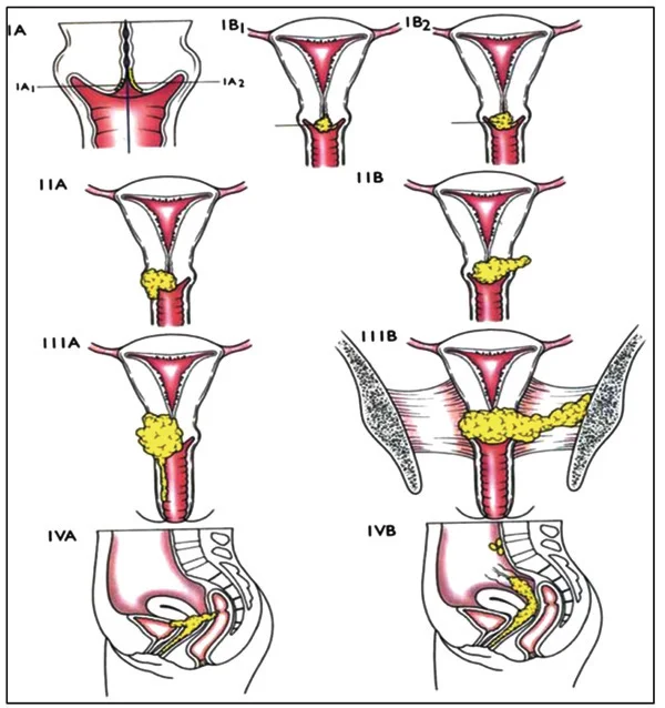
Figure 1Cervical Cancer Staging Image
Long description
Anatomical diagrams of each stage of cervical cancer, with a few sub-stages from the 2019 FIGO guidelines omitted.

- Cervical cytology (Pap test). Detects abnormal cells. The Bethesda system reports negative, ASC-US (equivocal squamous changes whose meaning is unclear), LSIL, ASC-H (cannot exclude HSIL), HSIL, AGC (atypical glandular cells) and invasive carcinoma.
- HPV testing (primary or cotest). Detects high-risk HPV DNA; more sensitive than cytology for CIN 3+ and provides longer reassurance after a negative result.
- Colposcopy with biopsy. Magnified examination of the cervix after acetic acid and Lugol iodine; biopsies of discrete acetowhite areas (usually 2 to 4 per examination) provide histology. A completely normal colposcopic impression without acetowhitening in the lowest-risk group does not require biopsy.[3]
- Endocervical curettage (ECC) when the transformation zone is not fully visible or glandular abnormality is suspected.
- Cone biopsy (LEEP or cold knife) is both diagnostic and therapeutic; it measures depth of invasion.
A screening test fixes risk, not a diagnosis. A woman with HPV-positive ASC-US after a negative screen last year has a low risk of CIN 3+ and can often be surveyed; a never-screened woman with HPV-positive HSIL has a high risk and qualifies for expedited treatment.[3]
- Expedited treatment without prior biopsy is preferred when the immediate risk of CIN 3+ is 60% or greater, and acceptable for risks between 25% and 60%.[3]
- Repeat HPV testing or cotesting at 1 year is recommended for minor abnormalities with low risk; colposcopy follows if results stay abnormal.[3]
- CIN 1 is observed rather than treated; treatment is acceptable for repeat diagnoses persisting 2 years or more.[3]
- After treatment of HSIL/CIN 2/CIN 3/AIS, surveillance continues with HPV testing or cotesting at 3-year intervals for at least 25 years.[3]
Clinical staging uses the FIGO 2018 system (most common worldwide). Work-up includes examination under anaesthesia, biopsy, MRI for local extent and lymph nodes, chest imaging and specialist gynaecological oncology review. The stage determines treatment. Key FIGO 2018 definitions:.[2]
| Stage | Description |
|---|---|
| IA | Invasive carcinoma diagnosable only by microscopy, depth of invasion 5 mm or less. IA1 = 3 mm or less; IA2 = greater than 3 mm and 5 mm or less. [2] |
| IB | Invasive carcinoma with depth greater than 5 mm, confined to the cervix, measured by maximum diameter. IB1 = 2 cm or less; IB2 = greater than 2 cm and 4 cm or less; IB3 = greater than 4 cm. [2] |
| IIA | Involvement of the upper two-thirds of the vagina without parametrial involvement. IIA1 = 4 cm or less; IIA2 = greater than 4 cm. [2] |
| IIB | Parametrial involvement, but not to the pelvic wall. |
| IIIA | Involvement of the lower third of the vagina, no pelvic-wall extension. |
| IIIB | Extension to the pelvic wall or hydronephrosis or non-functioning kidney. |
| IIIC | Involvement of pelvic (IIIC1) and/or para-aortic (IIIC2) lymph nodes, irrespective of tumour size or extent, with r/p notation for imaging or pathology. |
| IVA | Spread to adjacent pelvic organs (bladder or rectum mucosa). |
| IVB | Spread to distant organs. |
A useful prognostic anchor: in a large radiation series, the rate of distant metastases rose from 3% in stage IA to 75% in stage IVA.[2]
The principle is to match the intervention to the risk.
- CIN 1 (histologic LSIL): observe. Treatment is acceptable when CIN 1 persists 2 years or more.[3]
- CIN 2 and CIN 3 (histologic HSIL): excision (LEEP, cold-knife cone or laser) is preferred to ablation in the United States; ablation (thermal, cryotherapy) is acceptable in selected settings. Excision also provides tissue to exclude occult invasion.[3][1]
- AIS: excision is recommended (cold knife favoured over loop to avoid margin fragmentation), with hysterectomy for completed families.[2][3]
- Treatment failure / positive margins after excision: re-excite or hysterectomy according to age and fertility wishes.
- Pregnancy: in general, defer treatment of HSIL until postpartum; biopsy only if invasion is suspected, then plan delivery.
Stage IA1 without lymphovascular space invasion: conisation may be curative; simple hysterectomy if family complete.
Stage IA1 with lymphovascular invasion and stage IA2 to IB2, IIA1: modified or radical hysterectomy with pelvic lymphadenectomy, or, in women wishing fertility, radical trachelectomy. Surgery and radiation are equally effective for early, small-volume disease; surgery preserves ovarian function in younger women.[2]
Stages IB2-IB3 and IIA2 (bulky early disease): radical hysterectomy with lymphadenectomy, or definitive chemoradiation. Neoadjuvant chemotherapy is under evaluation.
Stages IIB, III and IVA: definitive concurrent chemoradiation is standard. The risk of death from cervical cancer was reduced by 30% to 50% with concurrent chemoradiation compared with radiation alone in phase III trials.[2]
- Regimen: concurrent cisplatin. A common weekly schedule is cisplatin 40 mg/m2 with radiation. Tri-weekly cisplatin 75 mg/m2 has similar survival but more grade 3-4 haematological toxicity is not its downside (weekly actually had more grade 3-4 haematological toxicity in the TACO trial, 49.3% versus 30.4%; weekly remains standard because it needs no hospitalisation).[6]
- Radiation: external beam (including intensity-modulated techniques) to the pelvis with or without para-aortic nodes, plus brachytherapy. Total dose and fractions follow radiotherapy protocols.
- Stage IVA may need interstitial brachytherapy or chemoradiation; palliative options for obstruction.
- Stage IVB and recurrent disease: palliative chemotherapy and radiotherapy, immunotherapy (checkpoint blockade), pelvic exenteration for isolated central recurrence, and enrolment in trials.
Concurrent cisplatin-based chemoradiation: weekly cisplatin 40 mg/m2 with pelvic radiation is the most common schedule used worldwide; evidence from five randomised phase III trials supports strong consideration of this approach.[2] [6]
Adjuvant treatment after surgery. Women with high-risk pathological features (positive margins, parametrial involvement, positive lymph nodes) gain a survival advantage from postoperative pelvic radiation with concurrent cisplatin.[2] Intermediate-risk factors (lymphovascular invasion, deep stromal invasion, large tumour size) may warrant radiation alone. This mirrors the shift from surgery to chemoradiation as disease volume grows.
Pretreatment investigations. Examination under anaesthesia, MRI of the pelvis for tumour size and parametrial extension, chest imaging, and renal function assessment. Cystoscopy and proctosigmoidoscopy are reserved for suspected invasion. Para-aortic nodal assessment is by imaging or surgico-pathological staging when it would change the radiation field.
- A multidisciplinary team (gynaecological oncology, medical oncology, radiation oncology, pathology, radiology) should confirm diagnosis and stage before treatment.
- Radiotherapy should not be delayed; completing treatment within the planned time improves locoregional control.
- Nephrotoxicity: cisplatin requires good renal function; carboplatin substitution or dose adjustment may be used when creatinine clearance is reduced.
- Fertility: discuss conisation, trachelectomy or oocyte preservation before treatment in women desiring future pregnancy.
- Local invasion: bladder or rectal fistula, ureteric obstruction and hydronephrosis, pelvic-wall and nerve-root pain, lymphoedema.
- Haematological and systemic: anaemia from bleeding; thrombosis; infection.
- Treatment complications:
- Surgery: haemorrhage, ureteric injury, fistula, lymphocyst, early menopause.
- Radiation: acute cystitis, proctitis, enteritis, vaginal stenosis; late fistula, stricture, secondary cancer.
- Chemotherapy: myelosuppression, nausea, renal and ototoxicity (cisplatin).
- Recurrent or metastatic disease most often presents within the first 2 years.[2]
Prognosis depends on stage, tumour volume and spread at diagnosis.
- Early-stage disease treated with surgery or radiation has high cure rates.
- Locally advanced disease treated with chemoradiation has markedly improved outcomes; concurrent therapy reduced the risk of death by 30% to 50% in clinical trials.[2]
- Stage, HIV status, tumour size, nodal status, and timeliness of treatment all influence outcome. HIV-positive women have more advanced disease and worse prognosis at presentation.[2]
- Post-treatment surveillance is usually every 3 to 4 months for the first 2 years, then every 6 months; most recurrences are found because of symptoms, so a careful history and examination matter. The benefit of routine cytology or imaging is uncertain.[2]
- After treatment of precancer, surveillance continues for at least 25 years because risk remains elevated.[3]
- Pain control for pelvic pain and nerve-root pain (opioids, neuropathic agents, nerve blocks).
- Renal drainage (stent or nephrostomy) for ureteric obstruction.
- Nutrition and blood transfusion for anaemia and cachexia.
- Vaginal brachytherapy complications (fistula, stenosis) need specialist aftercare; vaginal dilators preserve patency.
- Psychosocial and sexual health support (lymphoedema, stenosis, body image, fertility grief) should be offered throughout. Palliative chemotherapy, immunotherapy or radiotherapy can improve quality of life and control symptoms even when cure is not possible.
Exam trapA normal smear does not rule out cancer
A negative cytology tests the transformation zone sampled on that day; a rapidly progressive or inaccessible lesion (especially adenocarcinoma in the canal) can be missed. Persistent symptoms such as postmenopausal or postcoital bleeding always need examination and biopsy, regardless of recent smear results.
Exam trapTreating CIN 1
CIN 1 usually regresses. Observing avoids overtreatment and harm to future pregnancies; treating is for persistent or recurrent disease after 2 years.
Exam trapAblating without excluding invasion
HSIL on cytology may harbour occult cancer. Excisional methods (LEEP, cold knife) provide tissue and staged margins; blind ablation can miss invasive disease and delay treatment.
- All available vaccines protect against high-risk types 16 and 18, which cause around 76% of cervical cancers. Many also cover additional high-risk types and the low-risk types 6 and 11 that cause genital warts.[1]
- Who. Priority is girls aged 9 to 14 years, before sexual debut. Some countries also vaccinate boys. Immunocompromised individuals, including people living with HIV, should ideally receive 2 to 3 doses.[1]
- Dose schedule. Depending on the national programme and the vaccine, 1 or 2 doses are used for girls aged 9 to 14; 2 doses for immunocompromised individuals. WHO has added an HPV vaccine indication for single-dose use.[1]
- Effect. Vaccination prevents infection and the precancerous changes that follow, and is a central pillar of elimination. It does not treat existing infection.
The goal of screening is to detect precancer and treat it before invasion.
| Guideline / programme | Age range | Test and interval |
|---|---|---|
| WHO | 30 to 49 years (start at 25 for women living with HIV) | High-performance test every 5 to 10 years; at least two lifetime screens by age 35 and again by 45. Women living with HIV screened every 3 to 5 years. [1] |
| American Cancer Society (2020, updated 2026) | 25 to 65 years | Primary HPV testing every 5 years (preferred); cotesting every 5 years acceptable; cytology every 3 years acceptable. Self-collected vaginal specimens acceptable with repeat testing every 3 years after a negative result. [4] |
| NHS (England, public-facing) | 25 to 64 years | All women and everyone with a cervix invited every 5 years (HPV primary screening). |
| ASCCP (management) | Not a screening guideline | Risk-based management of abnormal test results and treated precancer. |
- Exit. ACS recommends screening exit only after negative primary HPV tests or negative cotesting at ages 60 and 65 years (or three consecutive negative cytology tests with the last at age 65, if HPV testing is unavailable). Women with prior abnormal results may need 10 years (after low-grade abnormalities) or 25 years (after treated high-grade precancer) of normal results, and immunosuppressed women do not qualify for exit at all.[4]
- Self-collection of vaginal samples for HPV testing increases uptake in underscreened groups and performs similarly to clinician sampling with PCR-based assays; a positive self-collected result requires a clinician-collected sample for cytology triage.[4]
- Low-resource settings (screen and treat). After a positive high-performance test, treatment may be offered in the same visit (thermal ablation, cryotherapy, LEEP or cone biopsy) or after visual assessment, which is especially useful where follow-up is uncertain. WHO encourages this for women living with HIV.[1]
United Kingdom (NICE)
In the UK programme, HPV is the primary screening test: samples are first tested for high-risk HPV and cytology is performed on HPV-positive samples. Practice details follow national programme guidance.
United States
In the United States, primary HPV testing from age 25 is the preferred strategy, with cotesting and cytology accepted; self-collection has been approved for use in clinical and, for one kit, home settings, with a 3-year interval after a negative self-collected result.
East Africa
In high-burden, lower-resource settings, a minimum of two lifetime screens with a high-performance test by ages 35 and 45, ideally with same-visit treatment, is the WHO-backed standard of care; services should be integrated with HIV care because women living with HIV need earlier and more frequent screening.
Pregnancy. Pregnancy does not cause cervical cancer but may be the time it is found. Screening continues in pregnancy when due (cytology preferred; colposcopy if abnormal, with biopsy for suspected invasion). Treatment of precancer is usually deferred to the postpartum period; conisation or trachelectomy may be considered for early invasive disease, ideally after specialist discussion and with planned delivery. Chemoradiation is contraindicated in pregnancy.
Children and adolescents. Cervical cancer is rare before age 20. Most adolescent HPV infection is transient and clears. Screening is not recommended under 21 (or 25 under ACS) in average-risk individuals; vaccination is the intervention. Screening of children is reserved for special circumstances such as in-utero DES exposure or immunosuppression, under specialist protocols.
Older adults. Incidence peaks in the mid-30s to 40s and falls, but cases and deaths in women older than 65 are increasingly recognised, which is why exit requires two negative tests at ages 60 and 65. Women who never screened should be offered at least one screen up to age 65.
Immunocompromised women (HIV, transplant, biologic therapy). They need screening earlier (from age 25), more frequently (every 3 to 5 years or as national guidance), and they do not qualify for self-collection or routine exit. Screen and triage, rather than screen and treat, is especially recommended for women living with HIV.[1]
Transgender men and non-binary people with a cervix. Screening policy should follow the organ present, not gender identity; barriers to care and the effects of testosterone therapy on the cervix need sensitive handling.
Approach to cervical cancer screening and management
- Average-risk woman with a cervixPrimary HPV test or cytology per programme
- Negative resultRepeat per interval (5 years primary HPV; 3 years cytology)
- Question: Positive or abnormal result
Minor (HPV+/ASC-US after recent negative screen; LSIL)
- SurveillanceRepeat HPV testing or cotesting in 1 year; colposcopy if still abnormal
High-grade (HSIL, ASC-H, AGC, HPV 16/18+)
- Colposcopy + biopsy2 to 4 targeted biopsies of acetowhite areas; endocervical curettage if needed
- HistologyCIN 1 observe; CIN 2/3 or AIS excise; cancer stage and treat
- Question: Invasive cancer
Early stage (IA-IB2, IIA1)
- Surgery or chemoradiationRadical hysterectomy + lymphadenectomy or trachelectomy; equal outcomes for small-volume disease
Locally advanced (IB2-IB3, IIA2, IIB-IVA)
- Concurrent chemoradiationCisplatin (weekly 40 mg/m2) with pelvic radiation plus brachytherapy
Metastatic / recurrent
- Palliative therapyChemotherapy, immunotherapy, radiotherapy, exenteration for isolated central recurrence
- Cervical cancer is almost always caused by persistent high-risk HPV infection; the progression from infection to cancer takes 15 to 20 years (5 to 10 in immunosuppression).
- Screening prevents cancer by finding and treating precancer (CIN), not by finding cancer early.
- HPV 16 and 18 cause around 76% of cervical cancers; vaccination of girls aged 9 to 14 years before sexual debut is the most effective prevention.
- A high-performance HPV test every 5 to 10 years from age 30 (every 3 to 5 years from age 25 in women living with HIV) is the WHO-preferred strategy; ACS supports primary HPV testing every 5 years from 25. [1][4]
- The NHS (England) invites everyone with a cervix aged 25 to 64 every 5 years under HPV primary screening.[5].- Abnormal results are managed by risk (2019 ASCCP): expedited treatment is preferred at a CIN 3+ risk of 60% or greater, colposcopy follows high-grade abnormalities, CIN 1 is observed, and treated HSIL needs 25 years of surveillance.
- Excision is preferred to ablation for HSIL/CIN 2 and CIN 3; excision is recommended for AIS.
- Locally advanced cervical cancer is treated with concurrent chemoradiation, usually weekly cisplatin 40 mg/m2 with pelvic radiation plus brachytherapy, which reduced the risk of death by 30% to 50%.
- FIGO 2018 staging uses size and spread (IA-IVB); stage III adds lymph-node involvement (IIIC1/2).
- WHO elimination requires the 90-70-90 targets by 2030: 90% of girls vaccinated by 15, 70% of women screened by 35 and again by 45, 90% treated.[1]- Immunocompromised women need earlier, more frequent screening and do not qualify for routine exit; older adults still need two negative tests at ages 60 and 65 to stop.
Answer each one in your head first, then open the answer.
Why does a persistent HPV infection cause cancer, and what two viral proteins drive the process?
Show answerHide answer
High-risk HPV persists in the transformation zone and integrates into the host genome; its E6 and E7 oncoproteins inactivate p53 and Rb, removing the controls on the cell cycle and DNA repair, so abnormal cells accumulate and progress from CIN to invasive cancer over 15 to 20 years. [1]
A 28-year-old, previously unscreened woman has HPV 16-positive HSIL cytology. What management does the ASCCP guideline support, and why?
Show answerHide answer
Expedited treatment without prior colposcopic biopsy is preferred: her immediate risk of CIN 3+ is 60% or greater, and never- or rarely-screened patients with HPV-positive HSIL are explicitly in that group. Colposcopy-directed biopsy or LEEP is then used to confirm stage.
A woman aged 62 has had two negative primary HPV tests, one at 60 and one at 65. A 68-year-old woman with a treated CIN 3 5 years ago asks to stop screening. What is the advice in each case?
Show answerHide answer
The first woman meets the ACS exit criteria and may stop. The second should continue surveillance: after treatment of HSIL at least 25 years of normal results are required, so she cannot stop yet and should also remain under the ASCCP surveillance schedule.
A 32-year-old woman living with HIV (CD4 400) has never been screened. Plan her screening and say why it differs from average risk.
Show answerHide answer
Start now with a high-performance HPV test and repeat every 3 to 5 years, beginning at age 25; she does not qualify for self-collection as her only modality nor for routine exit. Women living with HIV have a six-fold risk of cervical cancer and progress faster, so screening, triage and treatment should be integrated into her HIV care.
Stage a tumour: 3.5 cm, confined to the cervix, depth greater than 5 mm. Give its FIGO stage and first-line treatment in a 45-year-old.
Show answerHide answer
Stage IB2 (greater than 2 cm and 4 cm or less, with depth greater than 5 mm). First-line: either radical hysterectomy with pelvic lymphadenectomy or definitive concurrent chemoradiation; for bulky disease chemoradiation is often preferred, with cisplatin (weekly 40 mg/m2) and pelvic radiation plus brachytherapy. [2]
Most infections are transient and cleared, but the risk of high-grade disease is concentrated in the minority that persist. Longitudinal data show that infection with HPV 16 or 18 predicts high-grade CIN or greater for up to 18 years after the first test.[2]
Colposcopy should be performed to the ASCCP Colposcopy Standards. Optimising detection at the first visit matters because a low-grade impression leads to longer surveillance.
- Preparation: schedule outside menses; consider anxiolysis; use a water-based lubricant sparingly (some units avoid vaginal products beforehand).
- Examination: visualise the cervix after applying dilute acetic acid; look for acetowhite change, punctation, mosaic, and atypical vessels; apply Lugol iodine.
- Biopsy: two to four biopsies of every discrete acetowhite lesion; random biopsies are not recommended in the lowest-risk group with a completely normal impression.
- Endocervical sampling when the transformation zone is not fully seen or glandular disease is suspected.
- Documentation: colposcopic impression (normal, low-grade, high-grade, cancer), whether the squamocolumnar junction is fully visible, and biopsy sites.
Histology follows the Lower Anogenital Squamous Terminology (LAST)/WHO system: LSIL (CIN 1), HSIL (CIN 2, CIN 3) and AIS. p16 immunostaining may support the diagnosis of HSIL. Reporting CIN 2 versus CIN 3 separately matters because management of CIN 2 in very young women is sometimes more conservative.
A single negative HPV test therefore gives strong reassurance; a new abnormal result after a documented negative test within the previous 5 years carries roughly half the risk of CIN 3+ of the same result with unknown history, which is why the ASCCP tables incorporate screening history.[3] A negative cytology within 3 years of an abnormal result, by contrast, does not confer a comparable reduction in risk. This biological reality underlies the lengthening of intervals in modern screening programmes and the risk-based approach to management.
Cervical cancer is adjacent to topics on HPV infection and vaccination, vaginal and vulval disease, abnormal uterine bleeding, endometrial cancer, gynaecological oncology, and HIV in women. Planned (or already published) topics in these areas can be linked once verified; link with the topic slugs already present in the site.
- World Health Organization. Cervical cancer. WHO Newsroom fact sheet, 3 July 2026. Accessed .
- National Cancer Institute. Cervical Cancer Treatment (PDQ) – Health Professional Version. NCI PDQ, updated 13 May 2025. Accessed .
- Perkins RB, Guido RS, Castle PE, Chelmow D, Einstein MH, Garcia F, Huh WK, Kim JJ, Moscicki AB, Nayar R, Saraiya M, Sawaya GF, Wentzensen N, Schiffman M, for the 2019 ASCCP Risk-Based Management Consensus Guidelines Committee. 2019 ASCCP Risk-Based Management Consensus Guidelines for Abnormal Cervical Cancer Screening Tests and Cancer Precursors. J Low Genit Tract Dis 2020;24(2):102–131. Accessed .
- Perkins RB, Wolf AMD, Church TR, et al. Self-collected vaginal specimens for human papillomavirus testing and guidance on screening exit: An update to the American Cancer Society cervical cancer screening guideline. CA Cancer J Clin 2026;76(1):e70041. Accessed .
- National Health Service. Cervical screening. NHS website, accessed 2026. Accessed .
- Ryu SY, Nam BH, Kim MH, et al. A randomized phase III clinical trial of weekly versus tri-weekly cisplatin-based chemoradiotherapy for locally advanced cervical cancer: results of the TACO study. ESMO Open 2026;11(4):106102. Accessed .
Figure credits
Educational content for medical students and doctors. It is not a substitute for clinical judgement or local guidelines.