Endometrial cancer and hyperplasia
Endometrial cancer and hyperplasia: risk factors, postmenopausal bleeding work-up, hyperplasia with and without atypia, FIGO 2023 staging, molecular.
- Endometrial cancer is the commonest gynaecological malignancy in affluent settings: abnormal bleeding is the hallmark, and about 10% of postmenopausal bleeding is due to it [5][6]. [9]
- Long unopposed oestrogen is the central mechanism; obesity, anovulation, postmenopausal oestrogen-only therapy and tamoxifen (over 2 years) are the risks worth remembering [7].
- Postmenopausal bleeding is a suspected-cancer referral; the first tests are transvaginal ultrasound and endometrial sampling [3][6].
- Hyperplasia without atypia is usually benign and regresses with a progestin; atypical hyperplasia (EIN) acts like early cancer and needs surgery or closely supervised progestin therapy [4][14].
- FIGO 2023 staging now incorporates histotype and molecular subtype, and ESGO/ESTRO/ESP 2021 risk groups guide adjuvant therapy [2][1].
By the end of this page you should be able to:
- Define endometrial cancer and hyperplasia (EIN) and contrast the benign and premalignant forms.
- Trace why unopposed oestrogen leads to neoplasia, and use it to explain every risk factor.
- Work up postmenopausal bleeding and know the 4 mm endometrial thickness cut-off and when to sample.
- Stage endometrial cancer with the FIGO 2023 system and assign the molecular class.
- Explain the four molecular subgroups and why they stratify prognosis and adjuvant therapy.
- Manage hyperplasia with and without atypia, including progestin regimens and hysterectomy indications.
- Outline surgical staging, adjuvant radiotherapy/chemotherapy and advanced disease treatment per ESGO/ESTRO/ESP 2021, ESMO and NICE.
- Distinguish endometrial cancer from cervical cancer, fibroids, polyps and atrophy.
Endometrial cancer is a cancer of the lining of the uterus. It is the most common gynaecological malignancy in industrialised countries and usually presents with bleeding: irregular or heavy bleeding before the menopause, or any bleeding after it [5]. Endometrial hyperplasia is the disordered proliferation of endometrial glands caused by chronic unopposed oestrogen. In its benign (without atypia) form it rarely becomes cancer; with nuclear atypia it is the precursor of the commonest type of endometrial cancer (EIN, endometrial intraepithelial neoplasia) [4]. Because the two conditions share causes and bleeding symptoms, they are taught together.
DefinitionEndometrial cancer
A malignancy arising from the glandular epithelium of the endometrium, the inner lining of the uterus. The dominant histological type is endometrioid adenocarcinoma (about 80% of cases) [5].
- Classification in practice. Older teaching divided tumours into type 1 (endometrioid, oestrogen-driven, generally grade 1 to 2) and type 2 (serous, clear cell, carcinosarcoma, generally grade 3 and aggressive). Molecular profiling has largely replaced this split: the current classification is the molecular one (POLEmut, MMRd, NSMP, p53abn) introduced below, backed by FIGO 2023 [12].
- Histological types worth naming. Serous and papillary serous carcinomas account for 4% to 6% of endometrial carcinomas, and clear cell carcinomas for 1% to 2% [5]. The grade (1 well differentiated, 2 moderately, 3 poorly or undifferentiated) and the myometrial invasion depth determine prognosis and stage.
- Hyperplasia (WHO 2014). Two groups only: endometrial hyperplasia without atypia (benign enrichment) and atypical endometrial hyperplasia / EIN (premalignant) [4]. Nuclear atypia means nuclear enlargement, with or without prominent nucleoli.
- Cervical cancer on the same page. The book lists endometrial and cervical cancer on one page. Cervical cancer arises from the cervix, not the endometrium, and is a different disease with different causes and screening; it is mentioned here mainly as a differential when bleeding is evaluated (see below).
- Endometrial cancer is the commonest gynaecological malignancy in high-income countries. In the United States over 66,000 new uterine cancer cases and over 13,000 deaths were expected in 2023; endometrial carcinomas make up over 83% of uterine cancers [5].
- It is predominantly a postmenopausal cancer [5], and about 10% of postmenopausal bleeding is due to endometrial cancer [5].
- Incidence has risen with ageing and with the obesity epidemic [7].
- Type 1 cancers (endometrioid, about 80% of all) are the oestrogen-related ones; type 2 cancers are the aggressive serous/clear-cell forms [5].
The unifying mechanism is chronic unopposed oestrogen: oestrogen stimulates endometrial proliferation, and without progesterone's counterbalancing differentiation and shedding, glands pile up into hyperplasia and, eventually, cancer [4][7].
| Risk factor | Mechanism | Source |
|---|---|---|
| Obesity, adult weight gain | Adipose tissue converts androgens to oestrogen; insulin resistance; often anovulatory cycles | [7] |
| Postmenopausal oestrogen-only hormone therapy | Unopposed oestrogen acts directly on the endometrium; risk rises with duration of use | [7] |
| Early menarche or late menopause | More years of cumulative oestrogen exposure | [7] |
| Nulliparity | Less lifetime exposure to progesterone-dominant pregnancy | [7] |
| Chronic anovulation, PCOS | Persistent oestrogen without progesterone | [7] |
| Tamoxifen for over 2 years | Oestrogen-agonist effect on the endometrium; risk greater after the menopause | [7] |
| Lynch syndrome, Cowden syndrome, a parent, sibling or child with the same cancers | Genetic predisposition (Lynch in particular) | [7] |
| Type 2 diabetes, metabolic syndrome | Hyperinsulinaemia and oestrogen excess linked to obesity | [7] |
| Endometrial hyperplasia, especially atypical | Precursor lesion | [7] |
Protective factors: pregnancy and breastfeeding (lower oestrogen), combined oral contraceptives (protective; effect increases with duration used and persists for years after stopping), bariatric surgery, and physical activity [7]. Combining oestrogen with progestin in postmenopausal therapy does not increase endometrial cancer risk (though it increases breast cancer risk) [7]. Raloxifene does not have oestrogen-like effects on the uterus and is not linked to endometrial cancer [7].
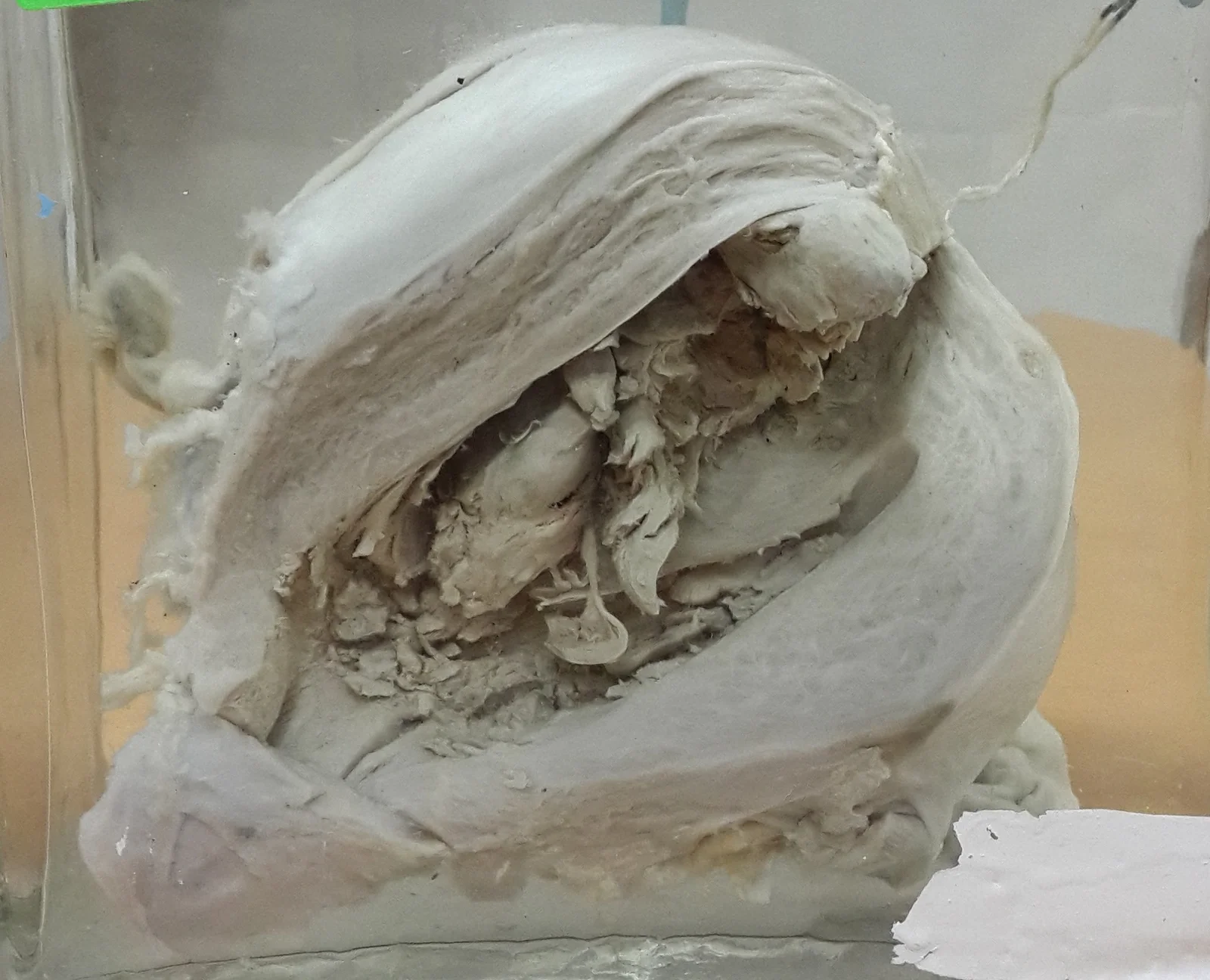
Figure 1Polypoidal endometrial carcinoma
Long description
A gross photograph of a polypoidal endometrial carcinoma in an opened uterus.

- Normal. In a normal ovulatory cycle, oestrogen rebuilds the endometrium in the proliferative phase and progesterone stabilises and then sheds it in the secretory phase and menses.
- Hyperplasia. When progesterone is absent or insufficient, oestrogen keeps the glands dividing. The gland-to-stroma ratio rises; glands become crowded. This is endometrial hyperplasia without atypia; it is a reversible hormonal state, not a clonal neoplasm, and often regresses if progesterone is restored [4].
- Atypical hyperplasia / EIN. With continuing stimulation, PTEN and other tumour-suppressor pathway changes accumulate and nuclei become atypical. This is the precursor of endometrioid (type 1) carcinoma; histologically it looks like early, well-differentiated endometrioid cancer [4].
- Invasion. Cancer is defined by stromal invasion. Spread begins within the uterine cavity, then into the myometrium, then across the serosa and to adnexa, cervix, vagina, parametrium, lymph nodes, peritoneum and distant sites; this sequence is what the FIGO stages record.
The second route (type 2) arises in atrophic endometrium with TP53 mutation and aneuploidy (serous carcinoma, clear cell), often in older women; it is more aggressive and less oestrogen-dependent, but it still shares many risk factors, including obesity [4][12].
- Abnormal uterine bleeding is the most frequent symptom [5]: heavy or prolonged menstrual bleeding, intermenstrual bleeding, or any bleeding after the menopause. Bleeding is painless. Bleeding after menopause, including a single episode, must be investigated for cancer [6].
- Pain or pressure from an enlarged uterus or extensive disease; watery, purulent or blood-stained discharge; and late systemic symptoms (weight loss, anaemia) with advanced disease.
- Risk story: early menarche, late menopause, nulliparity, obesity, PCOS, tamoxifen use, hormone therapy, family history of Lynch-related cancers.
- General: anaemia, wasting in advanced disease; signs of PCOS or metabolic syndrome.
- Abdominal: a pelvic mass in advanced disease.
- Pelvic: enlarged, irregular or fixed uterus; cervical lesions on speculum examination (do not assume all bleeding is uterine); adnexal or parametrial masses in advanced disease.
- Always exclude pregnancy in anyone of reproductive age, and ask about sources of bleeding outside the uterus (vagina, cervix, urethra, bladder, rectum, perineum) before calling bleeding "uterine" [6].
| Condition | Key distinguishing features |
|---|---|
| Endometrial hyperplasia (without atypia) | Same risk and bleeding profile; biopsy shows crowded glands without atypia; regress with progestin [4] |
| Atypical hyperplasia / EIN | Biopsy shows nuclear atypia; ~33% harbour concurrent cancer; treated as early cancer [4] |
| Cervical cancer | Arises in the cervix; postcoital bleeding, disordered cervical epithelium; speculum and cervical sampling diagnose it |
| Endometrial polyp | Focal bleeding, often intermenstrual; ultrasound/hysteroscopy shows a focal intracavitary lesion [4] |
| Uterine leiomyoma / adenomyosis | Heavy regular menses, bulk symptoms; imaging shows myometrial lesions [4] |
| Postmenopausal atrophy (vagina and endometrium) | Most common cause of postmenopausal bleeding (~60%); thin atrophic endometrium [6] |
| Hormone therapy breakthrough bleeding | Unscheduled bleeding in users of sequential or continuous HRT; limited evidence; refer if persistent [3] |
| Other genital malignancy (vulval, vaginal, ovarian) | Lesions seen on examination/imaging; different biopsy site |
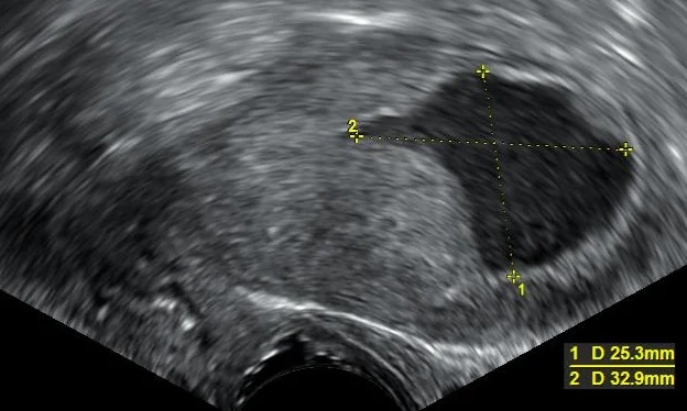
Figure 2Endometrial fluid accumulation, postmenopausal
Long description
Transvaginal ultrasonography showing a uterine fluid accumulation in a postmenopausal woman whose biopsy showed endometrioid adenocarcinoma.

Investigation is aimed at confirming cancer and excluding it. Hysteroscopy-guided sampling is preferred; blind endometrial biopsy can miss focal disease.
| Test | What it establishes | Cut-off / performance |
|---|---|---|
| Pregnancy test (urine hCG, then serum) | Exclude pregnancy as the cause of bleeding | Mandatory in any reproductive-aged woman [7] |
| Transvaginal ultrasound, endometrial thickness | First non-invasive step in postmenopausal bleeding; thickness assessment | Endometrial lining over 4 mm in a woman with postmenopausal bleeding is an indication for sampling; persisting bleeding or risk factors warrant sampling even with a thin stripe [6] |
| Endometrial biopsy | Histology: hyperplasia with/without atypia, type, grade | First-line sampling test; provides tissue for diagnosis of malignancy [6] |
| Hysteroscopy with directed biopsy | Visualise and biopsy focal lesions | Preferred to blind biopsy for persistent or recurrent bleeding [6] |
| MRI pelvis | Myometrial invasion, cervical extension, nodal disease, preoperative planning | Reserves ultrasound/MRI sequence as below |
| CT chest/abdomen/pelvis, PET-CT | Staging for advanced/recurrent disease | Used when stages III-IV suspected (NCI/ESGO) [11][1] |
| Mismatch repair immunohistochemistry, POLE testing | Molecular class | Encouraged in all cancers [2] |
| Serum CA125 | Adnexal involvement or recurrence | Sometimes measured; not a screening test [11] |
Special note on transvaginal ultrasound in premenopausal (cycling) women: a single measurement cannot reassure because endometrial thickness changes across the cycle, and sampling is indicated by age and risk rather than by any one thickness value.
NICE recommends suspected-cancer pathway referral for women aged 55 and over with unexplained postmenopausal bleeding not attributable to hormone replacement therapy; for women under 55 a suspected-cancer referral is to be considered [3]. For unexplained vaginal discharge in women aged 55 or over, direct-access ultrasound is appropriate, as are visible haematuria with low haemoglobin, thrombocytosis or high blood glucose [3].
Approach to postmenopausal bleeding
- Postmenopausal bleeding (any bleeding after 12 months of amenorrhoea)Confirm bleeding is vaginal and uterine in origin; check urethra, vulva, vagina, cervix and rectum
- History, risk factors and speculum/bimanual examinationObesity, PCOS, anovulation, tamoxifen, HRT, family history; cervical lesions, masses
- First-line investigationsTransvaginal ultrasound for endometrial thickness AND endometrial biopsy for tissue diagnosis
- Question: Endometrial thickness?
4 mm or less
- Cancer less likely on imagingStill sample if bleeding recurs or risk factors present
Over 4 mm
- Endometrial sampling indicatedHysteroscopy-directed biopsy preferred for focal lesions
- Question: Histology?
Atrophy or benign
- Treat cause (atrophy, polyp, etc.)Reinvestigate any recurrent or persistent bleeding
Hyperplasia
- Manage by type (see below)Without atypia: progestin; with atypia/EIN: hysterectomy usually
Cancer
- Staging work-up and oncology referralSurgical staging; see FIGO 2023 below
Endometrial cancer is surgically staged and pathologically examined. The grade, histological type and lymphovascular space invasion (LVSI) must be recorded in all stages. FIGO 2023 staging better reflects modern disease biology by separating aggressive from non-aggressive histotypes and adding molecular subtypes [2].
| Stage | Description |
|---|---|
| IA1 | Non-aggressive type limited to an endometrial polyp or confined to the endometrium |
| IA2 | Non-aggressive type invading under 50% of the myometrium, no or focal LVSI |
| IA3 | Low-grade endometrioid carcinoma limited to the uterus and ovary |
| IB | Non-aggressive type invading 50% or more of the myometrium, no or focal LVSI |
| IC | Aggressive type (serous, high-grade endometrioid, clear cell, carcinosarcoma etc.) limited to a polyp or confined to the endometrium, no myometrial invasion |
| IIA | Non-aggressive type invading cervical stroma |
| IIB | Non-aggressive type with substantial LVSI |
| IIC | Aggressive type with any myometrial invasion |
| IIIA1/2 | Spread to ovary/fallopian tube (except IA3) / involvement of uterine serosa |
| IIIB1/2 | Vagina/parametria / pelvic peritoneum |
| IIIC1i/ii | Pelvic lymph nodes, micrometastasis / macrometastasis |
| IIIC2i/ii | Para-aortic lymph nodes to renal vessels, with/without pelvic nodes |
| IVA | Invasion of bladder or bowel mucosa |
| IVB | Extrapelvic peritoneal metastasis beyond the pelvis |
| IVC | Distant metastasis |
Molecular notation: if the molecular subtype is known, add "m" and the subtype. In stages I and II, POLEmut downstages to IAm POLEmut and p53abn upstages to IICm p53abn, regardless of LVSI or histological type [2]. Stages III and IV are recorded as IIIm and IVm with the molecular class, without stage change.
The molecular classification derives from the TCGA genomic analysis and is now standard (WHO endorses a stepwise algorithm: POLE testing, then MMR immunohistochemistry, then p53 immunohistochemistry) [12]. It is encouraged in all cancers, especially high-grade tumours [1].
| Subgroup | Approx. share | Biology | Prognosis | Practical implication |
|---|---|---|---|---|
| POLEmut (polymerase epsilon-ultramutated) | 6-9% | Proofreading polymerase mutation; very high mutation burden | Excellent; 96% survival at 5 years | Adjuvant treatment may be de-escalated; low risk in ESGO groups [12][13] |
| MMRd (mismatch repair deficient) | 20-30% | MLH1/MSH2/MSH6/PMS2 loss; microsatellite instability; Lynch syndrome in ~3% of EC | Intermediate; associated with LVSI, high grade | Eligible for PD-1 blockade in advanced/recurrent disease [12] |
| NSMP (no specific molecular profile) | 40-50% | Copy-number low, usually endometrioid, ER/PR positive | Intermediate; highest BMI cluster | Hormone-responsive; CTNNB1/L1CAM mark worse subset [12][13] |
| p53abn (p53 abnormal) | 13-18% | TP53 mutation, copy-number high; 93% of serous, 85% of carcinosarcoma | Worst; responsible for 50-70% of EC mortality | Chemoradiotherapy; high risk in ESGO groups [12][13] |
NICE/ACOG note: women with endometrial cancer should be tested for Lynch syndrome (MMR IHC, then MLH1 promoter hypermethylation if MLH1/PMS2 loss, then germline testing) so that patients and relatives can enter surveillance and prevention [3].
Work is multidisciplinary, in a specialised centre with a dedicated team, with careful counselling about prognosis, risks and alternatives [1]. Fertility wishes must be clarified early, because they change the operation. Key general rules:
- Early, localised disease is treated surgically first. Hormonal therapy, chemotherapy and radiotherapy are selected by stage and risk group after surgery.
- Endometrial cancer cells are often progesterone-sensitive, which is why hormonal therapy works in some settings and why hormonal contraception lowers risk.
NoteRisk groups follow ESGO/ESTRO/ESP 2021.
ESGO/ESTRO/ESP 2021 (via its pocket guideline) defines low, intermediate, high-intermediate and high-risk groups by stage, grade, histotype, LVSI and molecular class, and assigns adjuvant treatment to each [1]. FIGO 2023 staging should be used to determine the anatomical group, and the molecular class modifies early-stage assignments [2].
This is the commonest reason an endometrial biopsy is taken. Management depends entirely on the presence of atypia.
Hyperplasia without atypia.
- Benign, reversible hormonal state; rarely progresses (under 5% over 10 years) [4]. Progesterone therapy gives 89% to 96% resolution versus 74% to 81% with expectant management [4].
- First-line drug: levonorgestrel-releasing intrauterine system. It concentrates progestin in the endometrium, avoids systemic side effects, and has higher histological regression than oral progestins for complex hyperplasia (92% versus 66%) and for atypical hyperplasia (90% versus 69%) [4][14].
- Traditional oral regimens: medroxyprogesterone acetate 10 to 20 mg daily, or norethisterone 10 to 15 mg daily, for 6 months, with repeat biopsy [4].
- Correct reversible causes (weight loss, ovulation induction) and repeat surveillance biopsy; hysterectomy is reserved for persistent disease after optimal medical therapy [4].
Atypical hyperplasia / EIN.
- Acts like early cancer: about 33% harbour concurrent cancer and the untreated progression risk is about 8.2% per year versus 2.6% for non-atypical hyperplasia [4].
- Definitive treatment is total hysterectomy (not supracervical) [14]; add bilateral salpingo-oophorectomy in postmenopausal women [4].
- Fertility sparing: after excluding coexisting cancer (hysteroscopy-directed sampling, imaging), use high-dose progestin (oral, intrauterine or a combination) with close histological follow-up every 3 to 6 months [14][4]. Levonorgestrel IUD has higher regression than oral progestins alone [14]. Recurrence after conservative therapy is well described, so surveillance continues long-term.
- Lifestyle counselling: weight loss and glycaemic control improve overall health and may reduce EIN and cancer risk [14].
| Drug/regimen | Typical dose | Use and notes |
|---|---|---|
| Levonorgestrel intrauterine system (52 mg) | 20 micrograms per day release | First-line for hyperplasia without atypia and many atypical cases needing fertility preservation; reduces heavy bleeding; check placement and remove/replace per product licence [4] |
| Medroxyprogesterone acetate | 10 to 20 mg daily (orally) for 6 months | Oral option for hyperplasia; continuous is preferred over cyclic therapy [4][14] |
| Norethisterone/norethindrone | 10 to 15 mg daily orally | Alternative oral progestin [4] |
| High-dose progestins for fertility sparing | Per oncology protocol | Higher oral progestin regimens (specialist-led, with imaging and hysteroscopic follow-up) after excluding cancer [14] |
| GnRH analogues | Standard hypogonism-inducing regimens | May be combined with progestins or hysteroscopic resection to increase regression; usually add-back therapy |
| Aromatase inhibitors | Anastrozole/letrozole per oncology protocols | Off-label adjuncts in selected recurrent/hormone-sensitive disease |
| Progestins/anti-oestrogens in recurrent cancer | Medroxyprogesterone acetate 200 to 1000 mg/day equivalents | Hormone-sensitive (ER/PR positive, usually NSMP), low-grade recurrent disease; doses are specialist |
| Tamoxifen | Not used for treatment of endometrial cancer | Agonist on endometrium; risk rises after over 2 years of use [7] |
Doses above beyond the three named progestin regimens are taken from the cited systematic and guideline sources; check your local formulary and oncology protocol before prescribing.
Surgery is the primary treatment [11][5].
- Early disease (confined to uterus): total hysterectomy with bilateral salpingo-oophorectomy and peritoneal washings, with pelvic (and para-aortic where indicated) lymph node assessment or sentinel lymph node mapping [5][1].
- Route: laparoscopic or robotic (minimally invasive) approaches are acceptable where expertise exists; laparotomy for large uterus, suspected extrauterine disease or contraindications. NICE supports laparoscopic hysterectomy for endometrial cancer in appropriate hands [3].
- Radical hysterectomy (parametrial resection) may be needed with cervical involvement; radical surgery is usually not required for endometrial primaries without cervical stromal invasion.
- Fertility preservation is possible in highly selected low-grade endometrioid cancer confined to the endometrium (stage IA) in young women: high-dose progestin plus hysteroscopy and imaging, then completion surgery after childbearing [1].
- Advanced disease: cytoreductive (debulking) surgery when feasible, aiming for no residual disease, followed by systemic therapy [1].
After surgery, adjuvant therapy is decided by risk group (ESGO/ESTRO/ESP 2021) [1]:
- Low risk: no adjuvant radiotherapy or chemotherapy; surveillance.
- Low-intermediate/intermediate: vaginal brachytherapy to reduce locoregional relapse; often enough to control vaginal recurrence.
- High-intermediate: pelvic radiotherapy plus or minus vaginal boost, particularly with substantial LVSI, grade 3 disease or age-related risk; ESGO risk tables integrate molecular class (stage I-II POLEmut is treated as low risk; p53abn stage I with myometrial invasion is treated as high risk) [1][12].
- High risk: combined chemoradiotherapy (pelvic radiotherapy plus platinum-based chemotherapy such as carboplatin/paclitaxel) for serous, clear-cell, carcinosarcoma and p53abn tumours; PORTEC-3 molecular subgroup analyses support adding chemotherapy in p53abn [12].
- Advanced/metastatic: systemic therapy first; radiotherapy for palliation and local control.
Post-operative radiotherapy reduces vaginal and pelvic recurrence but must be balanced against late bowel, bladder and sexual-function toxicity; modern intensity-modulated techniques reduce this.
- Chemotherapy: platinum plus taxane (most commonly carboplatin + paclitaxel) for advanced, metastatic or recurrent non-endometrioid/p53abn disease.
- Hormonal therapy: progestins, aromatase inhibitors or tamoxifen-based sequences (e.g. alternating tamoxifen and high-dose progestin) for low-grade, hormone receptor-positive recurrent disease; objective responses of months to years in selected patients.
- Immunotherapy/checkpoint inhibitors: PD-1 blockade (dostarlimab or pembrolizumab-based regimens) plus carboplatin/paclitaxel for first-line advanced or recurrent mismatch repair-deficient (MMRd/MSI-high) disease; and combinations with PARP inhibitors for selected non-MMRd cases per NICE and ESMO [3]. Pembrolizumab, dostarlimab-based and durvalumab-based combinations now have NICE technology appraisals [3].
- Targeted agents: everolimus (mTOR inhibition), bevacizumab (anti-VEGF) and signal-transduction inhibitors are used in recurrent disease, usually in clinical trials.
- Palliative radiotherapy: for vaginal bleeding, pelvic pain and bone metastases.
Strongly emphasise enrolment in clinical trials for advanced disease, where available.
After treatment, regular clinical review and imaging guided by risk. Recurrence surveillance is risk-stratified and focuses on pelvic, abdominal and pulmonary sites. Guide women on bleeding or pain red flags.
- Disease complications: severe bleeding and anaemia, invasion and obstruction of bladder/bowel, ureteric obstruction and hydronephrosis, pulmonary or distant metastases, malignant ascites.
- Treatment complications: operative bleeding, infection, ureteric or bladder injury, venous thromboembolism (prophylaxis needed), radiation enteritis/cystitis/proctitis, chemotherapy myelosuppression and neuropathy, premature menopause and oestrogen-deficiency symptoms after oophorectomy, sexual dysfunction.
- Fertility loss: total hysterectomy plus oophorectomy ends fertility; discuss storage or fertility-sparing pathways first.
- Prognosis depends on stage, grade, histotype, LVSI, molecular class and treatment.
- Stage I and II disease has an excellent prognosis; most women are cured because bleeding prompts early diagnosis [10].
- Overall 5-year survival for endometrial cancer is around 90%, far better than most solid tumours [6].
- Worst outcomes occur in p53abn, serous and clear-cell cancers and in stage III-IV disease; POLEmut tumours do very well even when high grade [12].
- Recurrence, where it happens, is usually pelvic, abdominal (peritoneal) or pulmonary.
- There is no standard screening test for endometrial cancer in asymptomatic women [8]. Transvaginal ultrasound has no proven mortality benefit as screening, and blind endometrial sampling is not justified routinely [8].
- Women at increased risk because of Lynch syndrome (hereditary non-polyposis colorectal cancer) should be offered yearly transvaginal ultrasound, beginning as early as age 25, alongside colonoscopic surveillance [8].
- (NCI summary) No standard or routine screening test exists; tamoxifen users should be investigated only if bleeding occurs; Pap smear is not a screen for endometrial cancer [8].
- Prevention: weight control, combined (oestrogen plus progestin) contraception, pregnancy/breastfeeding, physical activity and (where appropriate) bariatric surgery reduce risk; avoid unopposed oestrogen in women with a uterus; consider progestin or a levonorgestrel system in anovulatory/PCOS patients with persistent hyperplasia [7][4].
- New data suggest intrauterine hormone devices may reduce endometrial cancer risk; more studies are needed [7].
- Pregnancy/children not relevant for cancer (endometrial cancer is extremely rare before the menopause). For hyperplasia in young women with PCOS/anovulation, treat with weight control and cyclical progestin or levonorgestrel IUD, and avoid definitive surgery [4].
- Older adults: atrophy is the most common cause of postmenopausal bleeding; cancer must still be excluded. Fragility, comorbidity and frailty modify operative choice; pelvic radiotherapy or systemic therapy may replace surgery in very frail patients.
- Tamoxifen users: have higher endometrial cancer risk and need gynaecological review for any bleeding; routine TVU screening without symptoms is not useful [8].
- Lynch syndrome carriers: see prevention section; MMR testing of tumour plus germline counselling [3].
- Trans men and non-binary people with female reproductive organs: follow the same referral and investigation pathways [3].
Where hysteroscopy, MRI and molecular testing are unavailable, clinical diagnosis still holds: any postmenopausal bleeding warrants speculum examination, transvaginal ultrasound for thickness and endometrial biopsy (or D&C) for histology. Manage at the nearest oncology centre possible; the same hormonal thresholds (>4 mm) apply. Weight loss and progestin therapy are low-cost options in hyperplasia. The surgery (hysterectomy) and radiotherapy may require referral.
- Endometrial cancer's central risk is chronic unopposed oestrogen; obesity is the dominant modifiable driver.
- Any postmenopausal bleeding must be investigated: transvaginal ultrasound and endometrial sampling first; endometrium over 4 mm means sample or sample anyway if risk factors/recurrent bleeding [6].
- NICE: suspect cancer in women 55 and over (or under 55 with risk factors) with unexplained postmenopausal bleeding [3].
- Hyperplasia without atypia rarely progresses and regresses with progesterone (levonorgestrel IUD first line).
- Atypical hyperplasia/EIN carries ~33% concurrent cancer and ~8.2%/year progression; total hysterectomy is definitive [4].
- FIGO 2023 stages incorporate histotype and POLE/p53 molecular status.
- Molecular classes: POLEmut (excellent), MMRd (intermediate, immunotherapy target), NSMP (intermediate, hormone-sensitive), p53abn (worst).
- Surgery (TAH+BSO) is primary; adjuvant therapy by ESGO 2021 risk group.
- Advanced/recurrent: platinum + taxane, then immunotherapy in MMRd, hormonal therapy in low-grade receptor-positive disease.
- No routine screening exists; weight control and combined hormonal contraception are the main prevention.
Answer each one in your head first, then open the answer.
A 58-year-old woman presents with one episode of postmenopausal bleeding. She has obesity (BMI 36) and PCOS. What are your first investigations and what do the results mean?
Show answerHide answer
Transvaginal ultrasound (endometrial thickness) and endometrial biopsy at the same visit, because both are first-line in postmenopausal bleeding. If thickness is over 4 mm, tissue diagnosis is essential; if 4 mm or less but bleeding recurs or risk factors are present, still sample. Histology may show atrophy, polyp, hyperplasia with or without atypia, or cancer. Her obesity and PCOS raise pre-test probability for endometrial hyperplasia or cancer.
How does endometrial hyperplasia without atypia differ from atypical hyperplasia/EIN in behaviour and treatment?
Show answerHide answer
Hyperplasia without atypia is a reversible hormonal state that rarely progresses (under 5% over 10 years) and regresses with progestin; a levonorgestrel IUD is first line. Atypical hyperplasia/EIN has ~33% concurrent cancer and ~8.2% per year untreated progression; definitive treatment is total hysterectomy (add BSO postmenopausally), with high-dose progestin and close follow-up where fertility must be preserved.
What does FIGO 2023 staging add that the 2009 edition did not?
Show answerHide answer
It separates aggressive from non-aggressive histotypes (e.g. aggressive types upstage even without myometrial invasion), subdivides stages by LVSI, adnexal/peritoneal extent and node micro- versus macrometastasis, and incorporates molecular notation (POLEmut downstages early disease to IAm; p53abn upstages to IICm), while staging remains surgical and pathological.
Which molecular subgroup has the best prognosis and which the worst, and how does each change management?
Show answerHide answer
POLEmut has the best prognosis (often de-escalated adjuvant therapy). p53abn has the worst and receives chemoradiotherapy. MMRd and NSMP are intermediate; MMRd tumours are candidates for PD-1-based immunotherapy in advanced disease, while NSMP tumours are generally hormone receptor positive.
Explain why three apparently different drugs/choices and not just one: oral medroxyprogesterone, levonorgestrel IUD, and total hysterectomy, all appear in hyperplasia management.
Show answerHide answer
They suit different aims and severity: oral progestin is a cheap, accessible reversible option but has more systemic effects and lower regression; the levonorgestrel IUD concentrates hormone in the endometrium with better regression and suits women wishing to avoid systemic effects or preserve fertility; hysterectomy cures, eliminates concurrent cancer risk, and is favoured for atypical lesions and postmenopausal women.
A 45-year-old woman has taken tamoxifen for 4 years for breast cancer and now has vaginal bleeding. What do you do?
Show answerHide answer
Investigate, do not reassure: tamoxifen increases endometrial cancer risk after over 2 years of use. Perform speculum/pelvic examination and arrange transvaginal ultrasound and endometrial sampling. Routine TVU screening when asymptomatic is not useful in tamoxifen users.
A 32-year-old with stage IA grade 1 endometrioid cancer urgently wants to conceive. Is surgery always required?
Show answerHide answer
Not always: highly selected fertility-sparing management with high-dose progestin plus careful exclusion of extrauterine disease and close hysteroscopic follow-up is an option, with completion hysterectomy after childbearing. This is specialist-lead, and the patient must understand the oncologic trade-off.
Endometrial cancer and hyperplasia sit in the same pathway as abnormal uterine bleeding, polycystic ovary syndrome, uterine leiomyoma, cervical cancer, vaginal bleeding, postmenopausal problems, Lynch syndrome-related colorectal cancer, and gynaecological radiotherapy. These are listed as plain text until their own pages are verified.
- Concin N, Matias-Guiu X, Vergote I, et al. ESGO/ESTRO/ESP guidelines for the management of patients with endometrial carcinoma (2021). International Journal of Gynecological Cancer 2021;31:12-39. doi:10.1136/ijgc-2020-002230. Accessed .
- Berek JS, Matias-Guiu X, Creutzberg C, et al. FIGO staging of endometrial cancer: 2023. International Journal of Gynecology & Obstetrics 2023;162:383-394. Accessed .
- National Institute for Health and Care Excellence. Suspected cancer: recognition and referral. NICE guideline NG12 (2015; last reviewed 2026). Accessed .
- Singh G, Cue L, Puckett Y. Endometrial Hyperplasia. StatPearls [Internet]. Treasure Island (FL): StatPearls Publishing; 2026 Jan-. Last update 30 April 2024. Accessed .
- Mahdy H, Casey MJ, Vadakekut ES, Crotzer D. Endometrial Cancer. StatPearls [Internet]. Treasure Island (FL): StatPearls Publishing; 2026 Jan-. Last update 20 April 2024. Accessed .
- Sung S, Carlson K, Abramovitz A. Postmenopausal Bleeding. StatPearls [Internet]. Treasure Island (FL): StatPearls Publishing; 2026 Jan-. Last update 17 June 2026. Accessed .
- National Cancer Institute. Endometrial Cancer Causes, Risk Factors, and Prevention. NCI, last updated 06 May 2025. Accessed .
- National Cancer Institute. Endometrial Cancer Screening. NCI, last updated 06 May 2025. Accessed .
- National Cancer Institute. Endometrial Cancer Stages. NCI, last updated 13 November 2020. Accessed .
- National Cancer Institute. Endometrial Cancer Diagnosis & Prognosis. NCI, last updated 13 November 2020. Accessed .
- National Cancer Institute. Endometrial Cancer Treatment. NCI, last updated 13 November 2020. Accessed .
- PMC review article. Unsolved Issues in the Integrated Histo-Molecular Classification of Endometrial Carcinoma and Therapeutic Implications. Cancers (Basel) / open access review. Accessed .
- Journal review. Endometrial Cancer: Transitioning from Histology to Genomics. Frontiers in Oncology 2022. Accessed .
- ACOG Clinical Consensus. Management of Endometrial Intraepithelial Neoplasia or Atypical Endometrial Hyperplasia: ACOG Clinical Consensus No. 5. Obstet Gynecol 2023;142(3):735-744. Accessed .
Figure credits
- Figure 1: علاء, via Wikimedia Commons, CC BY-SA 4.0, CC BY-SA 4.0. resized.
- Figure 2: Mikael Häggström. When using this image in external works, it may be cited as: Häggström, Mikael (2014). "Medical gallery of Mikael Häggström 2014". WikiJournal of Medicine 1 (2). DOI:10.15347/wjm/2014.008. ISSN 2002-4436. Public Domain. or By Mikael Häggström, used with permission., via Wikimedia Commons, CC0, CC0.
Educational content for medical students and doctors. It is not a substitute for clinical judgement or local guidelines.