Acute leukaemias
Acute lymphoblastic and myeloid leukaemia: classification, presentation, diagnosis, risk stratification, treatment and emergencies for students.
- Acute leukaemia is a clonal overgrowth of immature blood cells: blasts fill the marrow and stop normal blood production. Acute lymphoblastic leukaemia (ALL) comes from lymphoid blasts, acute myeloid leukaemia (AML) from myeloid blasts; both present with marrow failure and are fatal within weeks if untreated.
- The two are separated by immunophenotype and genetics, not appearance. ALL more often affects children and young adults; AML more often older adults. In ALL there is B-cell and T-cell disease, and in AML there is treatment-fit (intensive) and less-fit disease. [13]
- Diagnosis needs a bone marrow with at least 20 percent blasts, immunophenotyping by flow cytometry, cytogenetics and molecular tests. The blast threshold can be 10 percent or more in certain genetically defined AML under the 2022 International Consensus Classification. [1]
- Treatment is risk-adapted: induction to remove blasts, consolidation to deepen remission, then transplant or maintenance. Targeted drugs (imatinib, FLT3 and IDH inhibitors, venetoclax, blinatumomab, CAR-T) have changed outcomes, especially in fit younger patients.
- Emergencies dominate early mortality: tumour lysis syndrome, leukostasis with a very high white count, neutropenic fever and, in APL, catastrophic bleeding. These must be anticipated before the first dose of chemotherapy.
By the end of this page you should be able to:
- Define acute leukaemia and distinguish ALL from AML by lineage, epidemiology and genetics.
- Describe the classification of AML by WHO 2022 and ICC 2022, and of ALL by B-cell and T-cell lineage, and apply the European LeukemiaNet 2022 risk groups.
- Explain the clinical picture of marrow failure, infiltration and hyperleucocytosis, and recognise the emergencies of tumour lysis and leukostasis.
- Interpret the diagnostic pathway: blood film, bone marrow with blast count, flow cytometry, cytogenetics and molecular tests.
- Outline induction, consolidation and post-remission choices in ALL and AML, including the role of transplant and targeted agents.
- Adapt management when curative therapy, targeted drugs or transplant are not available.
Acute leukaemia is the end result of a genetic event that traps a blood-cell precursor in an immature state. The cell stops differentiating but keeps dividing, so blasts pour into the marrow and, in ALL and AML alike, they are not made fast enough to replace the normal cells the marrow should be producing. The reader is left with a marrow packed with useless cells and blood that contains too few red cells, neutrophils and platelets. The disease is usually fatal within weeks without treatment, yet it is also one of the most curable cancers in children and many adults.
For a student meeting the topic for the first time, the organising idea is simple. There is one disease pattern (blasts causing marrow failure) and two lineages that matter most (lymphoid for ALL, myeloid for AML). Within each, the prognosis is set less by how sick the patient looks on day one than by what the blasts are (cytogenetics and mutations), how many cells remain after induction (measurable residual disease), and whether the patient is fit enough for intensive treatment.
A blunt but useful rule is that an acute leukaemia is a neoplasm of blasts, and its name describes the lineage of those blasts. In ALL the blasts have committed to the lymphoid line (B-cell or T-cell precursors, often called lymphoblasts); in AML the blasts belong to the myeloid line (myeloblasts, monoblasts, erythroblasts). Age, the height of the white count and even how ill the patient is do not settle the diagnosis; the immunophenotype does.
ALL is traditionally divided into B-ALL and T-ALL. B-ALL is the commoner in both children and adults and subdivides by genetics: Philadelphia-chromosome (Ph)-positive ALL with BCR::ABL1, Ph-like ALL, KMT2A-rearranged ALL, hypodiploid TP53-mutated disease, and ETV6::RUNX1 or hyperdiploid disease in children, which is usually favourable. T-ALL, including T-lymphoblastic lymphoma when a mediastinal mass dominates, is about 15 percent of childhood acute lymphoblastic leukaemia and 20 to 25 percent of adult cases, shows a male predominance of roughly 2 to 1, and is driven in more than 60 percent of cases by NOTCH1 activation. [12] [9]
AML is now classified genetically rather than by morphology. The fifth edition of the World Health Organization Classification of Haematolymphoid Tumours and the 2022 International Consensus Classification of Myeloid Neoplasms and Acute Leukemias, both published in 2022, replaced the old French-American-British morphological types with genetically defined entities. [1] A comparison of the two systems appears in the table below. The International Consensus Classification lowers the blast threshold to 10 percent or more if a recurring genetic abnormality is present, while the WHO 2022 classification has no fixed blast threshold and asks for correlation between morphology and molecular genetics to confirm that the abnormality is driving the disease. [1] For the student, the practical message is that blast percentage is no longer the whole story: a patient with a defining abnormality and fewer blasts may still be treated as AML.
| Feature | WHO 2022 (5th edition) | ICC 2022 |
|---|---|---|
| Blast threshold | No fixed threshold; correlate morphology with molecular genetics | At least 10 percent blasts for most subtypes, with at least 20 percent for BCR::ABL1 AML |
| Recurring genetic abnormalities | Expanded list | Expanded list |
| Therapy-related myeloid neoplasms | Diagnostic qualifier, not a separate entity | Diagnostic qualifier, not a separate entity |
| Prior MDS or MDS/MPN | Criterion for AML-myelodysplasia-related | Diagnostic qualifier |
| Secondary mutation lists | 8 genes (ASXL1, BCOR, EZH2, SF3B1, SRSF2, STAG2, U2AF1, ZRSR2) | 9 genes, adding RUNX1 |
| TP53-mutated AML | No separate entity | Separate entity in the International Consensus Classification (VAF threshold not specified here) [1] |
| NPM1-mutated AML | Distinct entity | Distinct entity |
| BCR::ABL1 AML | Distinct entity with at least 20 percent blasts | Distinct entity with at least 20 percent blasts |
The European LeukemiaNet (ELN) criteria supply the risk framework for adults. ELN 2022 (from the international panel on AML in adults) keeps three groups: favourable, intermediate and adverse, based on cytogenetics and molecular genetics. [1][2] A notable change from ELN 2017 is the removal of the FLT3-ITD allelic ratio: any FLT3-ITD-mutated AML without adverse lesions is intermediate risk, regardless of ratio or NPM1 status. [1] Patients with NPM1 mutation without FLT3-ITD, in-frame bZIP CEBPA mutations, or core binding factor translocations (RUNX1::RUNX1T1 or CBFB::MYH11) sit in the favourable group. Adverse risk covers complex or monosomal karyotype, TP53 mutations at a variant allele fraction of at least 10 percent, several myelodysplasia-related gene mutations, and MECOM or BCR::ABL1 rearrangements. [1][2] [3][4] [7][13]
AML also has subtype names that are not risk labels. Acute promyelocytic leukaemia (APL) is AML with t(15;17) and PML::RARα, roughly 5 to 8 percent of AML in series, and behaves so differently that it is treated as its own disease. [2][8] Core binding factor AMLs (t(8;21) or inv(16)/t(16;16)) make up 12 to 15 percent of adult AML and 25 to 30 percent of paediatric AML and are generally favourable. [2] Therapy-related AML and secondary AML after a myelodysplastic syndrome or myeloproliferative neoplasm are defined by clinical history or cytogenetics, and AML with TP53 mutation is a separate entity in the International Consensus Classification.
| Risk | Genetic lesions |
|---|---|
| Favourable | t(8;21)/RUNX1::RUNX1T1; inv(16)/CBFB::MYH11; mutated NPM1 without FLT3-ITD; in-frame bZIP CEBPA mutation |
| Intermediate | Mutated NPM1 with FLT3-ITD; wild-type NPM1 with FLT3-ITD; t(9;11)/MLLT3::KMT2A; cytogenetic/molecular abnormalities not favourable or adverse |
| Adverse | t(6;9)/DEK::NUP214; KMT2A-rearranged (excluding partial tandem duplication); t(9;22)/BCR::ABL1; inv(3)/MECOM; monosomy 5/del(5q), monosomy 7, monosomy 17; complex or monosomal karyotype; mutations in ASXL1, BCOR, EZH2, RUNX1, SF3B1, SRSF2, STAG2, U2AF1, ZRSR2; TP53 mutation |
Note that the ELN and NCCN risk definitions above and in the management section are largely built from younger patients treated with 7 plus 3, and may not carry the same weight in older patients or in those receiving lower-intensity therapy. [2]
ALL is the commonest malignancy of childhood, and childhood ALL has cure rates above 80 to 90 percent with modern regimens. [11] Adolescents and young adults (defined here as 15 to 39 years) have lagged; historically their 5-year overall survival was only 30 to 45 percent with adult-type regimens, although pediatric-inspired regimens improve this. [11] Adults then do worse again, with 5-year event-free survival ranging from 40 to 70 percent. [12] AML has a median age at diagnosis over 65 years, and the median age is about 68 years. [2][5] It is rare in children. APL is 5 to 8 percent of AML and arises at about 0.28 per 100,000 people each year in series from the United States. [8] Ph-positive ALL is about 20 to 30 percent of adult ALL, 5 percent of childhood ALL, and approaches 50 percent of patients older than 50 years. [14] Ph-like ALL is 20 to 30 percent of adult B-ALL. [9] [3][13]
The strongest population contrast is age. ALL peaks in early childhood; AML peaks in late adulthood. Underlying these numbers is also geography and resource: cancer occurs disproportionately in low- and middle-income regions, where 70 percent of newly diagnosed cancer cases arise, cancer survival is 30 to 50 percent lower than in high-income countries, and practical barriers (laboratory, drug access, infrastructure) drive outcomes more than biology. [14] In one systematic review of leukaemia in sub-Saharan Africa, acute leukaemias accounted for 64.8 percent of reported cases, with AML 37.3 percent and ALL 17.7 percent of all leukaemias, and survival ranged from 2 weeks to 5 years. [14]
For ALL, no environmental cause is firmly established for most cases. Known factors include: genetic syndromes (Down syndrome, Fanconi anaemia, ataxia-telangiectasia, Li-Fraumeni syndrome), ionising radiation, and certain inherited variants (ARD5B, IKZF1, CDKN2A, PAX5, ETV6). [14] KMT2A rearrangements are common in infant ALL. Ph-positive ALL increases with age as above.
For AML, risk factors that appear repeatedly in paediatric and adult series are:
- Age. The median age is about 68 years; older patients more often carry complex karyotype and TP53 mutations. [2]
- Prior chemotherapy or radiotherapy (therapy-related AML): alkylating agents, topoisomerase II inhibitors, and radiotherapy induce AML, often after a myelodysplastic phase. [1][2]
- Pre-existing myelodysplasia or another myeloid disorder (secondary AML), which carries a worse prognosis. [1][2]
- Exposure to benzene and other solvents.
- Inherited predisposition: Li-Fraumeni (germline TP53, enriched in therapy-related AML), DDX41 mutations, RUNX1 mutations, and inherited bone marrow failure syndromes. [2]
- Antecedent or concurrent myeloproliferative neoplasms.
Several of these converge through the TP53 pathway: TP53 mutations occur in 2 to 20 percent of AML, are frequent in therapy-related and secondary disease, are associated with complex karyotype in about 90 percent of cases, and carry a very poor prognosis. [2]
The final common pathway of acute leukaemia is simple to state and worth keeping in mind: the blast clone expands, normal haematopoiesis is squeezed out, and the blood can no longer do its three jobs (carry oxygen, fight infection, clot). A useful student habit is to map every symptom and sign onto one of those three failures, or onto a rare specific effect of infiltration.
In ALL, lymphoblasts first accumulate in marrow and then spill into blood; from there they seed extramedullary sites. T-ALL in particular has a tropism for mediastinal (thymic) tissue, producing a mediastinal mass in most cases and sometimes superior vena cava syndrome, and for the central nervous system and testes, which are sanctuary sites that chemotherapy reaches poorly. [12] B-ALL more often affects lymph nodes and, in children, bone marrow. The KMT2A-rearranged subtypes and high-count disease carry a high incidence of central nervous system and extramedullary disease. [2]
In AML the leukaemic myeloblasts and their progeny accumulate in marrow; in monocytic and myelomonocytic subtypes they more readily infiltrate organs (gums, skin, liver, spleen, central nervous system). APL is a special case: promyelocytes loaded with granules release procoagulant material that produces a consumptive coagulopathy, so the presentation is as much a bleeding emergency as an infiltration emergency. [8] Blast counts can be very high in AML with FLT3 mutations or KMT2A rearrangements, and hyperleucocytosis is then a problem in itself.
The marrow-failure triad is the same in both: anaemia from reduced erythropoiesis, neutropenia from reduced granulopoiesis, thrombocytopenia from reduced megakaryopoiesis. Blasts also raise the metabolic stakes: they are big consumers of purines, and when they break down they release uric acid, potassium and phosphate, which is the origin of tumour lysis syndrome.
Figure 1Acute Myeloid Leukemia (M2 type)
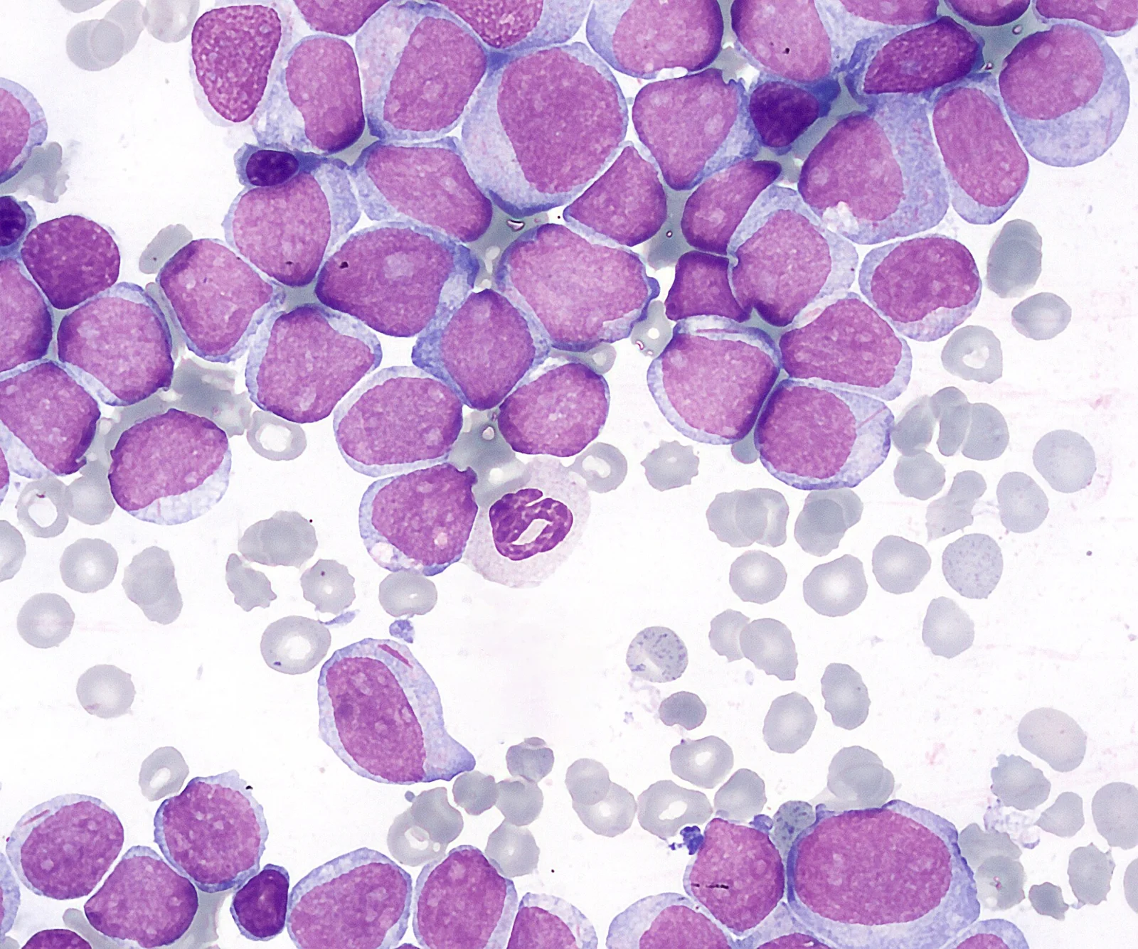
Long description
Bone marrow smear in acute myeloid leukaemia (M2 type): many large myeloblasts with prominent nucleoli and little normal marrow.

The bone marrow in acute leukaemia is hypercellular and packed with blasts. In ALL the marrow is replaced by lymphoblasts: small to medium cells with high nucleocytoplasmic ratio, fine chromatin and small nucleoli, staining for terminal deoxynucleotidyl transferase and for lineage markers (B-ALL: CD19, CD22, CD79a, often CD10; T-ALL: cytoplasmic CD3, CD7, with CD1a and dual CD4/CD8 in cortical types). [12] T-ALL and T-lymphoblastic lymphoma differ by extent: conventionally, bone marrow blasts below 25 percent alongside a mass lesion is called lymphoma, while marrow involvement of at least 25 percent is leukaemia. [12] Auer rods in the cytoplasm of blasts point to myeloid lineage and are a classic hook for AML in a bone marrow aspirate or peripheral blood film. [1]
Cytochemistry and immunophenotype fix the diagnosis: myeloperoxidase, Sudan black, CD13, CD33, CD117 and CD34 in AML (with monocytic markers CD14, CD64, CD36 in monocytic disease), and lymphoid markers in ALL. Molecular and cytogenetic studies then assign the genetic subtype. The presence of mixed-phenotype acute leukaemia (T/myeloid), which meets criteria for both lineages, reminds students that lineage assignment has edge cases. [12]
The history is the marrow-failure triad translated into the patient's words: fatigue and breathlessness (anaemia); fever, infections and failure to clear them (neutropenia); bruising, petechiae, epistaxis, bleeding gums and heavy periods (thrombocytopenia). In children, bone pain from marrow expansion and periosteal involvement is a classic ALL symptom and is often missed because it looks like growing pains or osteomyelitis. Fever and bone pain together should raise leukaemia early in the differential. Infection can be the first sign in a neutropenic patient whose white count is deceptively high because of circulating blasts; the neutrophil count, not the total white count, is what matters.
Examination should look at the whole patient. Pallor, petechiae, purpura. Hepatosplenomegaly and generalised lymphadenopathy are common in ALL, particularly T-ALL; gum hypertrophy suggests monocytic AML. A mediastinal mass in T-ALL can produce cough, dyspnoea, tracheal deviation, and superior vena cava syndrome. Chloroma (granulocytic sarcoma) is a solid infiltration of myeloblasts in soft tissue or bone, occasionally the presenting sign of AML. Central nervous system involvement presents with headache, cranial nerve palsies (especially the sixth and seventh), papilloedema and, in late disease, signs of raised intracranial pressure.
Hyperleucocytosis deserves its own mention because it is a laboratory value with clinical consequences. A white cell count above 100 x 10^9 per litre is commonly used to define it. At this level leukostasis can occur: sludging of poorly deformable blasts in small vessels, producing hypoxia, dyspnoea, visual and neurological symptoms, and sometimes pulmonary or cerebral haemorrhage. Leukostasis is most dangerous in AML, where the blasts are larger and less deformable than lymphoblasts; T-ALL with a very high count can also be symptomatic. [13] In one paediatric series, 44.4 percent of children with ALL had hyperleucocytosis above 100 x 10^9 per litre, and it was seen in more than half of children with T-ALL but not in any children with B-ALL in that small series. [13]
Extramedullary sanctuaries matter for relapse. The central nervous system and testes are sanctuary sites in ALL; central nervous system involvement at diagnosis is more common in B-ALL with high-count disease and in T-ALL than in standard-risk B-ALL. [10][12] T-ALL and T-lymphoblastic lymphoma in particular seed the central nervous system compared with B-cell disease. [12]
Leukaemia mimics several other diseases and, conversely, is mimicked by them. A table maps common presentations.
| Condition | What points to it | How it differs from acute leukaemia |
|---|---|---|
| Aplastic anaemia | Pancytopenia, empty marrow, no blasts | Hypocellular marrow without blast infiltration; reticulocytes low; no blasts on film |
| Myelodysplastic syndrome | Dysplasia, cytopenias, older adults | Hypercellular/dysplastic marrow, blast count below 20 percent, clonal changes such as monosomy 7 or complex karyotype |
| Transient myeloproliferative disorder / Down syndrome | Down syndrome, neonatal period | Polyclonal in neonates; regresses; not acquired mutations of adult leukaemia |
| Acute infection / sepsis | Fever, cytopenias, reactive marrow | Blasts absent; marrow shows reactive change, not a clone |
| Lymphoma with marrow involvement | Mass, staging marrow | Blasts not dominant; lineage markers of mature lymphoma |
| Immune thrombocytopenia / autoimmune haemolysis | Isolated cytopenia | Marrow normal or reactive, no blasts |
| Acute mononucleosis | Atypical lymphocytes, fever | Reactive atypical cells, Epstein-Barr virus serology positive |
| Hairy cell leukaemia / chronic lymphocytic leukaemia | Mature lymphocytes, older adults | Insidious; distinct immunophenotype |
| Myeloid sarcoma / chloroma | Soft-tissue mass | Myeloblasts in tissue; marrow may be spared early |
The most dangerous clinical pitfall is to call a drug-induced or infection-induced marrow suppression "aplastic" or to call a reactive leukocytosis "leukaemia". The marrow biopsy with blast percentage, immunophenotype and cytogenetics removes the ambiguity.
The pathway moves from screen to confirm to characterise, because the treatment that follows depends on characterisation rather than on the diagnosis alone.
First-line and screening tests:
- Full blood count with differential and reticulocyte count. Look at the film yourself. Blasts on the film are the clue; a low reticulocyte count with anaemia and pancytopenia points to marrow failure. Neutropenia with a total white count raised by blasts is common.
- Coagulation screen, especially in any patient with suspected APL, because disseminated intravascular coagulation can be florid before treatment starts.
- Lactate dehydrogenase, uric acid and electrolytes, because they set the tumour lysis baseline and risk.
- Chest radiograph or computed tomography for a mediastinal mass in T-ALL, and ultrasound if the neck, testes or abdomen are involved.
Confirmatory tests:
- Bone marrow aspiration and trephine biopsy, with cellularity, morphology, immunophenotype by flow cytometry, cytogenetics (karyotype and FISH for translocations such as t(9;22), t(15;17), t(8;21), inv(16), KMT2A) and next-generation sequencing for mutations (NPM1, FLT3, IDH1, IDH2, TP53, CEBPA, and the myelodysplasia-related genes). [2]
- Lumbar puncture only when central nervous system involvement is suspected, or as prophylaxis later in treatment; in ALL it is deferred if the platelet count is very low and may be performed after induction counts recover.
- Testicular examination and, in adults, ultrasound of the testes to exclude sanctuary-site disease.
The single most important rule for a student is that immunophenotype and genetics, not blast morphology, classify the disease and set treatment. Blasts in ALL and AML can look identical on a Romanowsky stain.
What each result means:
- In ALL, flow cytometry distinguishes B from T lineage and the subtypes; in B-ALL the common subclones (pro-B, pre-B, mature) are less important than genetics. [9]
- In AML, the immunophenotype separates myeloid from monocytic; cytochemistry confirms myeloperoxidase positivity.
- Cytogenetics put the patient into an ELN or NCCN risk group. [2]
- Next-generation sequencing not only refines risk but increasingly directs therapy (FLT3, IDH, TP53, KMT2A, NPM1).
A marrow with at least 20 percent blasts defines an acute leukaemia in most classifications. [1][12] With certain defining abnormalities, AML with 10 to 19 percent blasts is treated as AML (MDS/AML) under the International Consensus Classification. [1] Mixed-phenotype acute leukaemia is diagnosed when immunophenotype meets criteria for more than one lineage.
Beyond the diagnosis, staging in acute leukaemia is genetic. The European LeukemiaNet 2022 criteria are the standard risk framework for adults, with NCCN providing a parallel cytogenetic-molecular classification. [2] By gene mutations, APL and core binding factor AML are favourable; FLT3-ITD moves intermediate; NPM1 with adverse lesions is adverse; and TP53-mutated AML is very adverse. [2] A flowchart for suspected acute leukaemia sits below.
Suspected acute leukaemia: from cytopenia to treatment plan
- Cytopenia or blasts on filmanaemia, neutropenia and/or thrombocytopenia; infection or bleeding; bone pain
- Blood film and historyexclude sepsis, drug exposure and inherited marrow failure; look for blasts, Auer rods, film morphology
- Question: What do marrow and flow cytometry show?
Lymphoid blasts
- Acute lymphoblastic leukaemiaB-cell vs T-cell by immunophenotype; cytogenetics and molecular tests (including BCR::ABL1, KMT2A, hypodiploidy)
- Check sanctuary sitesCNS (lumbar puncture) and testes imaging; Ph testing before any tyrosine kinase inhibitor use
Myeloid blasts
- Acute myeloid leukaemiamyeloblasts or monoblasts; look for Auer rods; coagulation screen in every case
- Question: Is t(15;17) / PML::RARα present?
Yes
- Acute promyelocytic leukaemiatreat as APL emergency: ATRA plus arsenic, with coagulopathy management
No
- Genotype the leukaemiaNPM1, FLT3, IDH1/2, TP53, CEBPA, RUNX1::RUNX1T1, CBFB::MYH11, KMT2A, and ELN risk assignment
- Assign risk and fitnessEuropean LeukemiaNet or NCCN risk group + patient fitness; plan induction, consolidation and post-remission therapy
Management is built on three questions: is the patient fit for intensive chemotherapy, what is the genetic risk, and is there a targeted target? In practice all three are decided in parallel once the marrow result returns.
Induction aims for complete remission with multi-agent chemotherapy (corticosteroids, vincristine, an anthracycline, asparaginase, plus intrathecal prophylaxis). For adolescents and young adults (15 to 39 years), pediatric-inspired, asparaginase-based regimens improve 5-year overall survival (relative risk 1.40 compared with adult regimens) and are now preferred. [11] Early incorporation of blinatumomab in Ph-negative B-ALL, including MRD-negative disease, increases 3-year overall survival (from 68 to 85 percent in the E1910 trial data). [9]
Ph-positive ALL is treated with a tyrosine kinase inhibitor (imatinib historically; dasatinib or ponatinib now), combined with chemotherapy or with blinatumomab; chemotherapy-free ponatinib plus blinatumomab regimens produce complete molecular response above 85 percent with estimated 3-year event-free and overall survival of about 77 and 91 percent. [9] IKZF1-plus (IKZF1 deletion plus CDKN2A/B or PAX5 deletions) identifies Ph-positive patients with higher relapse risk on dasatinib-based regimens, but this adverse effect is partly offset by ponatinib plus blinatumomab. [9]
Ph-like ALL is targeted when ABL-class fusions are present (tyrosine kinase inhibitors), while the CRLF2/JAK pathway disease is still being studied. [9] KMT2A-rearranged ALL has a high risk of central nervous system and extramedullary relapse, benefits from allogeneic stem cell transplant in first remission, and is a target for menin inhibitors; revumenib is approved for relapsed or refractory KMT2A-rearranged leukaemia aged 1 year and older. [2] Blinatumomab and inotuzumab ozogamicin (an anti-CD22 drug conjugate) can bridge to transplant in relapsed T-cell or B-cell disease; CD19-directed CAR-T is standard salvage for relapsed B-ALL in children and young adults. [9]
Post-remission, most patients receive 2 to 3 years of maintenance therapy (mercaptopurine, methotrexate). Allogeneic stem cell transplant in first remission is now reserved for defined high-risk subtypes (TP53-mutated, KMT2A-rearranged, Ph-like, or persistent measurable residual disease) or Ph-positive disease that does not achieve deep remission on a tyrosine kinase inhibitor. [9][11] In adolescents and young adults with Ph-negative ALL, transplant did not improve overall survival compared with pediatric regimens in available series, so transplant-free protocols are preferred for most. [11]
Acute promyelocytic leukaemia must be treated as an emergency distinct from other AML. Induction is all-trans retinoic acid (ATRA) 45 mg per square metre daily in two divided doses plus arsenic trioxide 0.15 mg per kilogram intravenously daily. [2] ATRA plus arsenic trioxide gives 10-year overall survival above 90 percent versus 75 percent for the older AIDA (ATRA plus idarubicin) regimen and reduced induction mortality to under 5 percent. [2] In high-risk APL (white blood cells above 10 x 10^9 per litre, or rising above this during induction), gemtuzumab ozogamicin 6 to 9 mg per square metre is added; arsenic is capped at 15 mg per day because higher doses can cause renal failure. [2] Consolidation is 9 months of ATRA (given for 2 weeks every month) plus arsenic (daily for 5 days, weekly for 4 weeks, every other month), totalling 110 doses including induction, with gemtuzumab used for persistent PML::RARα measurable residual disease. [2] Supportive cautions: granulocyte colony-stimulating factor should never be given in APL because it can worsen it; differentiation syndrome is prevented with dexamethasone 10 to 20 mg daily for 1 to 2 weeks during induction; ATRA headaches are managed with acetazolamide 125 to 250 mg two to four times daily; and ATRAs fluid retention should be distinguished from differentiation syndrome by stopping treatment and diuresing aggressively. [2] ATRA-ATO improves remission, disease-free, event-free and overall survival compared with ATRA plus chemotherapy (meta-analysis of recent trials: complete remission relative risk 1.04, event-free survival relative risk 1.25, overall survival relative risk 1.07), with QTc prolongation as the characteristic excess toxicity of arsenic (relative risk 3.79). [8] Disseminated intravascular coagulation in APL can be dominantly thrombotic, and ATRA may worsen that setting; idarubicin or gemtuzumab is preferred there. [2] [1][3][7][13] [6]
Other AML is treated by risk and fitness.
Fit patients receive intensive 7 plus 3 induction (daunorubicin 60 mg per square metre, idarubicin 12 mg per square metre, or mitoxantrone 12 mg per square metre intravenously days 1 to 3 plus cytarabine 100 to 200 mg per square metre daily by continuous infusion days 1 to 7) with options such as FLAG-IDA plus venetoclax or CLIA plus venetoclax (CRc above 90 percent, MRD negativity 71 to 90 percent). [1][3] Genotype-specific points: [2]
- NPM1-mutated AML (favourable without FLT3-ITD) responds to high-dose cytarabine and 7 plus 3; consolidation is cytarabine 1.5 to 3 g per square metre for 3 to 4 cycles; no transplant in first remission unless measurable residual disease is positive. [3]
- FLT3-mutated: add midostaurin 50 mg twice daily days 8 to 21, or quizartinib 40 mg daily, to 7 plus 3 and continue through consolidation; allogeneic transplant in first remission is preferred; post-transplant FLT3 inhibitor maintenance is used in MRD-positive patients. [3] Midostaurin improved median overall survival from 35.6 to 74.7 months in the RATIFY trial. [3]
- Core binding factor AML: 7 plus 3 with gemtuzumab ozogamicin (3 mg per square metre days 1, 4 and 7, capped at 5 mg; one dose on day 1 is also widely used) plus 2 to 4 cycles of intermediate-dose cytarabine (1000 to 1500 mg per square metre, or 500 to 1000 if aged at least 60 years) every 12 hours days 1 to 3; transplant is not recommended in first remission; gemtuzumab is used, but watch veno-occlusive disease if transplant is planned within 3 months, and disease relapses in up to 35 percent. [1][3]
- KMT2A-rearranged AML: conventional chemotherapy plus menin inhibitors emerging; CNS prophylaxis with 2 to 4 intrathecal injections is recommended because of central nervous system and extramedullary disease. [2] [12]
- IDH1-mutated: ivosidenib 500 mg daily added to 7 plus 3 (phase 1: complete remission 70 percent, overall survival at 3 and 5 years 67 and 61 percent) or, for unfit patients, ivosidenib with azacitidine (AGILE: median overall survival 29.3 months versus 7.9 months, hazard ratio 0.42, transfusion independence 53.8 versus 17.1 percent), deep molecular clearance in 30 percent. [7][3] [4][6]
- IDH2-mutated: enasidenib 100 mg daily with chemotherapy or venetoclax-based combinations. [3]
- TP53-mutated: poor response to any therapy; use CPX-351 (liposomal daunorubicin and cytarabine, 100 units per square metre days 1, 3 and 5, with consolidation on days 1 and 3), or lower-intensity regimens; allogeneic transplant in first remission is common practice despite poor data. [1][2]
Fit patients with adverse or complex disease, secondary or therapy-related AML have also been treated with CPX-351, although CPX-351 label indications use older nomenclature. [1]
Unfit or older patients (generally 75 years and older, or younger with comorbidity) receive lower-intensity therapy. Azacitidine plus venetoclax is the standard: median overall survival 14.1 versus 9.6 months, complete remission 36.7 versus 17.9 percent, complete remission with incomplete count recovery 66.4 versus 28.3 percent in VIALE-A. [4][5] Venetoclax (a BCL-2 inhibitor) is ramped up with tumour lysis monitoring; doses follow local protocols. Alternatives include low-dose cytarabine plus venetoclax, glasdegib plus low-dose cytarabine, and for IDH1-mutated disease azacitidine plus ivosidenib (AGILE median survival 29.3 versus 7.9 months). [4][5] Therapy should continue at least six cycles, with response assessed after two cycles and dose modification for cytopenias. [4] Measurable residual disease is used to refine risk and decide transplant. [2][3] Best supportive care alone (plus or minus hydroxyurea for count control) is appropriate for pre-terminal or declining patients; in one review 36 percent of patients aged 70 to 79 and 59 percent aged 80 and older received only supportive care. [4] [6][7]
- Infection and sepsis during neutropenia, the leading cause of death in the first 60 days. Fungal infection (invasive aspergillosis, candidaemia) and neutropenic enterocolitis; viridans streptococci can break through in high-dose cytarabine patients. [3]
- Bleeding: thrombocytopenia below 10 x 10^9 per litre; petechiae, epistaxis, gastrointestinal and intracranial haemorrhage; in APL, a consumptive coagulopathy with fibrinogen fall demands immediate platelet and cryoprecipitate support and rapid start of ATRA before chemotherapy. [2][8]
- Tumour lysis syndrome (below).
- Leukostasis and symptomatic hyperleucocytosis.
- Chemotherapy toxicities: anthracycline cardiotoxicity, high-dose cytarabine cerebellar ataxia, veno-occlusive disease after transplant or gemtuzumab, and cytarabine syndrome (fever, rash, myalgia 6 to 12 hours after a dose, treated with corticosteroids). [3]
- Differentiation syndrome in APL (fever, dyspnoea, weight gain, effusions, renal impairment, occurring 2 to 21 days after ATRA or arsenic starts) treated with dexamethasone 10 mg twice daily and temporary ATRA pause if severe. [2]
- Venetoclax adverse effects in AML include febrile neutropenia (about 42 percent in VIALE-A versus 19 percent for azacitidine alone) and failure of low-grade nausea. [4]
- In APL, differentiation syndrome can be insidious fluid retention rather than classic cytokine storm; stopping drug and aggressive diuresis is needed. [2]
Tumour lysis syndrome is the metabolic emergency of rapid blast death: hyperkalaemia, hyperphosphataemia, secondary hypocalcaemia and hyperuricaemia, with uric acid and calcium phosphate crystallising in renal tubules and producing acute kidney injury. It occurs 3 to 30 percent in haematological malignancies, with clinical TLS in about 20 to 40 percent and TLS-related acute kidney injury in 5 to 40 percent of children, up to 75 percent in high tumour burden. [13] Spontaneous TLS is more common in children than adults (about 30 to 72 percent in some paediatric series) and reached 56 percent in one Egyptian cohort of high-risk disease. [13] Risk is highest with bulky, fast-growing, treatment-sensitive disease such as Burkitt lymphoma, high-count aggressive leukaemia and, at induction, any acute leukaemia with a high white count or large marrow burden. [13]
Prevention, the priority:
- Hydration is the single most important step; aim for high urine output before chemotherapy starts.
- Stratify TLS risk with Cairo-Bishop laboratory or Howard-Pui clinical criteria and local protocols. In the Elshafey cohort every patient in the non-spontaneous TLS group was high risk. [13]
- Hypouricemic agents: in low- or intermediate-risk disease the British Society for Haematology recommends xanthine oxidase inhibitors (allopurinol). In high-risk disease, rasburicase (recombinant urate oxidase) rapidly lowers uric acid to under 1 mg per decilitre within 4 hours but carries risk of haemolysis and methaemoglobinaemia, so glucose-6-phosphate dehydrogenase deficiency should be excluded before use in at-risk populations. [13]
- Where rasburicase is unavailable, xanthine oxidase inhibitors are a pragmatic fallback (febuxostat in the Egyptian cohort), with the acknowledged risks of xanthine accumulation and xanthine nephropathy, and drug safety and efficacy data in children are limited. [13]
- Avoid acidification or over-alkalinisation; modern practice rarely uses alkalinisation because it favours calcium phosphate deposition.
Management of established tumour lysis: stop the causative drug if practical, aggressive hydration, electrolyte management, phosphate binders such as sevelamer, calcium for symptomatic hypocalcaemia, anti-hyperkalaemic measures, diuretics, and kidney replacement therapy when indicated. Rasburicase if not already given. Monitoring should track uric acid, phosphate, potassium, calcium and creatinine serially; in the cohort above, a daily serum phosphate rise above 2.6 mg per decilitre was the strongest early predictor of severe acute kidney injury (AUC 0.839), outperforming uric acid change. [13] Hyperphosphataemia was the most common laboratory criterion (94 percent) and hyperkalaemia the least common (14 percent); clinical TLS (most patients) carried a 60 percent incidence of severe kidney failure requiring dialysis in 62 percent of children. [13] Outcomes among survivors were favourable: all surviving children had complete renal recovery by 3 months. [13]
Hyperleucocytosis is defined as a white cell count above 100 x 10^9 per litre. [13] Leukostasis is the end result: blasts are large, rigid and poorly deformable, so they plug pulmonary and cerebral capillaries and produce hypoxia, respiratory distress, visual disturbance and neurological symptoms. It is more dangerous in AML than ALL. [13] Management is not by chemotherapy alone while the count is critical: slow single-agent cytarabine or hydroxyurea for controlled cytoreduction, careful oxygenation and fluid balance, avoid diuretics that haemoconcentrate, and therapeutic leukapheresis or exchange transfusion where available. [2] Definitive chemotherapy starts once cytoreduction has begun. Prophylaxis of tumour lysis is required throughout.
A single oral temperature of at least 38.3 degrees Celsius (101 degrees Fahrenheit) or sustained 38 degrees for an hour with an absolute neutrophil count below 0.5 x 10^9 per litre (or expected to fall below that). Risk stratification (low-risk versus high-risk) determines whether outpatient oral antibiotics or inpatient intravenous broad-spectrum therapy is appropriate, using local febrile-neutropenia protocols. In ALL induction the risk is high and admission is usual; empiric antipseudomonal beta-lactam therapy (cefepime, piperacillin-tazobactam or a carbapenem per local protocol) should be started immediately on triage and at least two sets of blood cultures taken. [4] Consider antifungal coverage for persistent fever on antibiotics. Granulocyte colony-stimulating factor is used selectively to shorten neutropenia; evidence shows it reduces infection and hospitalisation without improving survival or marrow response in aplastic anaemia, and in APL it can worsen disease. [2]
Prognosis is set by genetics and by measurable residual disease more than by any presenting feature.
- Childhood ALL: cure above 80 to 90 percent. [11]
- AYA ALL improves to approach childhood rates with pediatric regimens. [11]
- Adult ALL: 5-year event-free survival 40 to 70 percent, with Ph-positive patients now approaching 80 to 90 percent overall survival on ponatinib plus blinatumomab in recent series. [9][12]
- AML overall survival is about 30 percent at 5 years; fit younger patients on intensive therapy do better and older patients fare worse (about 10 percent 5-year survival if older than 60 years receiving intensive therapy). [2]
- APL: above 90 percent 10-year overall survival on ATRA plus arsenic; induction mortality below 5 percent. [2]
- AML risk by genotype: favourable (CBF, NPM1 without FLT3, bZIP CEBPA), intermediate (FLT3-ITD, t(9;11)), adverse (TP53, complex karyotype, MECOM, defined mutations). [1][2] But ELN risk groups were derived from intensively treated younger patients and do not predict outcomes well in patients treated with venetoclax and azacitidine, for which a four-gene (TP53, FLT3-ITD, NRAS, KRAS) model gives median overall survival of 5.5, 12.1 and 26.5 months for lower-, intermediate- and higher-benefit groups. [5][6]
- In IDH1-mutated AML, ivosidenib with azacitidine gives median overall survival 29.3 versus 7.9 months in unfit patients. [7]
- Venetoclax with azacitidine, a standard for unfit AML, gives median survival 14.7 versus 9.6 months and improves complete remission to 66.4 versus 28.3 percent. [5]
Measurable residual disease is the best predictor of relapse, independent of baseline genetics, and can reclassify patients during treatment. [9] Because central nervous system and testicular disease can cause late relapse, prophylactic intrathecal therapy is standard in ALL, and testicular imaging is part of long-term follow-up.
There is no population screening. What can be done is early recognition of the genetic syndromes with predisposition (Down syndrome, inherited bone marrow failure, Li-Fraumeni) and early testing for fusion transcripts at presentation, since targeted therapy starts at induction and delays waste time. Prevention also means avoiding unnecessary exposure to alkylating or topoisomerase chemotherapy, and not assuming that an isolated high white count in a generally well child excludes leukaemia.
Children: ALL is largely curable; the ETV6::RUNX1 and hyperdiploid subtypes are favourable, hypodiploid TP53-mutated disease is adverse. T-ALL affects children commonly, with mediastinal mass in most cases and more central nervous system involvement than B-ALL. [12]
Adolescents and young adults: use pediatric-inspired asparaginase-based regimens; 5-year survival improves from 30 to 45 percent to around 60 percent. [11]
Pregnancy: leukaemia in pregnancy requires urgent haematology, obstetric and neonatal input; treatment is given in the second and third trimesters, with asparaginase and anthracyclines having relative safety data; ATRA in APL carries teratogenicity concerns requiring individualised decisions. [2]
Older adults: AML dominates; use fitness assessment (not age alone) and lower-intensity combined therapy with venetoclax; transplant when fit and in remission. [4]
Inherited syndromes: germline TP53 and DDX41 change donor selection and tolerance; Li-Fraumeni enriches therapy-related AML. [2]
The reality in many low- and middle-income regions is that the gap is not biology but diagnosis, drug access and follow-up. [14] Practical priorities:
- Diagnose first with whatever is available. A full blood count and blood film diagnose most ALL and AML; a single bone marrow aspirate establishes the blast percentage and distinguishes leukaemia from aplastic anaemia and myelodysplasia. [1][14]
- Immunophenotyping by flow cytometry and cytogenetics may be unavailable outside teaching hospitals. Where they are missing, clinical-rather-than-molecular risk stratification is unavoidable, and the choice is between pragmatic ALL regimens and older AML regimens.
- Medicines: imatinib, all-trans retinoic acid and arsenic trioxide are relatively available; venetoclax, ivosidenib, blinatumomab, CAR-T and transplant are often not. Regimens adapted for low-resource settings exist, such as the Hunger protocol for ALL (vincristine, prednisone, cyclophosphamide, intrathecal methotrexate, mercaptopurine, dexamethasone and asparaginase) with cranial prophylaxis and maintenance. [14] Older protocols with vincristine, prednisone, asparaginase and methotrexate remain the backbone in many African centres. [14]
- Tumour lysis: rasburicase is often unavailable and prohibitively expensive; xanthine oxidase inhibitors are used, with careful hydration and monitoring, recognising xanthine nephropathy risk. In one resource-limited paediatric cohort using universal xanthine oxidase inhibitors, 60 percent reached severe (failure-stage) acute kidney injury and 62 percent needed renal replacement therapy, versus 32 and 29 percent where rasburicase was used in half of patients. [13] Early serial phosphate monitoring (a daily rise above 2.6 mg per decilitre predicts severe kidney injury) is cheap and practical. [13]
- Access gaps apply to immunotherapy: in Morocco, blinatumomab is limited by cost, regulatory delays and reimbursement, so many patients receive conventional chemotherapy despite trial evidence. Strategies such as shortened, vial-sharing or outpatient regimens have been proposed to improve feasibility in low- and middle-income countries. [10]
- Nutritional status and infection control (including tuberculosis and HIV co-infection in some regions) strongly influence both chemotherapy tolerance and leukaemia outcome. [14]
- Loss to follow-up is a major cause of apparent mortality; tracking systems and community health workers improve retention. [14]
- Acute leukaemia is a clone of blasts causing marrow failure and, without treatment, death within weeks; ALL is lymphoid, AML is myeloid, and both are classified by immunophenotype and genetics rather than morphology.
- ALL is 15 percent of childhood ALL in the T-cell form and the majority B-cell; cure in childhood ALL is above 80 to 90 percent.
- AML has a median age around 68 years; 5-year overall survival is around 30 percent, lower still in the over-60s (about 10 percent), and favourable subtypes (CBF, NPM1 without FLT3, bZIP CEBPA) do far better. [2]
- APL (t(15;17)) is 5 to 8 percent of AML but has the best prognosis: above 90 percent 10-year survival on ATRA plus arsenic trioxide; it is a coagulation emergency first.
- Marrow blasts of at least 20 percent usually define acute leukaemia, but genetically defined AML may be treated at 10 to 19 percent. Flow cytometry and genetics, not the film alone, settle diagnosis and risk. [1]
- Treatment is risk- and fitness-based: ALL uses multi-agent induction with paediatric-style regimens in younger patients and targeted therapy (imatinib or ponatinib plus blinatumomab); AML uses 7 plus 3 in fit patients, CPX-351 in some secondary or therapy-related cases, lower-intensity venetoclax-based therapy in unfit patients, and targeted agents such as midostaurin, quizartinib, ivosidenib, enasidenib, revumenib and venetoclax according to mutation.
- Prognosis in AML is set by European LeukemiaNet 2022 genetics in intensively treated patients and by a four-gene (TP53, FLT3-ITD, NRAS, KRAS) model in patients treated with venetoclax and azacitidine, ranging from 5.5 (low benefit) to 26.5 (higher benefit) months median survival.
- Measurable residual disease is the best predictor of relapse; central nervous system and testes are sanctuary sites in ALL, so prophylactic intrathecal therapy and, in adults, sanctuary-site surveillance are standard.
- Emergencies dominate early death: tumour lysis syndrome (prevent by hydration and hypouricemic therapy, monitor phosphate daily), leukostasis above 100 x 10^9 per litre, neutropenic fever needing immediate antibiotics, and the APL coagulopathy.
- In low-resource settings, a film, marrow aspirate, basic FBC and pragmatic regimens with ATRA, imatinib, xanthine oxidase inhibitors and energetic follow-up preserve most of the benefit that biology allows.
Answer each one in your head first, then open the answer.
A 4-year-old child has 3 weeks of bone pain, pallor and bruising. Blood count shows pancytopenia and 6 percent blast-equivalent cells on the film. Which test confirms the diagnosis and what must it not miss?
Show answerHide answer
Bone marrow aspiration and trephine biopsy with immunophenotyping by flow cytometry, cytogenetics and molecular testing. Morphology alone cannot separate ALL from AML; the film may look identical. The biopsy must also rule out myelodysplasia, inherited marrow failure and infection. A hypercellular marrow packed with blasts supports leukaemia.
A 62-year-old woman has AML with NPM1 mutation (without FLT3-ITD) and is fit. Using current European LeukemiaNet guidance, what is the expected risk group, consolidation choice and transplant plan?
Show answerHide answer
Favourable risk. Induction with 7 plus 3 (daunorubicin or idarubicin plus cytarabine) with gemtuzumab ozogamicin; consolidation with 3 to 4 cycles of high-dose cytarabine (1.5 to 3 g per square metre). Allogeneic transplant is not recommended in first remission; reserve it for relapse or persistent measurable residual disease. If measurable residual disease is positive after consolidation, reconsider transplant.
A 58-year-old man with APL (PML::RARα positive) presents with a platelet count of 12 x 10^9 per litre and prolonged prothrombin time. Before any chemotherapy, what must you start immediately and why?
Show answerHide answer
Start all-trans retinoic acid immediately (45 mg per square metre daily in two doses) plus arsenic trioxide 0.15 mg per kilogram daily: this chemotherapy-free regimen gives 10-year survival above 90 percent and reduces induction mortality below 5 percent. Do not wait for confirmatory genotyping or for a class switch; in APL ATRA comes first and chemotherapy comes later. Treat the consumptive coagulopathy with platelets, cryoprecipitate or fibrinogen and fresh frozen plasma as clinically needed. Avoid granulocyte colony-stimulating factor, which worsens APL, and beware dominant thrombotic disseminated intravascular coagulation, where idarubicin or gemtuzumab ozogamicin is preferred.
An older unfit patient with AML has mutated TP53. What trial evidence guides the regimen and what survival should you quote?
Show answerHide answer
Azacitidine plus venetoclax gives a higher complete remission rate in TP53-mutated AML (55.3 versus 0 percent in trial data) than azacitidine alone, but median overall survival is similar (5.2 versus 4.9 months) and such patients fall in the lowest-benefit group of the four-gene model (5.5 months). A reasonable approach is azacitidine plus venetoclax for induction to optimise remission, then azacitidine monotherapy for maintenance to reduce cytopenia-related toxicity, or supportive care with hydroxyurea. Enrollment in trials of novel combinations is encouraged, because no current strategy has clearly improved survival in this group. [5]
A child with Burkitt lymphoma starts induction. Baseline uric acid is mildly high, creatinine normal. How do you prevent and what monitoring predicts severe kidney injury?
Show answerHide answer
Begin generous hydration before chemotherapy and risk-stratify tumour lysis with Cairo-Bishop or Howard-Pui criteria. For low or intermediate risk use a xanthine oxidase inhibitor such as allopurinol; for high risk use rasburicase if available and glucose-6-phosphate dehydrogenase status permits. Keep urine alkaline neutral; avoid over-alkalinisation that precipitates calcium phosphate. Monitor daily: uric acid, phosphate, potassium, calcium and creatinine. A daily rise in serum phosphate above 2.6 mg per decilitre is the strongest early predictor of severe acute kidney injury (area under the curve 0.839). Be aware that 5 to 40 percent of children develop tumour lysis-related kidney injury, up to 75 percent with a large tumour burden, and that hypouricemic therapy is not universally accepted as kidney-protective.
A 28-year-old has T-cell ALL with white cells 180 x 10^9 per litre and cough. What emergencies do you anticipate and how does T-cell disease differ from B-cell disease?
Show answerHide answer
Anticipate mediastinal mass with superior vena cava syndrome or tracheal compression, leukostasis from the high count, tumour lysis syndrome and central nervous system and testicular sanctuary relapse. T-ALL is about 20 percent of adult ALL, has a male predominance of roughly 2 to 1, a mediastinal mass in 70 to 90 percent of cases, and more central nervous system involvement than B-ALL; white count often exceeds 50 x 10^9 per litre. It is treated with intensive multi-agent protocols (often augmented Berlin-Frankfurt-Münster or Children's Oncology Group schedules with nelarabine and pegylated asparaginase) plus intrathecal prophylaxis. Allogeneic transplant is an important curative arm for adults with high-risk disease. [12]
Related MedGrasp topics are named here in plain text because their pages are written separately: anaemia (approach and iron deficiency), aplastic anaemia (bone marrow failure, a key differential of pancytopenia), anticoagulant and haemostatic drugs, disseminated intravascular coagulation, hypercoagulable states, myelodysplastic syndromes, normal haemostasis and thrombosis, polycythaemia (another myeloproliferative disease), transfusion reactions (blood product support during marrow failure), and tumour lysis syndrome would sit naturally as a linked topic once written. In the meantime this page stands alone.
- Platzbecker U, Larson RA, Gurbuxani S. Diagnosis and treatment of AML in the context of WHO and ICC 2022 classifications: Divergent nomenclature converges on common therapies. HemaSphere 2025;9(2):e70083. doi:10.1002/hem3.70083. Accessed .
- Kantarjian HM, DiNardo CD, Kadia TM, Daver NG, Altman JK, Stein EM, Jabbour E, Schiffer CA, Lang A, Ravandi F. Acute myeloid leukemia management and research in 2025. CA Cancer J Clin 2025;75(1):46-67. doi:10.3322/caac.21873. Accessed .
- Gangat N, Ravandi F. Newly diagnosed acute myeloid leukemia in Fit patients: 2026 treatment algorithms. Blood Cancer J 2026;16:183. doi:10.1038/s41408-026-01578-9. Accessed .
- Schuh AC, Chornenki N, Elemary MA, Elsawy M, Houston BL, Leber B, Mozessohn L, Sabloff M, Salter B, Sanford D, Brandwein J. Management of Acute Myeloid Leukemia in Older Patients: An Updated Canadian Consensus. Curr Oncol 2026;33(7):429. doi:10.3390/curroncol33070429. Accessed .
- Pelosi E, Castelli G, Testa U. The Spectrum of Venetoclax-Based Treatments in Acute Myeloid Leukemia. Cancers (Basel) 2026;18(8):1201. doi:10.3390/cancers18081201. Accessed .
- Döhner H, Pratz KW, DiNardo CD, Wei AH, Jonas BA, Pullarkat VA, Thirman MJ, Récher C, Schuh AC, Babu S, Li X, Ku G, Liu Z, Sun Y, Potluri J, Dail M, Chyla B, Pollyea DA. Genetic risk stratification and outcomes among treatment-naive patients with AML treated with venetoclax and azacitidine. Blood 2024;144(21):2211-2222. doi:10.1182/blood.2024024944. Accessed .
- Montesinos P, Marchione DM, Recher C, Heuser M, Vives S, Zarzycka E, Wang J, Riva M, Calado RT, Schuh AC, Yeh SP, Tron AE, Hui J, Gianolio DA, Choe S, Patel P, De Botton S, DiNardo CD, Döhner H. Long-term results from the AGILE study of azacitidine plus ivosidenib vs placebo in newly diagnosed IDH1-mutated AML. Blood Adv 2025;9(20):5177-5189. doi:10.1182/bloodadvances.2025016399. Accessed .
- Ahmad H, Henna F, Khurram S, Khan A, Ahmed A, Ali MH, Hassan MA, Abbas M, Farooq D, Muhammad S, Basit AA, Ahmad A. Efficacy and safety of ATRA plus arsenic trioxide versus ATRA plus chemotherapy in newly diagnosed acute promyelocytic leukemia: a grade-assessed systematic review and meta-analysis. BMC Cancer 2026;26:597. doi:10.1186/s12885-026-15932-4. Accessed .
- Baek DW. High-risk features in adult patients with B-cell acute lymphoblastic leukemia: redefining risk in the era of targeted immunotherapy. Blood Res 2026;61(1):55. doi:10.1007/s44313-026-00175-w. Accessed .
- Eibschutz LS, Crow S, Tatum C, Reed D, Keng M. Breaching the Blood-Brain Barrier: Evolving Strategies for Central Nervous System Disease in Adult Acute Lymphoblastic Leukemia. Curr Hematol Malig Rep 2026;21:14. doi:10.1007/s11899-026-00782-5. Accessed .
- Sereda Y, Mai HJ, Kanaan G, Melson JW, Terasawa T, Cheung MC, Stock W, Wolfson JA, Saldanha IJ, Balk EM. Pediatric-inspired regimens and HSCT for adolescents and young adults with acute lymphoblastic leukemia. Blood Adv 2026;10(10):3664-3675. doi:10.1182/bloodadvances.2025018736. Accessed .
- Danielson D, Lagerstrom IT, Rogers M, Simon K, Aguilera NS, Auerbach A. T-lymphoblastic leukemia/lymphoma: a comprehensive review of pathology, molecular features, and differential diagnosis. J Pathol Transl Med 2026;60(5):502-515. doi:10.4132/jptm.2026.08.01. Accessed .
- Elshafey SA, Essa L, Zeid MY, Wali Y, El Chazli Y. Tumor lysis syndrome in children with hematological malignancies: a nephrology perspective in resource-limited settings. Front Oncol 2026;16:1778776. doi:10.3389/fonc.2026.1778776. Accessed .
- Ayirebi AA, Twumasi S, Olaga LK, Sackey B, Antonio DNM, Ansah RO, Ansah EA, Sah JA, Anto EO, Anang-Quartey Y, Koomson A, Opoku A, Donkor GA, Duah P, Boadu AG, Essien-Baidoo S, Boadu WIO, Boateng LA. Acute and Chronic Leukemia in Sub-Saharan Africa: A Systematic Review. Cancer Rep (Hoboken) 2026;9(8):e70635. doi:10.1002/cnr2.70635. Accessed .
Figure credits
- Figure 1: Makysm, via Wikimedia Commons, CC0, CC0. resized.
Educational content for medical students and doctors. It is not a substitute for clinical judgement or local guidelines.