Chronic lymphocytic leukaemia
CLL: epidemiology, diagnosis, staging, prognosis and modern treatment from watchful waiting to targeted therapy.
- Chronic lymphocytic leukaemia (CLL) is a chronic, usually slow-growing cancer of mature B lymphocytes that accumulate in the blood, marrow and lymphoid tissue.
- It is a disease of older adults, typically diagnosed around 70 years, and about twice as common in men as in women.
- Most people have no symptoms at diagnosis: the condition is often found incidentally on a routine blood count showing lymphocytosis.
- It is a single disease with small lymphocytic lymphoma (SLL): the name depends on whether the abnormal cells predominate in the blood or in the tissues.
- There are now excellent biological markers (IGHV mutation status, TP53 changes) and usually effective, well-tolerated therapy when treatment becomes necessary, but many patients remain well for years on no treatment at all.
- Treatment is started not by the number on the blood count alone, but only when international criteria for active disease are met.
By the end of this page you should be able to:
- Recognise CLL clinically and on a blood count, blood film and immunophenotyping.
- Distinguish CLL from SLL and from monoclonal B-cell lymphocytosis.
- Apply the Rai and Binet staging systems.
- Explain how IGHV status, TP53 changes, del(17p), del(11q) and complex karyotype alter prognosis and treatment choice.
- Apply the International Workshop on CLL (iwCLL 2018) criteria for when to treat.
- Outline modern first-line treatment by biological profile and compare time-limited and continuous therapy.
- Recognise complications, including Richter transformation, hypogammaglobulinaemia, autoimmune haemolysis and immune thrombocytopenia.
- Plan assessment of relapsed or refractory disease and know where NCCN and EHA guidance converge and differ.
- Outline the management of CLL where specialist drugs are unavailable.
DefinitionChronic lymphocytic leukaemia
A clonal neoplasm of small, mature B lymphocytes that accumulate progressively in the peripheral blood, bone marrow and lymphoid tissues. In practice the diagnosis requires at least 5 x 10^9/L clonal B lymphocytes with a characteristic immunophenotype. Together with small lymphocytic lymphoma, it forms one entity in the WHO 2022 classification.
At its core, CLL is a slow failure of apoptosis rather than rapid proliferation: the cells survive too long. That explains why the count can stay high for years without producing acute symptoms, and why, unlike acute leukaemia, the abnormal lymphocytes look mature on the blood film.
The disease spans a spectrum. At one end is monoclonal B-cell lymphocytosis (MBL), in which a small clone of CLL-phenotype lymphocytes circulates at a concentration too low for the disease to be called CLL. At the other is transformation to an aggressive lymphoma (Richter transformation), discussed below. Knowing where a patient sits on this spectrum is more useful than any single label.
CLL is the commonest chronic leukaemia in adults in European populations. Reported incidence in Europe is 4.7 cases per 100,000 per year.[1] The median age at diagnosis is 70 years, and men are affected nearly twice as often as women.[1] It is rare under 40 years of age. Population figures depend on how many asymptomatic cases are detected, so the apparent incidence is rising in countries where routine blood counts are common.
Mortality from CLL has fallen steadily in Western countries, from about 2.0 deaths per 100,000 to about 1.0 per 100,000 in the 2018-2022 period.[1] This improvement predates most modern targeted therapy and reflects both earlier detection and better supportive care.
Geography and ethnicity matter. CLL is less common in people of Asian and African ancestry and in the Middle East than in white populations of European descent. In parts of East Africa and southern Asia overrepresentation of older men in hospital series may reflect referral rather than true incidence; truly incidence data are sparse. In one recent Tanzanian report, outcomes were worse where access to targeted therapy and supportive care were limited, an observation consistent with what is known in the literature.[1]
No modifiable risk factor is convincingly established. The aetiology is multifactorial:
- Age is the dominant risk factor, with incidence rising steeply with each decade past 50.
- Sex: men carry a consistent excess, and this holds across populations.
- Family history. CLL shows one of the strongest familial tendencies of the leukaemias. First-degree relatives have an estimated two- to eight-fold increased risk compared with the general population, and relatives with other B-cell lymphoid malignancies may share predisposition. (This general statement is widely quoted; the precise figure should be read from the original familial aggregation study you rely on.)
- Genetics. Large genome-wide association studies have identified many low-risk susceptibility loci, but no single inherited gene accounts for most cases. Rarely, families carry high-penetrance mutations, most notably in TP53-associated syndromes.
- Environment. Pesticides and other occupational exposures are recurrently suggested, but no causal agent is confirmed. Infection and smoking are not established causes.
Unlike chronic myeloid leukaemia, there is no single acquired molecular event that defines the disease; initiation is polygenic and appears to follow acquisition of one of several epigenetic or genetic landscapes in long-lived B cells.
CLL cells are antigen-experienced, mature CD5-positive B cells. They usually carry mutated or, in a substantial minority, unmutated immunoglobulin heavy-chain variable (IGHV) genes, a distinction that is both a biological marker and a clinical predictor. They are not lazy: even a slowly progressive leukaemia may proliferate briskly in the bone marrow and, crucially, in the lymph node proliferation centres, where cells undergo somatic evolution. That node-based proliferation explains why the blood count can suddenly rise after years of stability.
CLL cells depend on signals from their microenvironment: contact with stromal cells, T-helper cells, nurse-like cells and cytokines (such as BAFF, APRIL and interleukin-6) that activate survival pathways including NF-kappaB and BCL-2. Overexpression of BCL-2 is a hallmark; venetoclax, a BCL-2 inhibitor, works by stripping that survival signal. Defective apoptosis is the central mechanism.
Tumour cells progressively infiltrate marrow, spleen and nodes. The marrow eventually fails to make sufficient red cells, neutrophils and platelets. Autoimmune cytopenias may arise, in which the immune system damages red cells or platelets independent of infiltration. Many patients develop hypogammaglobulinaemia, which explains the recurrent infections characteristic of later disease.
Each clinical finding maps to a mechanism:
- lymphocytosis reflects accumulation of long-lived cells in blood
- lymphadenopathy reflects nodal infiltration, often in neck, axilla and groin
- splenomegaly reflects splenic infiltration with or without hypersplenism and cytopenias
- anaemia may reflect marrow infiltration, hypersplenism, bleeding, or (in a minority) immune haemolysis
- thrombocytopenia may reflect marrow infiltration or immune destruction
- infections reflect hypogammaglobulinaemia, defective cellular immunity, and the immunosuppression of treatment
- systemic symptoms reflect cytokine release and tumour burden
- Richter transformation reflects acquisition of further genetic lesions, particularly involving TP53 and MYC
CLL is usually diagnosed by the blood rather than biopsy. The marrow is hypercellular and infiltrated by small, round B lymphocytes that may form aggregates or interstitial patterns. Lymph nodes are effaced by sheets of small, uniform lymphocytes with condensed chromatin and scant cytoplasm, with paler proliferation centres.
Figure 1Chronic Lymphocytic Leukaemia (lymphocytosis and smear cells)
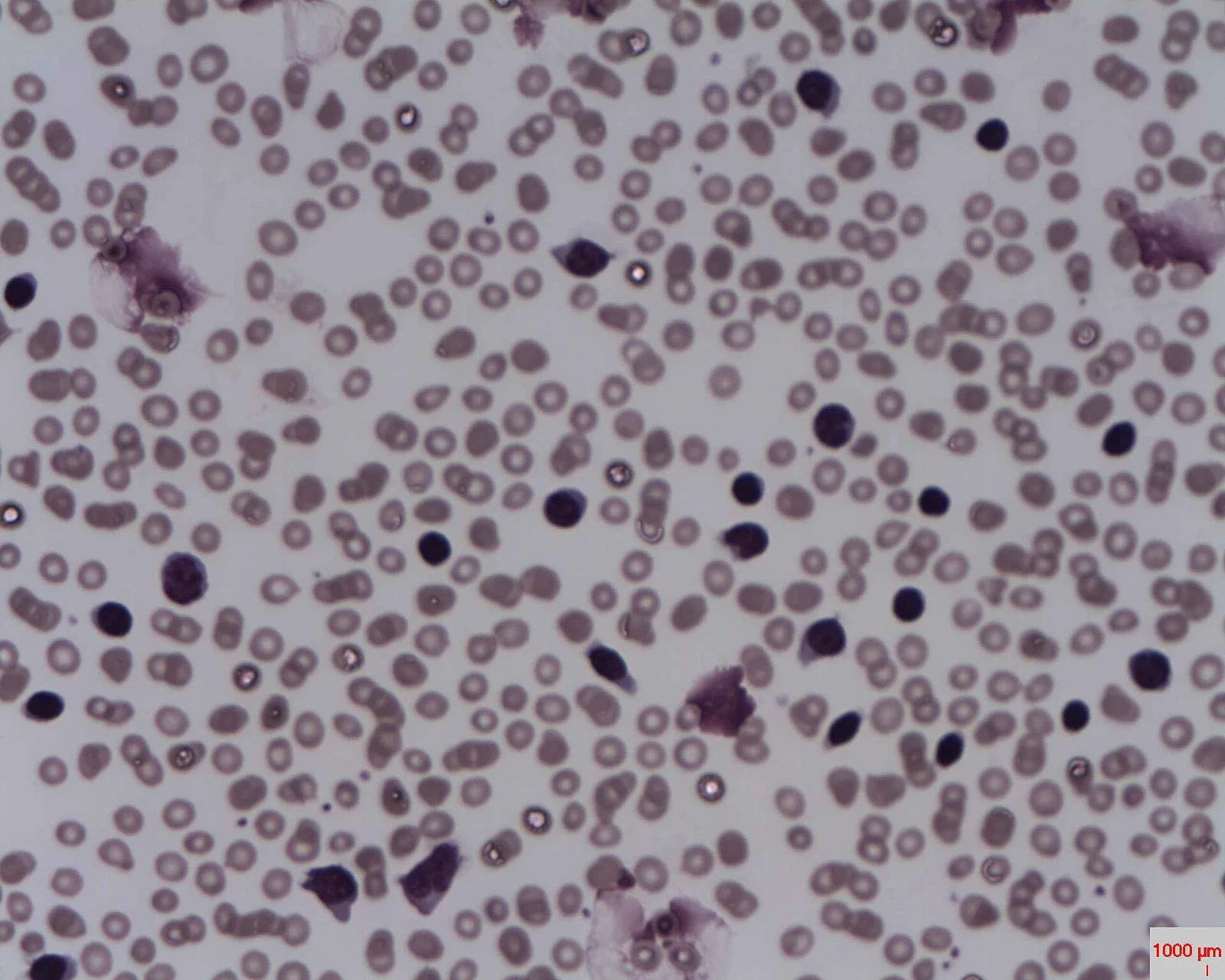
Long description
Blood film in chronic lymphocytic leukaemia showing lymphocytosis and smudge cells, the fragile lymphocytes crushed during film preparation.

Figure 2B-cell chronic lymphocytic leukemia (B-CLL)
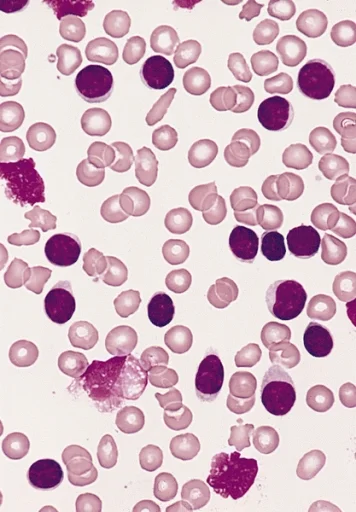
Long description
Blood film in chronic lymphocytic leukaemia showing an excess of small, mature lymphocytes with dense round nuclei.

The characteristic on a peripheral blood film is a modest to gross lymphocytosis of small, mature lymphocytes with scant cytoplasm and clumped chromatin. Smudge cells (also called basket cells) are a classic but non-specific artefact, produced when fragile leukaemic lymphocytes are broken during the physical preparation of the smear. They point towards CLL but are not a diagnosis on their own. Red cells and platelets are usually normal until late or complicated disease.
Flow cytometry establishes the diagnosis. The cells are clonal B lymphocytes that are characteristically: [1]
- positive for CD19, CD5 and CD23
- positive for CD20, but at low density
- positive for CD79b at low density
- with surface immunoglobulin (IgM or IgD) at low density and light-chain restriction (kappa or lambda)
- often positive for CD43, CD200 and ROR1, which help in distinguishing CLL from mantle cell lymphoma and other B-cell neoplasms
CD5 marks a B cell that behaves very differently from a conventional B cell, which is why a clinician must not assume CD5-positivity means a T cell. The combination of CD5 and CD23 on a B cell is strongly suggestive of CLL and has largely displaced older markers in the initial work-up. The differential on an atypical film remains with mantle cell lymphoma (CD5+, CD23-), marginal zone lymphoma (CD5-, CD23-) and hairy cell leukaemia (CD103+, CD11c+, CD25+).
About a third to a half of patients have no symptoms and are diagnosed when a blood count performed for another reason shows lymphocytosis. The "B symptoms" of fever, night sweats and weight loss occur but may be less dramatic than in aggressive lymphoma, and when marked they should raise suspicion of transformation. Infections may be the presenting problem in patients whose disease has been present but unrecognised.
- tiredness, which may be vague and out of proportion to the count
- lumps in the neck, axilla or groin, often discovered incidentally when shaving or dressing
- pressure, early satiety or abdominal discomfort when the spleen is enlarged
- repeated coughs, colds, sinus or skin infections
- unintended weight loss, night sweats, or low-grade fevers, particularly in more advanced or transformed disease
- breathlessness due to anaemia
- easy bruising or petechiae when platelets are low
Health-anxious well patients can be diagnosed while still asymptomatic and then struggle with the idea of having a "leukaemia" that is not being treated. The explanation of watchful waiting is part of treatment.
- generalised, non-tender lymphadenopathy, often painless and mobile
- splenomegaly, sometimes large, with a left-sided abdominal mass crossing into the midline
- hepatomegaly is less common
- signs of anaemia
- signs of infection, including skin and mucous membrane evidence
- signs of autoimmune haemolysis (jaundice, splenomegaly, dark urine)
- petechiae and purpura when platelets are low
- skin infiltration or Richter lesions in occasional cases
Implications of stage matter. Low-stage disease may have no palpable nodes at all; Binet C or Rai III-IV disease typically shows large nodes or splenomegaly with cytopenias.
The key alternatives depend on whether the presenting problem is lymphocytosis, lymphadenopathy or splenomegaly.
| Presentation | Likely alternative | What separates it from CLL |
|---|---|---|
| Persistent lymphocytosis (mature) | Monoclonal B-cell lymphocytosis | Fewer than 5 x 10^9/L clonal B cells, no cytopenias, normal nodes |
| Pertussis or recent viral infection | Polyclonal lymphocytosis that resolves; atypical mononuclear cells | |
| Aplastic or post-clearance marrow | Low counts, not a clonal B expansion | |
| Lymphadenopathy | Non-Hodgkin lymphoma | Histology; CD23/CD5 profile; bulkier, localised disease |
| Hodgkin lymphoma | B symptoms, systemic disease, histology | |
| Chronic infection or sarcoidosis | Non-clonal, biopsy, systemic features | |
| Splenomegaly | Marginal zone or hairy cell leukaemia | CD103, CD11c profile; splenic villous lymphocytes |
| Myelofibrosis | Teardrop cells, left shift, marrow fibrosis | |
| Portal hypertension | Liver disease; non-clonal | |
| Cytopenias | Acute leukaemia | Immature blasts, marrow failure, acute presentation |
| Myelodysplastic syndrome | Dysplasia, ring sideroblasts, IPSS risk |
A rising or persistent absolute lymphocyte count with otherwise normal or mildly abnormal blood counts is the usual starting point.
First line
- Full blood count with differential and an absolute lymphocyte count, repeated after six to twelve weeks to confirm persistence.
- Peripheral blood film.
- Flow cytometry on blood for immunophenotype (CD5, CD19, CD20, CD23, light chain restriction) is the confirmatory test.
- Biochemistry: renal and hepatic function, lactate dehydrogenase, uric acid.
- Hepatitis B and C and HIV serology before any systemic therapy.
- Direct antiglobulin test if anaemia develops (up to a third of patients have a positive DAT at some stage, although overt warm autoimmune haemolytic anaemia is less frequent).[1]
- Measurement of serum immunoglobulins (IgG, IgA, IgM) if infection occurs or treatment is planned.
- Bone marrow is not routinely required; reserve it for unexplained cytopenias, ambiguous morphology, or research protocols.
Staging and extended profiling
- Physical examination with measurement of lymph-node sizes and spleen size below the costal margin. Neither Rai nor Binet uses imaging. CT scanning of the neck, chest, abdomen and pelvis is reserved for specific circumstances, such as measuring bulky disease when venetoclax is planned, or for assessing response in a trial.[1]
- FISH or array comparative genomic hybridisation for del(17p); sequencing for TP53 mutations; IGHV sequencing according to ERIC recommendations. These are done before a first line of treatment and before any subsequent line.
- Beta-2 microglobulin is sometimes used in prognostic scores.
- Cardiac evaluation (ECG, cardiovascular risk assessment) is indicated before a BTK inhibitor, particularly when hypertension or arrhythmia coexist.
What each result means
- A clonal B-cell count of 5 x 10^9/L or more with the characteristic phenotype confirms CLL.
- A count of less than 5 x 10^9/L with no nodes, cytopenias or B symptoms is MBL, not CLL.
- del(17p) or a TP53 mutation redirects therapy away from chemoimmunotherapy.
- IGHV "unmutated" status predicts shorter intervals to treatment and shorter response to treatment.
The diagnosis is made by flow cytometry in an otherwise consistent clinical context, as above. Imaging is not part of staging.
Two clinical staging systems are used, both derived from physical examination and full blood count; neither requires imaging.[1] The modified Rai system describes five stages condensed into three risk groups; the Binet system, more commonly used in Europe, splits disease into three groups depending on the number of involved lymphoid areas and on blood counts.
| Binet | Rai | Risk | Lymphocytosis | Lymph nodes / spleen / liver | Haemoglobin | Platelets |
|---|---|---|---|---|---|---|
| A | 0 | Low | Present | 0-2 lymphoid areas, no large nodes, spleen and liver not enlarged | at least 110 g/L (Rai), at least 100 g/L (Binet) | at least 100 G/L |
| A or B | I | Intermediate | Present | 3 or more lymphoid areas or palpable nodes | at least 110 g/L | at least 100 G/L |
| II | Intermediate | Present | 3 or more lymphoid areas or palpable splenomegaly (liver may be involved) | at least 110 g/L | at least 100 G/L | |
| C | III | High | Present | irrelevant | under 110 g/L (Rai), under 100 g/L (Binet) | at least 100 G/L |
| C | IV | High | Present | irrelevant | irrelevant | under 100 G/L |
Interpretation:
- Binet A / Rai 0: early disease, often observed.
- Binet B: intermediate disease with widespread lymphadenopathy or splenomegaly but preserved blood counts.
- Binet C: advanced disease with anaemia or thrombocytopenia due to marrow infiltration, warranting treatment in almost all cases (unless the cytopenia is clearly autoimmune).
- Importantly, a patient may have a high count (for example over 30 x 10^9/L) yet remain Binet A and be well: the count alone does not stage, and early-stage disease is not treated simply because the count is high.
No single staging system now captures every patient's risk. Over the last two decades a second layer of biological profiling has been added, particularly IGHV mutation status and TP53 changes.
| Marker | What it is | Clinical effect |
|---|---|---|
| IGHV mutation status | Sequence of the rearranged immunoglobulin heavy-variable gene compared with germline; "unmutated" when at least 98% identity | Unmutated (U-CLL) disease typically runs faster, with shorter time to first treatment and shorter response to treatment; mutated disease is usually indolent.[1] Unmutated status persists over time; chromosome and TP53 changes may evolve. |
| del(17p) / TP53 mutation | Loss or mutation of TP53 | The classic chemoimmunotherapy-resistant subgroup. Frequency is 5-10% at diagnosis, 10-20% when first-line therapy is started, and up to 50% in advanced or transformed disease.[1] In targeted therapy, allele burden and clone size still have uncertain cut-offs. |
| del(11q) | Deletion involving ATM on chromosome 11q | Associated with bulky lymphadenopathy, unmutated IGHV and worse outcome in the chemoimmunotherapy era; its adverse effect is substantially mitigated by targeted therapy. |
| Complex karyotype | Three or more clonal chromosomal abnormalities | Associated with TP53 changes and worse progression-free survival, especially with venetoclax-based therapy when five or more abnormalities are present (so-called high-complexity karyotype).[1] Routine testing outside clinical trials is not recommended because of limited reproducibility. |
| Beta-2 microglobulin | Serum surrogate of tumour burden | Included in International Prognostic Index for CLL (CLL-IPI) scores. |
| Whole-genome score / stereotyped subsets | #2 and #8 subsets in particular | Associated with aggressive disease; still mainly research tools. |
The EHA 2026 guideline supports determining IGHV status before first-line therapy and TP53 status before every line, and reserves other genomic markers (such as SF3B1, NOTCH1) for research rather than routine practice.[1] Early-stage, asymptomatic disease does not require biological profiling, because no study has shown a survival benefit from treating on these markers alone.[1]
Time-limited therapy combines venetoclax with a CD20 antibody (obinutuzumab), possibly with a BTK inhibitor. In a phase III study of 432 previously untreated patients with comorbidity, venetoclax plus obinutuzumab produced a median progression-free survival (PFS) of 76.2 months, with shorter PFS in the unmutated IGHV group and median PFS not reached in the mutated group.[1] Five-year PFS was 81.8% with venetoclax plus obinutuzumab and 85.5% with the triple combination (venetoclax plus obinutuzumab plus ibrutinib), although infection rates were higher (grade 3-4 infections 21%) with the triple. Venetoclax plus ibrutinib given as 15 months of treatment produced 5-year PFS of 59.9% in older or less fit patients and 66% (97% OS) in younger patients, with 55% (95% CI 45-64) at 5.5 years in the unmutated IGHV group versus 79% (95% CI 68-87) in the mutated group. Time-limited treatment is generally preferred, when feasible (bearing in mind renal function and the venetoclax ramp-up), in patients without TP53 disruption, particularly those with mutated IGHV, because continuous therapy is avoided whenever possible in that subgroup.[1]
The AMPLIFY study established acalabrutinib plus venetoclax with or without obinutuzumab for 14 cycles versus FCR or BR, with an estimated 36-month PFS of 76.5% (doublet), 83.1% (triplet) and 66.5% (chemoimmunotherapy), and an OS advantage for the doublet (hazard ratio 0.33).[1]
Continuous targeted therapy. For patients in whom time-limited therapy is not advisable, covalent BTK inhibitors (acalabrutinib, zanubrutinib, historically ibrutinib) provide long-term disease control, including in both IGHV subgroups. Ten-year follow-up of ibrutinib versus chlorambucil in RESONATE-2 gives a median PFS of 8.9 years.[1] Second-generation BTK inhibitors had fewer off-target effects and fewer cardiac toxicities than ibrutinib, so acalabrutinib or zanubrutinib are preferred for new patients; patients tolerating ibrutinib well need not be switched.[1] Continuous acalabrutinib plus obinutuzumab was more effective but more toxic than acalabrutinib alone and is not generally preferred. EHA advises a cardiovascular work-up before initiation of any BTK inhibitor and avoidance where there is ventricular arrhythmia or uncontrolled heart failure; if cardiovascular disease or anticoagulant use coexists, BCL-2-based treatment is preferred.[1]
TP53 disruption. Continuous BTK inhibition has generally been preferred in this high-risk group until longer follow-up data arrive, because time-limited data are shorter, although triple combinations (acalabrutinib or ibrutinib plus venetoclax plus obinutuzumab) show encouraging results and may be considered in fit patients who can tolerate the additional infection risk. Venetoclax plus obinutuzumab produced a shorter PFS in the TP53 subgroup in a head-to-head comparison, so time-limited combinations in TP53-aberrant disease are reserved for shared decision-making.[1] Idelalisib plus rituximab or venetoclax monotherapy remain approved fallback if BTK inhibitors are unsuitable or unavailable.[1]
Treatment again starts only when iwCLL 2018 criteria are met. The choice rests on the prior drug class and duration of response:
- after chemoimmunotherapy: venetoclax plus (often) rituximab as time-limited therapy, or a second-generation covalent BTK inhibitor as continuous therapy; most patients do not receive further chemotherapy.[1]
- progression on a covalent BTK inhibitor: switch to a venetoclax-based regimen or to the non-covalent BTK inhibitor pirtobrutinib; re-expose to a BTK inhibitor class only if the reason for stopping was intolerance rather than progression.[1]
- after time-limited venetoclax: re-treat with venetoclax if the prior response lasted more than about two years, otherwise switch drug class (to a covalent BTK inhibitor, or to pirtobrutinib if these have been used).[1]
- venetoclax-rituximab gives 72-80% responses and 23-25 months median PFS in this setting.[1]
- pirtobrutinib gives 69% responses and a median PFS of 14 months after covalent BTK failure, including in double-exposed patients.[1]
Double-exposed and double-refractory disease is the new high-risk area. "Double-exposed" means both BTK and BCL-2 inhibitors have been used but not for progression; "double-refractory" means progression after both. For double-refractory disease pirtobrutinib is preferred, or (if unavailable) idelalisib plus rituximab, with allogeneic stem cell transplantation and clinical trials of BTK degraders, bispecific antibodies or CAR T cells considered in eligible patients. About half of double-refractory patients with controlled disease before transplant remain in long-term remission.[1] Other double-refractory approaches include venetoclax-r (re-challenge) in selected cases. Newer NCCN options in this space include pirtobrutinib and CD19-directed CAR T cells for relapsed or refractory disease after both classes.[2]
About 2-10% of patients with CLL transform to an aggressive lymphoma, usually diffuse large B-cell lymphoma (Richter transformation, RT). Loss of p53 function and MYC involvement are central. Suspicion should rise with sudden rapid node growth, new extranodal disease, rising pain, B symptoms or LDH. Diagnosis requires a biopsy, ideally excisional.[1]
Key points from EHA 2026 and NCCN 2026:
- Clonal relationship to the underlying CLL is established by IGHV sequencing and determines prognosis: clonally-related RT behaves more aggressively than apparently de-novo DLBCL arising in a CLL patient.[1][2]
- FDG-PET/CT helps select a biopsy site, although the optimum SUV threshold is debated in the targeted-therapy era.[1]
- del(17p)/TP53 aberrations are found in 50-60% of RT cases.[1]
- Clonally-unrelated RT-DLBCL is treated as de novo DLBCL. For clonally-related disease, clinical trials are preferred; outside trials, standard DLBCL chemotherapy is used, with complete remission in 5-38% and median survival rarely exceeding one year.[1]
- Targeted approaches (BTK or BCL-2 inhibitors, pirtobrutinib) show limited but real activity and may bridge to allogeneic stem cell transplantation, which should be considered in fit patients after a good response.[1]
- RT presenting as classical Hodgkin lymphoma is treated under Hodgkin guidelines.
- Whenever possible, enroll these patients in a trial.
Hypogammaglobulinaemia and infection. Immune dysfunction is present to some degree from diagnosis and worsens with therapy. Immunoglobulin (IgG) replacement is advised for patients with recurrent or acute infections who have total IgG below 4 g/L; subcutaneous preparations are associated with higher trough levels, fewer adverse events and better quality of life than intravenous.[1] In practice, treat the treatable (non-CLL infections, dental sources, sinus disease) before starting replacement. Early detection of infections, prompt antibiotics, and vaccination (avoiding live vaccines) are the ordinary measures. Avoid antibiotics for suspected viral infection; reserve them for confirmed bacterial sources.
Autoimmune haemolytic anaemia (AIHA). Up to a third of patients have a positive direct antiglobulin test at some stage, but overt warm AIHA is less common. For warm AIHA without an iwCLL indication to treat the CLL itself, immunosuppression (steroids, with rituximab if refractory) is tried before CLL-directed therapy; when CLL treatment is indicated, BTK or BCL-2 inhibitors plus an anti-CD20 antibody can control both. Steroid plus rituximab is superior to steroids alone in randomised studies of warm AIHA.[1]
Immune thrombocytopenia (ITP). Approximately 2-5% of patients develop ITP, mostly when not on CLL-directed therapy; about 30% also have a history of AIHA (Evans syndrome). First-line response to steroids or intravenous immunoglobulin is 50-60%; weekly rituximab for four weeks is effective, eltrombopag has shown response in a phase II study, and splenectomy is reserved for refractory cases. Before escalating ITP therapy, treat the CLL if it meets criteria.[1]
Pure red-cell aplasia and autoimmune neutropenia are rare, under 1%, and treated on the same lines as AIHA.[1]
Second malignancies. CLL patients have excess skin, lung and other cancers; screening and strong anti-smoking advice are part of care. Family counselling is restricted to patients with strong family history or suspected constitutional cancer syndromes.
The full targeted-therapy pathway above is not available in many settings. The principles that still apply:
- For diagnosis, immunophenotyping (flow cytometry) is the confirmatory test; where unavailable, the diagnosis rests on a consistent clinical picture, a mature lymphocytosis, and marrow morphology. Treatable causes of lymphocytosis should still be excluded.
- Rai/Binet staging requires only a physical examination, a full blood count, and a smear: no imaging.
- Chemoimmunotherapy (chlorambucil alone or combined with rituximab where rituximab exists; bendamustine where affordable; FCR at specialist centres) remains effective, albeit with more toxicity and less durable remission than targeted agents; chemoimmunotherapy should be used when modern drugs are unavailable, and targeted agents obtained on compassionate or trial access whenever possible.
- The commonest avoidable harm is premature treatment: the iwCLL criteria can be applied without advanced resources or any imaging.
- Infections dominate outcomes: prophylaxis, prompt antibiotic therapy, careful transfusion practice, and adequate nutrition matter more in these settings than marginal differences between targeted regimens.
- Richter transformation cannot be treated radically without access to aggressive chemotherapy and possibly transplantation; early recognition and referral to a specialist centre is the priority.
- Hypogammaglobulinaemia should be identified clinically and, where replacement is unavailable, infections should be treated aggressively and patients vaccinated against preventable disease.
- Direct family and patient support (transport, cost) often determine more of the outcome than the drug chosen.
CLL and SLL are one entity: the difference in name is anatomical. SLL accounts for around 5% of cases and presents with nodal, splenic or other tissue involvement with under 5 x 10^9/L clonal B cells in blood; absent significant leukaemic phase. SLL is treated by nodal and lymphoma staging principles at presentation but uses the same iwCLL criteria for treatment and the same drugs. MBL, characteristically a low-count clone of CLL-phenotype cells without cytopenias, nodes, organomegaly or B symptoms, is a precursor rather than a cancer and is not treated. CLL-type MBL with 0.5-5 x 10^9/L cells may progress to treatment-requiring CLL at about 1% per year; low-count MBL is essentially non-progressive. A lymphocyte count below 5 x 10^9/L with no B symptoms or cytopenia requires no treatment beyond routine follow-up.[1]
Read alongside acute leukaemias, non-Hodgkin lymphoma, Hodgkin lymphoma, multiple myeloma, chronic myeloid leukaemia, autoimmune haemolytic anaemia, immune thrombocytopenia and the approach to lymphadenopathy.
- CLL is the commonest chronic leukaemia of adults in European populations: incidence about 4.7 per 100,000 per year, median age 70, male predominance roughly 2:1, mortality falling.
- Diagnosis rests on at least 5 x 10^9/L clonal B cells with CD5+, CD23+, low-intensity CD20 and light-chain restriction, detected by flow cytometry.
- Smudge cells are a characteristic artefact of the blood film, not a diagnosis.
- SLL and CLL are the same disease; MBL is the sub-threshold precursor and accounts for a rising detection rate.
- Rai and Binet staging use history, examination and a blood count only, not imaging.
- IGHV unmutated status and TP53 disruption (del(17p) or mutation) redirect therapy away from chemoimmunotherapy; del(11q) and complex karyotype modify prognosis; targeted therapy has made the latter much less relevant.
- TP53 abnormalities occur in 5-10% at diagnosis, 10-20% at first-line and up to 50% later; test before every line.
- IGHV is largely stable; test once, before first-line treatment.
- Treat by iwCLL 2018 criteria only; asymptomatic early-stage disease gains nothing from immediate therapy.
- Time-limited BCL-2-based combinations and continuous BTK inhibitors have displaced chemoimmunotherapy where available.
- Hypogammaglobulinaemia is the driver of infection: replace Ig only when IgG is under 4 g/L and infections are frequent; subcutaneous preparations are preferred.
- Autoimmune cytopenias: conventional immunosuppression first, CLL-directed therapy when criteria are met.
- Richter transformation (2-10%) is the principal fatal complication; biopsy is mandatory and clonal relation predicts outcome.
Answer each one in your head first, then open the answer.
A 72-year-old man has a routine count showing lymphocytes 22 x 10^9/L, haemoglobin 128 g/L, platelets 180 x 10^9/L, and no lymphadenopathy. What is the most likely diagnosis and management?
Show answerHide answer
CLL, Binet A / Rai 0. He has fewer than three lymphoid areas and normal counts, so no treatment is indicated; watch and monitor with blood counts every 3-6 months. He will not benefit from biological profiling while asymptomatic.
The same man returns 18 months later with haemoglobin 92 g/L, platelets 70 x 10^9/L, bilateral cervical nodes 4 and 5 cm, and weight loss 3 kg beyond normal. What changes and why?
Show answerHide answer
He now meets iwCLL 2018 criteria: progressive marrow failure (haemoglobin under 100 g/L, platelets under 100 G/L) and nodes over 1 cm that are enlarging, plus systemic symptoms. Binet C / Rai III-IV. Treat only after pre-treatment assessment, including full blood count, liver and kidney function, hepatitis/HIV serology, cardiac review before any BTK inhibitor, and TP53 plus IGHV testing, which guide the choice of modern first-line therapy.
Why must chemoimmunotherapy be avoided in a patient with 35% of leukaemia cells carrying del(17p)?
Show answerHide answer
del(17p) disrupts p53, the key pathway that cytotoxic drugs rely on. Targeted agents acting outside the p53 pathway (BTK inhibitors, BCL-2 inhibition, especially in combinations) produce better outcomes. Test TP53 before every line, because its frequency rises from 5-10% at diagnosis to 10-20% before first-line and up to 50% in later disease.
A patient diagnosed with CLL, Binet A, has frequent chest infections and an absolute lymphocyte count of 65 x 10^9/L but feels well. Can you start treatment for the count?
Show answerHide answer
No. A high lymphocyte count alone is not an iwCLL criterion (LDT is used rather than absolute count), and early-stage asymptomatic disease does not benefit from treatment even with high-risk biology. Investigate and treat the infections, measure IgG; if IgG is under 4 g/L with recurrent infections, give immunoglobulin replacement. Re-stage at the next review.
Up to a third of CLL patients have a positive DAT at some stage. Does a positive DAT alone demand treatment of the CLL?
Show answerHide answer
No. Treat the AIHA with immunosuppression (steroids, with rituximab if refractory) when warm haemolysis is present; only treat the CLL itself if the haemolysis is refractory or if other iwCLL criteria also apply. BTK or BCL-2 inhibition with anti-CD20 therapy can control both when treatment is indicated.
Which drug class is preferred when the patient has uncontrolled atrial fibrillation and needs first-line therapy for CLL without TP53 disruption?
Show answerHide answer
A BCL-2-based (venetoclax) time-limited regimen, because BTK inhibitors are generally avoided in the setting of arrhythmia or uncontrolled cardiovascular disease, in favour of BCL-2 inhibitors. EHA 2026 places time-limited therapy first in unmutated and mutated IGHV alike when the comorbidity balance allows.
A 60-year-old with CLL develops rapidly enlarging bulky cervical nodes, fever, and a sudden rise in LDH. What is the next diagnostic step?
Show answerHide answer
Suspect Richter transformation. Biopsy (preferably excisional) is required, not just a blood test. Once confirmed as DLBCL, determine clonal relation to the CLL by IGHV sequencing, look for TP53 disruption, and stage under PET-CT, then manage per transformation guidance: clonally-unrelated disease as de novo DLBCL, clonally-related disease usually on trial or with DLBCL-type chemotherapy with consolidation if fit.
In settings where BTK inhibitors, venetoclax, or fludarabine are unavailable, oral chlorambucil plus prednisolone can control disease in many older patients for long periods; rituximab should be added if affordable. Splenic irradiation helps local symptoms. AIHA responds to prednisolone. Palliative whole-body or targeted radiotherapy can relieve bulky disease discomfort. None of these choices should be dressed up as equivalent to targeted therapy, but they are real options and, for a frail patient, may be preferable to toxicity-intensive modern combinations.
Read plainly: some of the statements in this topic summarise 2026 guidance; older or non-European regimens may differ, and local practice takes precedence.
- Eichhorst B, Ghia P, Bosch F, et al. EHA Guidelines on management of chronic lymphocytic leukaemia and Richter transformation. HemaSphere 2026; article 42293463. Accessed .
- Wierda WG, Brown J, Abramson JS, et al. NCCN Guidelines Insights: Chronic Lymphocytic Leukaemia/Small Lymphocytic Lymphoma, Version 2.2026. Journal of the National Comprehensive Cancer Network 2026;24(3):68-80. Accessed .
Figure credits
Educational content for medical students and doctors. It is not a substitute for clinical judgement or local guidelines.