Chronic myeloid leukaemia and hairy cell leukaemia
Chronic myeloid leukaemia (BCR-ABL1, TKIs) and hairy cell leukaemia (BRAF V600E, purine analogues): diagnosis and treatment.
- Chronic myeloid leukaemia (CML) is an acquired clonal disorder of a pluripotent haematopoietic stem cell. Its hallmark is the Philadelphia chromosome, a t(9;22)(q34;q11) translocation that creates the BCR-ABL1 fusion gene and a constitutively active tyrosine kinase.
- The disease begins in chronic phase, with granulocytic leucocytosis, a left shift, basophilia, often thrombocytosis, splenomegaly and a characteristically low leukocyte alkaline phosphatase. It may progress to accelerated phase and then to blast crisis, which behaves like acute leukaemia.
- Tyrosine kinase inhibitors (TKIs) directed at BCR-ABL1 have transformed the outlook: most chronic-phase patients now have a life expectancy close to that of the general population, and some with a durable deep molecular response can safely stop treatment and enter treatment-free remission (TFR).
- Hairy cell leukaemia (HCL) is an indolent mature B-cell neoplasm of red pulp and bone marrow, defined by the BRAF V600E mutation in most cases, pancytopenia with monocytopenia, massive splenomegaly and a dry marrow tap. [5]
- The purine nucleoside analogues cladribine and pentostatin are first-line treatment for HCL; they induce durable complete remissions in most patients.
By the end of this page you should be able to:
- Explain how the Philadelphia chromosome produces BCR-ABL1 and why it drives CML.
- Recognise the chronic, accelerated and blastic phases and outline their criteria.
- Interpret the blood film, leukocyte alkaline phosphatase, marrow cytogenetics and BCR-ABL1 quantitative PCR.
- Choose and dose first-line therapy for chronic-phase CML, assess response against ELN milestones, and manage resistance, blast crisis and TFR.
- Recognise HCL by its clinical picture, blood film, immunophenotype, dry tap and BRAF V600E, and choose between cladribine and pentostatin schedules.
- Distinguish CML, HCL and other chronic leukaemias and leukemoid reactions, and adapt care in low-resource settings.
DefinitionChronic myeloid leukaemia (CML)
CML is a myeloproliferative neoplasm arising in a haematopoietic stem cell, characterised by the Philadelphia chromosome t(9;22)(q34;q11) and the BCR-ABL1 fusion gene, leading to uncontrolled granulocytic proliferation.
CML is traditionally described in three phases. Chronic phase is the presenting state for most patients: the marrow is hypercellular with granulocytic proliferation and the blood shows a left shift. Accelerated phase is an intermediate stage of rising blasts and basophils, failing haematopoiesis and emerging new abnormalities. Blastic phase (blast crisis) is the terminal transformation, resembling acute leukaemia. Classification has changed between WHO editions, which matters for trials and for what is called "accelerated". [2]
DefinitionHairy cell leukaemia (HCL)
HCL is a rare, indolent mature B-cell neoplasm that infiltrates bone marrow, spleen and liver. Classic HCL is defined by the BRAF V600E mutation, pancytopenia with monocytopenia, splenomegaly without significant lymphadenopathy, and characteristic "hairy" cytoplasmic projections.
Classic (classical) HCL is the common form and carries BRAF V600E in 90-95 percent of cases. [5] Variant HCL (HCL-v) is biologically distinct, lacks BRAF V600E, is usually CD25-negative, carries a poorer response to purine analogues and needs different management. [4][5]
CML accounts for roughly 15 to 20 percent of adult leukaemias. [2] With TKI therapy, the life expectancy of a newly diagnosed chronic-phase patient now approaches that of age-matched people in the general population in Western countries. [1] Before the TKI era it was invariably fatal within a few years of progression.
HCL is uncommon, accounting for 2 to 3 percent of adult leukaemias. [4] It is more frequent in men, with a male to female ratio of about 4 to 1, and the median age at onset is about 52 years. [4] Untreated, median survival used to be around 4 years. [4] HCL is rare in children and in younger women. [4]
CML is an acquired somatic stem-cell disease. The t(9;22) and BCR-ABL1 fusion are found in pluripotent cells; there is no common inherited form. The clinical message is that once BCR-ABL1 is present, the fusion protein itself drives the disease through its tyrosine kinase activity, so blocking that kinase is the targeted treatment. [1][3]
For HCL, the dominant molecular event is the BRAF V600E somatic mutation, which (through MEK/ERK signalling) confers the characteristic hairy-cell phenotype in 90-95 percent of classic cases. [5] Variant HCL lacks this mutation. [5] A causative environmental exposure has not been firmly established. [4]
CML. The BCR-ABL1 fusion tyrosine kinase is constitutively active. It activates downstream signalling that expands the granulocytic lineage (myelocytes, promyelocytes and metamyelocytes), impairs apoptosis and produces genomic instability over time. [1][3] This explains the massive leucocytosis with a left shift, basophilia and eosinophilia, and the extramedullary haematopoiesis that enlarges the spleen and sometimes the liver. Platelet counts may rise. With progressive disease, blasts accumulate and haematopoiesis fails, producing the anaemia and thrombocytopenia of accelerated or blastic phase.
Leukocyte alkaline phosphatase (LAP), an enzyme in mature neutrophils, is characteristically low in CML, whereas it is raised in infection and in leukaemoid reactions. [3] LAP is a supportive, not a definitive, marker today; the diagnosis rests on genetic studies.
HCL. Hairy cells are clonal late-memory B cells that infiltrate red pulp and marrow. The BRAF V600E-driven proliferation, together with marrow fibrosis, underlies the typical picture. [5] The bone marrow is heavily infiltrated and reticulin fibrosis is marked, which is why aspiration often yields a dry tap. Cytopenias result from marrow replacement, splenic sequestration and suppression; monocytopenia in particular drives infection risk even when neutrophils are near normal. [4][5]
CML. Many patients are asymptomatic at diagnosis, identified by an incidental full blood count. Typical symptoms are fatigue, weight loss, night sweats, abdominal fullness or left upper quadrant pain from splenomegaly, and bone pain. Massive splenomegaly is characteristic. [1][3] Hepatomegaly can occur. Bleeding or thrombosis may appear, and priapism occasionally. The blood film shows granulocytic leucocytosis with a full left shift, basophilia and sometimes eosinophilia and thrombocytosis. Blast crisis presents with fever, bone pain, bleeding and rapidly falling counts, resembling acute leukaemia.
HCL. Typical features are fatigue, recurrent infections, bruising or bleeding, and dragging left upper quadrant discomfort from splenomegaly. On examination the spleen is often massively enlarged; significant lymphadenopathy is uncommon, and this contrast helps distinguish HCL from lymphoma. [4] Blood counts commonly show pancytopenia (anaemia, neutropenia, thrombocytopenia) with an absolute monocytopenia. [4][5] Leucocytosis occurs in a minority. Weight loss and fever usually signal infection or advanced disease rather than the tumour itself.
Table 1 contrasts the chronic leukaemias and leukaemoid reactions that CML and HCL are most often confused with. The first discriminator is the cell lineage that predominates.
| Condition | Cell that predominates | Common associated findings | Point that settles it |
|---|---|---|---|
| Chronic phase CML | Granulocytes with full left shift | Leucocytosis, basophilia, splenomegaly, low LAP | Philadelphia chromosome t(9;22); BCR-ABL1 by PCR [1][3] |
| Hairy cell leukaemia | Mature "hairy" B cells (often scarce in blood) | Pancytopenia with monocytopenia, splenomegaly, dry tap | BRAF V600E; bright CD20 with co-expression of CD11c, CD25 and CD103 on flow cytometry; TRAP [4][5] |
| Chronic lymphocytic leukaemia | Small mature lymphocytes | Lymphadenopathy, lymphocytosis, smudge cells | CD5/CD23/CD19 flow profile; no Ph chromosome |
| Chronic neutrophilic leukaemia | Mature (segmented) neutrophils | Neutrophilia without left shift, splenomegaly | No BCR-ABL1; marker not in sources used |
| Chronic myelomonocytic leukaemia | Monocytes and dysplastic granulocytes | Monocytosis, cytopenias, dysplasia | No BCR-ABL1; WHO criteria apply |
| Leukaemoid reaction | Mature neutrophils with toxic granulation | Infection, inflammation, tissue necrosis | Raised leukocyte alkaline phosphatase, no Ph chromosome, resolves with the trigger [3] |
Exam trapLeukaemoid reaction versus CML
A note on the HCL row: TRAP denotes tartrate-resistant acid phosphatase, an enzyme stain or immunohistochemical stain that (together with the BRAF V600E stain) supports the diagnosis, although IHC for TRAP is more likely to be negative in HCL variants. [5]
Approach to suspected CML
- Leucocytosis with left shiftSymptoms, splenomegaly, basophilia, low LAP
- Bone marrow aspirateblasts and basophils for phase; core biopsy for fibrosis
- Cytogenetics and RT-PCRPhiladelphia chromosome; identify BCR-ABL1 transcript
- Question: BCR-ABL1 found?
Yes: CML
- Start a TKI in chronic phaseimatinib 400 mg daily or selected 2GTKI; baseline qPCR
- Monitor counts and qPCRcounts every 2 weeks; qPCR at least every 3 months
No: consider leukaemoid reaction or other MPN
- Treat the trigger; repeat testinghigh LAP points to reactive disease
Figure 1HD.17.076 (11966201853)
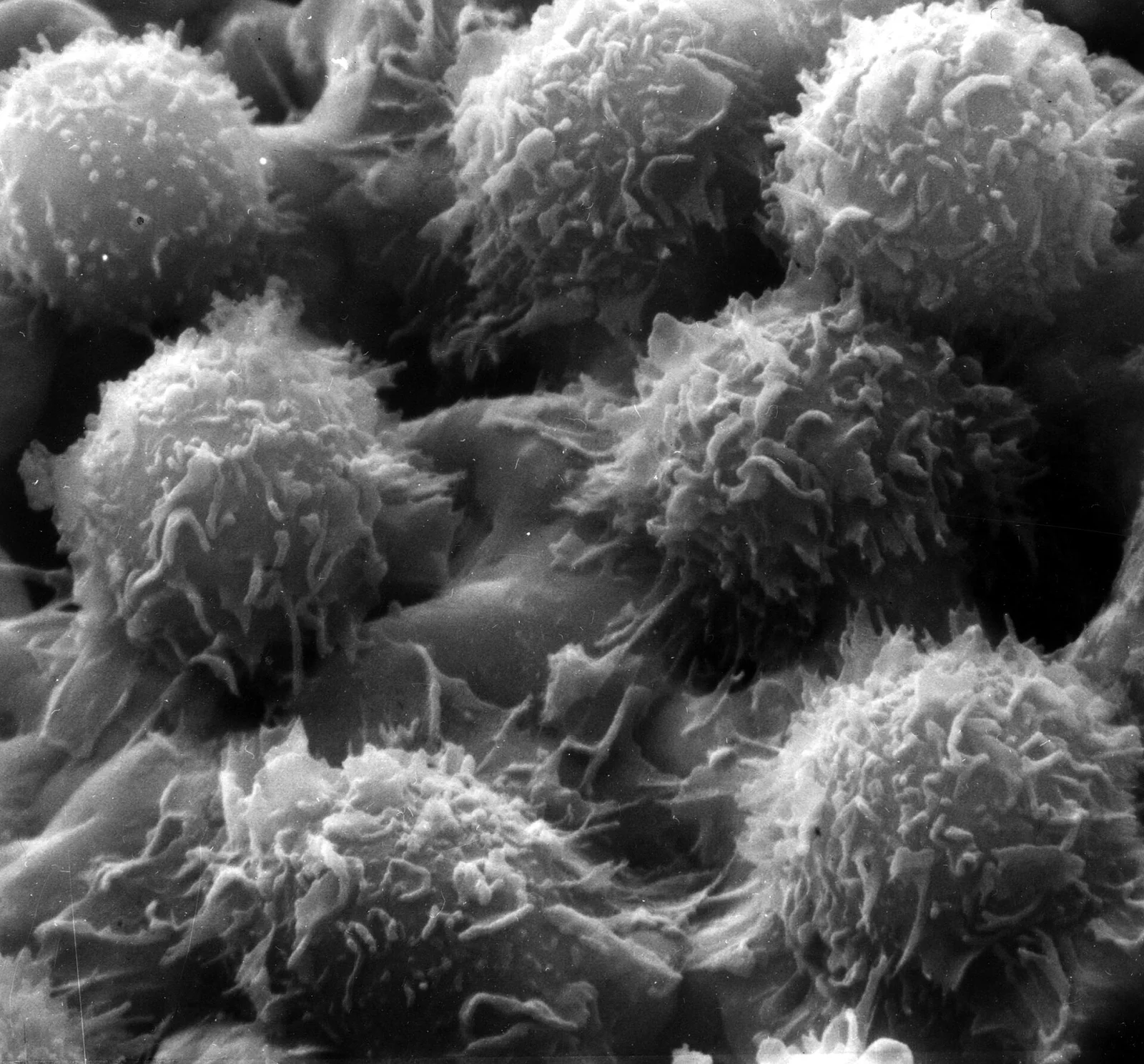
Long description
Scanning electron micrograph of hairy cells, leukaemic B cells covered in fine hair-like cytoplasmic projections.

CML. A full blood count with differential and a careful film are first. Bone marrow is needed at diagnosis: the European LeukemiaNet (ELN) recommends an aspirate for morphology, because the blast and basophil percentages distinguish chronic from accelerated and blastic phase, and for cytogenetics. [1] Cytogenetics should be performed by conventional banded analysis (CBA) of Giemsa-stained metaphases. [1] Fluorescence in situ hybridisation can detect cryptic or variant translocations. RT-PCR must be done on peripheral blood to identify the BCR-ABL1 transcript type, and quantitative RT-PCR (qPCR) on the International Scale is the backbone of molecular monitoring. [1] LAP is not required to make the diagnosis but explains the old clinical distinction from leukaemoid reactions.
Baseline assessment by phase includes blast and basophil counts; marrow fibrosis assessment by core biopsy, which has prognostic value. [1]
HCL. The blood film may show hairy cells, but they can be scanty. Flow cytometry of blood or marrow cells demonstrates the characteristic profile (CD19, CD20, CD22, with co-expression of CD11c, CD25 and CD103). [4] Bone marrow aspiration often fails (dry tap) because of fibrosis, so trephine biopsy is essential; biopsy shows interstitial infiltration and reticulin fibrosis. [4][5] BRAF V600E testing (by PCR or by VE1 immunohistochemistry) is now considered part of the defining diagnosis. [5] Immunohistochemistry for TRAP can also help, though it is more often negative in variants. [5]
CML phases (2017 WHO, summarised from the 2022 comparison). [2]
- Chronic phase: blasts under 10 percent in blood and bone marrow. [2]
- Accelerated phase (2017 WHO criteria): 10-19 percent blasts in blood or marrow, at least 20 percent basophils in blood, persistent thrombocytopenia (100 x 10^9/L or below) unrelated to therapy, persistent or increasing splenomegaly, or new clonal cytogenetic abnormalities in Philadelphia-positive cells. [2]
- Blastic phase: 20 percent or more blasts in blood or marrow, extramedullary (organ) blast infiltrates, or any lymphoblasts in blood or marrow regardless of number. [2]
Classifications differ between editions: the 2022 WHO classification no longer recognises accelerated phase as a separate category and uses higher blast thresholds for blast crisis under ICC 2022 criteria. [2] When reading older trials, check which definition was used.
The ELN recommends that risk stratification at baseline uses the EUTOS long-term survival (ELTS) score rather than the older Sokal score; it uses age, spleen size, peripheral blood blasts and platelet count, all pre-treatment. [1]
HCL. There is no universally accepted staging system for prognosis or treatment. [4] The diagnosis rests on (i) morphology of hairy cells, (ii) a characteristic immunophenotype, (iii) BRAF V600E and (iv) characteristic marrow fibrosis. Response after treatment is assessed on peripheral counts and marrow (4 to 6 months after cladribine, or after 8 to 9 courses of pentostatin). [4]
First-line treatment is a TKI. The standard dose of imatinib is 400 mg once daily. [1] Second-generation TKIs (2GTKIs) are also first-line options: dasatinib 100 mg once daily, nilotinib 300 mg twice daily and bosutinib 400 mg once daily. [1] All target BCR-ABL1 but differ in potency, side-effect profile and contraindications. They have been compared against imatinib but not against each other. [1]
Practical selection:
- Generic imatinib is the cost-effective initial choice, especially where resources are limited. [1]
- Dasatinib, nilotinib and bosutinib produce deeper molecular responses faster than imatinib in trials, but without an overall survival advantage. [1] They are useful first-line when imatinib is unsuitable or when a deep response matters most (younger patients, women planning pregnancy). [1]
- Match the drug to comorbidity: nilotinib has vascular and metabolic risks; dasatinib has pleuro-pulmonary toxicity and pleural effusion; bosutinib has a different toxicity profile; ponatinib has cardiovascular toxicity. [1]
- Imatinib can be reduced to 300 mg once daily if 400 mg is not tolerated and the response is optimal; dasatinib can be used at 50 mg daily with similar efficacy in chronic phase. [1]
Pregnancy. TKIs are teratogenic and contraindicated throughout pregnancy. [1] Men on imatinib or a 2GTKI can safely father children; women need individualised planning. If pregnancy is desired, eligible women may discontinue TKI after a durable deep molecular response, and treatment during pregnancy (if needed) can use interferon alpha or leucapheresis; TKI should be stopped in the first trimester. [1] Women who are pregnant and lose molecular response usually remain clinically stable to term. [1]
ELN treats molecular monitoring by qPCR on the International Scale as central. [1] Blood counts and differential should be done every 2 weeks until complete haematologic response (or more frequently with toxicity), and qPCR at least every 3 months even after major molecular response. [1] Molecular targets:
- BCR-ABL1 above 10 percent at 3 months is the failure threshold when confirmed. [1]
- Major molecular response (MMR, MR3) is BCR-ABL1 at or below 0.1 percent. [1]
- MR4 is at or below 0.01 percent, or undetectable disease with more than 10,000 ABL1 transcripts; MR4.5 at or below 0.0032 percent (or undetectable with more than 32,000 transcripts). [1]
A bone marrow aspiration for cytogenetics and mutation testing is required if resistance or progression is suspected; BCR-ABL1 kinase domain mutation analysis should be sent at failure. [1]
Assess adherence and drug interactions first. If response remains suboptimal, sequence by mutation:
- No mutation or common mutations: dasatinib, nilotinib or bosutinib are options after imatinib failure; 2GTKIs have activity in many resistant cases. [1]
- T315I mutation: ponatinib is the TKI active against it; the approved dose is 45 mg once daily, though lower doses (15-30 mg) may be used for lesser resistance to reduce cardiovascular risk. [1]
- Ponatinib cardiovascular toxicity occurs in about 30 percent of recipients and may be dose related. [1]
- Bosutinib is used at 400 mg daily first-line and 500 mg daily second-line; dasatinib 70 mg twice daily is the approved dose in advanced phase. [1]
Treat accelerated phase as high-risk disease with early consideration of allogeneic stem cell transplantation if the response is not optimal. [1] For blast crisis, the aim is to return to a second chronic phase: imatinib (or a 2GTKI chosen by mutation profile), combined with acute leukaemia-type chemotherapy, followed by allogeneic transplantation if possible. [1] Outcomes in blast crisis remain poor, with survival generally under a year once blast crisis has occurred. [1]
ELN recognises TFR as an important goal after sufficient treatment and deep response. [1] Consider discontinuing TKI only when:
- CML is in first chronic phase only.
- The patient is motivated and has reliable access to high-quality qPCR with rapid turnaround.
- First-line therapy (or second-line only if intolerance was the reason for change). [1]
- Duration of TKI therapy greater than 5 years (greater than 4 for 2GTKIs in the minimal group).
- Sustained deep molecular response (MR4 or better) for at least 2 years (minimal), optimally 3 years for MR4 or 2 years for MR4.5. [1]
- No prior treatment failure. [1]
Monitoring after stopping is intensive: monthly for the first 6 months, every 2 months for months 6-12 and every 3 months thereafter. [1] More than 80 percent of recurrences happen within the first 6-8 months. [1] Restart the TKI when major molecular response is lost; a polymyalgia-like withdrawal syndrome can occur but is usually mild and self-limited. [1]
Refer urgently for suspected blast crisis, uncontrolled counts, resistance mutations such as T315I, and all cases being considered for transplantation. Early haematology involvement also allows TKI selection by comorbidity and pregnancy planning.
Most asymptomatic HCL with minimal cytopenias is observed every 3 to 6 months with full blood count and clinical review. [4] Treatment is driven by symptomatic cytopenias, painful splenomegaly or infection risk. [4]
Purine analogues are first-line, and cladribine and pentostatin are equally effective for inducing and maintaining remission. [4] Both achieve complete remission in more than 80 percent of patients, with median disease-free survival over 10 years. [4] Typical schedules: [4] [1]
- Pentostatin: 4 mg/m^2 intravenously or intramuscularly every 2 weeks until maximum response, plus one or two extra injections. [4]
- Cladribine (2-chlorodeoxyadenosine): 0.1 mg/kg/day as a continuous intravenous infusion for 7 days; or 0.14 mg/kg/day intravenously over 2 hours for 5 consecutive days; or weekly/subcutaneous schedules with similar efficacy. [4]
Bone marrow should be reassessed 4 to 6 months after cladribine, or after 8 to 9 courses of pentostatin; a second course is given if complete remission is not achieved. [4] If complete remission is short (under 2 years), switch purine analogue at relapse; if longer, retreat with the same drug. [4] Adding rituximab (for example 375 mg/m^2 weekly for 4 to 8 doses) deepens responses and targets minimal residual disease. [4]
Interferon alpha has a limited role: for severe pancytopenia with a pressing need for count recovery, and in pregnancy, where it is acceptable throughout gestation. [4][1]
Splenectomy is now largely reserved for splenic rupture, resistant massive splenomegaly or variant HCL where standard therapy fails. [4]
Emerging agents include BRAF inhibitors (such as vemurafenib) and B-cell receptor signalling inhibitors, aimed at relapsed or molecularly high-risk disease. [4][5]
CML. Progression to accelerated and blast phase despite or because of resistance is the major complication. TKI toxicities are common but manageable: cardiovascular events with ponatinib (about 30 percent), pleural effusion with dasatinib, QT prolongation with nilotinib and chronic low-grade fatigue and cramps on any TKI. [1] Marrow fibrosis can complicate advanced disease. [1]
HCL. Pancytopenia and infection (including opportunistic infections driven by monocytopenia) dominate morbidity. [4] Purine analogues are myelosuppressive and immunosuppressive in the short term. [4] Failure to achieve complete remission, minimal residual disease and relapse, especially with variant HCL, are the long-term risks. [4][5]
CML. Untreated CML progresses within 3 to 5 years, but with TKIs the median survival in chronic phase now exceeds 10 years and in some series reaches normal life expectancy. [1][3] Deep molecular response is the gateway to TFR. High-risk additional chromosomal abnormalities and delayed response predict blast transformation. [1]
HCL. Complete remission rates with purine analogues exceed 80 percent; median disease-free survival is over 10 years. [4] Overall survival is excellent once durable remission is achieved, though 30 to 40 percent of patients relapse within 5 to 10 years. [4] Minimal residual disease predicts shorter relapse-free survival, which is why many clinicians add rituximab to reach MRD-negative remission. [4]
Children. CML is rare in childhood but management principles are similar; paediatric-specific data are limited in the sources used. HCL is not seen in children.
Older adults. Age is a prognostic variable but not a contraindication to TKI therapy. Because comorbidity is common, ELN stresses comorbidity-matched TKI selection and dose reduction when response remains optimal. [1]
Pregnancy. See CML pregnancy section. For HCL, pregnancy is managed by haematology and obstetrics together; interferon alpha is the safer systemic option when counts require treatment. [4][1]
Immunocompromised patients. Monocytopenia in HCL warrants vigilance for opportunistic infection; prophylaxis and early treatment of infection are appropriate.
The ELN guideline explicitly addresses resource-poor settings, where the goal shifts from TFR to survival. [1] Practical measures:
- Generic imatinib is the affordable, cost-effective backbone of CML therapy. [1]
- If qPCR is unavailable, diagnose with cytogenetics plus conventional monitoring where possible, and escalate when counts or symptoms suggest failure; send samples to a reference laboratory if feasible.
- Blood counts can be monitored cheaply; symptoms, splenomegaly and count recovery are useful proxies where molecular testing is delayed.
- For HCL, the practical priorities are a trephine biopsy and immunophenotyping for diagnosis, then access to cladribine or pentostatin: both are short, fixed-duration courses, which is attractive where follow-up is difficult. [4]
- Splenectomy, though now uncommon, remains a fallback where purine analogues or supportive care are unavailable. [4]
- Build referral links to centres able to deliver qPCR, TKI resistance testing and transplantation.
- CML arises from the t(9;22) Philadelphia translocation, which creates the BCR-ABL1 tyrosine kinase; it progresses from chronic through accelerated to blast phase. [1][2]
- Chronic-phase CML presents with granulocytic leucocytosis, a left shift, basophilia, splenomegaly and a low leukocyte alkaline phosphatase. [1][3]
- LAP is low in CML but raised in leukaemoid reactions; cytogenetics is definitive. [3]
- First-line chronic-phase therapy is a TKI (imatinib 400 mg daily or a 2GTKI at standard doses); generic imatinib is the cost-effective choice. [1]
- Monitor with blood counts every 2 weeks until response, then qPCR at least every 3 months; failure is more than 10 percent BCR-ABL1 at 3 months. [1]
- MMR is BCR-ABL1 at or below 0.1 percent; MR4 and MR4.5 are deeper thresholds that enable TFR. [1]
- TFR requires years of deep molecular response and close post-stop monitoring. [1]
- T315I is managed with ponatinib; allogeneic transplantation matters in advanced or multiply resistant disease. [1]
- HCL is a BRAF V600E-driven B-cell neoplasm with pancytopenia, monocytopenia, splenomegaly and a dry tap. [4][5]
- First-line HCL treatment is a purine analogue, cladribine or pentostatin; both give long remissions; add rituximab for residual disease. [4]
- Low-resource care leans on generic imatinib for CML and short-course purine analogues for HCL. [1][4]
Answer each one in your head first, then open the answer.
A 55-year-old man has a white cell count of 120 x 10^9/L with myelocytes, metamyelocytes, basophils and eosinophils, a platelet count of 450 x 10^9/L and a 12 cm spleen. His leukocyte alkaline phosphatase is very low. What is the diagnosis and the definitive confirmatory test?
Show answerHide answer
Chronic-phase CML. The blood film and low LAP are classic, but the definitive test is demonstration of the Philadelphia chromosome t(9;22), by conventional cytogenetics, with BCR-ABL1 confirmed by reverse-transcriptase PCR. A leukaemoid reaction would have a high LAP and no Philadelphia chromosome. [1][3]
Which of the following is the European LeukemiaNet-preferred baseline risk score for CML, and what variables does it use?
Show answerHide answer
The EUTOS long-term survival (ELTS) score. It uses pre-treatment age, spleen size, peripheral blood blasts and platelet count, with different weights from the older Sokal score. [1]
A chronic-phase CML patient on imatinib has BCR-ABL1 of 12 percent at 3 months (confirmed). What does this mean and what is the next step?
Show answerHide answer
This meets the definition of treatment failure. Confirm adherence and interactions, test for BCR-ABL1 kinase domain mutations, move to a second-generation TKI selected by mutation profile (or ponatinib for T315I), and consider allogeneic stem cell transplantation in younger or resistant disease. [1]
List the criteria ELN gives for considering stopping TKI to attempt treatment-free remission.
Show answerHide answer
First chronic phase only; motivated patient with access to rapid high-quality qPCR; first-line therapy (or second-line only for intolerance); TKI for more than 5 years (more than 4 for a 2GTKI) in the minimal group; sustained deep molecular response (MR4 or better) for at least 2 years (optimal: 3 years for MR4 or 2 years for MR4.5); and no prior treatment failure. Post-stop monitoring is monthly for the first 6 months. [1]
A 55-year-old man has a 6-month history of fatigue, recurrent infections and a 20 cm spleen. His full blood count shows pancytopenia with an absolute monocytopenia. Flow cytometry shows bright CD20 with co-expression of CD11c, CD25 and CD103; bone marrow aspiration is dry and biopsy shows fibrosis. What is the diagnosis and the molecular lesion?
Give one standard cladribine schedule for HCL and the standard pentostatin dose, and say when the marrow should be reassessed.
Show answerHide answer
Cladribine: 0.1 mg/kg/day as a continuous intravenous infusion for 7 days (or 0.14 mg/kg/day intravenously over 2 hours for 5 consecutive days). Pentostatin: 4 mg/m^2 every 2 weeks until maximum response, plus one or two extra doses. Reassess the marrow 4 to 6 months after cladribine, or after 8 to 9 courses of pentostatin. [4]
How can CML be distinguished from a leukemoid reaction on the laboratory results?
Show answerHide answer
In CML the leukocyte alkaline phosphatase is low, basophils are increased and cytogenetics shows the Philadelphia chromosome. In a leukaemoid (reactive) process, LAP is high and the Philadelphia chromosome is absent; the blood picture settles as the infection or trigger resolves. [3]
Plain-text associations only: myeloproliferative neoplasms; other leukaemias (acute leukaemia, chronic lymphocytic leukaemia); myelofibrosis; tyrosine kinase inhibitor drugs as a class; bone marrow transplantation. Navigate via the haematology-oncology hub. No topic links are embedded here.
- Hochhaus A, Baccarani M, Silver RT, et al. European LeukemiaNet 2020 recommendations for treating chronic myeloid leukemia. Leukemia. 2020. doi:10.1038/s41375-020-0776-2. PMCID:PMC7214240. Accessed .
- Emiliano EG, Santos GEAA, Miranda ECM, et al. Impact of the changes in chronic myeloid leukemia classification proposed by the 2022 World Health Organization: a single-center Brazilian study. Hematol Transfus Cell Ther. 2026. doi:10.1016/j.htct.2026.106470. PMCID:PMC13197710. Accessed .
- Tungjitviboonkun S, Daran L, Unsuwan S. Chronic Myeloid Leukemia: Historical Perspective, Pathophysiology, and Treatment Advances. 2026. doi:10.1159/000551493. PMCID:PMC13186551. Accessed .
- Lopez-Rubio M, Garcia-Marco JA. Current and emerging treatment options for hairy cell leukemia. OncoTargets and Therapy. 2015;8:2147-2156. doi:10.2147/OTT.S70316. PMCID:PMC4548752. Accessed .
- Shah PV, Ng A, Shih A, et al. Classic Hairy Cell Leukemia and Related Disorders: An Updated Review of Molecular Features and Personalized Therapies. Journal of Personalized Medicine. 2026;16(6):276. doi:10.3390/jpm16060276. PMCID:PMC13301818. Accessed .
Figure credits
Educational content for medical students and doctors. It is not a substitute for clinical judgement or local guidelines.