Multiple myeloma
Multiple myeloma: clonal plasma cell disease, CRAB and SLiM criteria, diagnosis, staging, treatment and low-resource care.
- Multiple myeloma is a clonal malignancy of plasma cells in the bone marrow. The malignant cells secrete a monoclonal protein and its light chain, usually into the blood and sometimes into the urine, and they crowd out normal blood cell production.
- It is most often seen in older adults. Fatigue, bone pain, recurrent infections and a raised "paraprotein" on a blood test are the common ways it first appears.
- A diagnosis needs at least 10 percent clonal plasma cells in the marrow (or a plasmacytoma) plus evidence of organ damage: the CRAB features (hypercalcaemia, renal failure, anaemia, bone lesions) or one of the newer SLiM biomarkers.
- Treatment has changed out of recognition in the last twenty years: bortezomib, lenalidomide and dexamethasone-based triplets, autologous stem cell transplant, and increasingly the anti-CD38 antibodies such as daratumumab and newer immunotherapies.
- The disease is not curable in most patients, but it is controllable for years, and most patients are managed by a haematology team with strong supportive care for bone, kidney and infection.
By the end of this page you should be able to:
- Define multiple myeloma and explain how it differs from MGUS and smouldering myeloma.
- Explain the CRAB and SLiM criteria and say why each SLiM biomarker counts as a myeloma-defining event.
- Describe the pathophysiology from clonal plasma cell through monoclonal protein, light chain cast nephropathy and osteoclast activation.
- Plan the diagnostic work-up (serum and urine protein studies, marrow, imaging) and interpret the results.
- Stage the disease with the ISS and R-ISS and explain what high-risk cytogenetics add.
- Outline modern first-line therapy for transplant-eligible and transplant-ineligible patients, bone-targeted therapy, relapse therapy and CAR-T/bispecific approaches, and adapt care when resources are limited.
Multiple myeloma is a malignant proliferation of clonal plasma cells that live in, and depend on, the bone marrow. Plasma cells are late-stage B lymphocytes whose normal job is to make antibodies. In myeloma a single clone expands and produces a single immunoglobulin species, the monoclonal (M) protein, or only its light chain. The consequences follow in three directions: the marrow stops making enough red cells, white cells and platelets; the antibodies and the cells that make them disrupt bone; and the light chains are filtered by the kidneys and damage them.
The disease is best understood as the end of a spectrum. It usually starts as monoclonal gammopathy of undetermined significance (MGUS), a small clone with under 10 percent marrow plasma cells and no organ damage. It may then sit in smouldering (asymptomatic) myeloma, with 10 to 60 percent plasma cells and a higher paraprotein but still no damage. Only when there is organ or tissue impairment, the myeloma-defining events, does treatment begin. That transition point is the single most important decision in the disease and it is defined precisely by the International Myeloma Working Group (IMWG) 2014 criteria.[2]
This page follows the whole teaching arc: normal plasma cells and bone, what goes wrong, how patients present, how the diagnosis is made and staged, what treatment now looks like, and what to do when the ideal drugs and tests are not available.
Multiple myeloma is defined by four linked facts. First, there is a clonal population of plasma cells in the bone marrow (or, rarely, a solitary plasmacytoma in bone or soft tissue). Second, this clone makes a monoclonal protein: a complete immunoglobulin, most often IgG or IgA, and/or its light chain. Third, the clone has caused damage that can be measured, either the traditional CRAB features or validated biomarkers. Fourth, the proportion of clonal plasma cells in the marrow is usually at least 10 percent.[1][5]
The spectrum of clonal plasma cell disorders, in order of increasing tumour burden and risk, is:
- MGUS: under 10 percent clonal marrow plasma cells, serum monoclonal protein under 30 g/L (equivalent to 3 g/dL), and no myeloma-defining events and no amyloidosis. MGUS is asymptomatic and is a premalignant condition, not a disease requiring treatment. It is found in about 3 percent of people over 50 years and 5 percent over 70.
- Smouldering (asymptomatic) multiple myeloma (SMM): 10 to 60 percent clonal marrow plasma cells, and/or serum monoclonal protein at least 30 g/L, or Bence Jones protein at least 500 mg in 24 hours, with no myeloma-defining events and no amyloidosis.[1]
- Active multiple myeloma: the same or greater plasma cell burden with one or more myeloma-defining events (below).
- Solitary plasmacytoma: a single localised collection of clonal plasma cells in bone or soft tissue, with no significant marrow involvement and no systemic disease.
- Monoclonal immunoglobulin deposition disease and amyloid light-chain amyloidosis are related clonal plasma cell disorders in which the light chain deposits in tissues and causes organ damage; they are managed as part of the same pathway.
Multiple myeloma is uncommon but not rare. It accounts for roughly 1 percent of all cancers and is the second most common haematological malignancy in high-income countries, although other solid cancers dominate overall. The age-standardised incidence and mortality reported in recent global estimates are about 1.8 and 1.1 per 100,000 population respectively, with Asia carrying a large share of incident cases worldwide.[1] It is a disease of older adults: the median age at diagnosis is around 65 to 70 years, and it is more common in men than in women and in people of African ancestry.[5]
In Malaysia the incidence rose from about 0.98 to 1.04 per 100,000 between 2005 and 2019, and prevalence from 2.05 to 2.39 per 100,000, with a five-year prevalence of about 1,010 cases (3.0 per 100,000).[1] These figures are not just statistics: they explain why the Malaysian consensus re-classifies international recommendations into "limited resource" and "enhanced resource" tiers, a distinction that applies in many other settings too. There is no established risk factor that a person can modify: ionising radiation and certain industrial solvents have been associated, but most cases have no identifiable cause.
No single cause is known. The clone almost certainly begins during the germinal-centre reactions of class-switch recombination and somatic hypermutation of B cells, which is where the characteristic chromosomal translocations of myeloma arise: translocations involving the immunoglobulin heavy-chain locus at 14q32, most often t(11;14), t(4;14) and t(14;16), in non-hyperdiploid disease, or hyperdiploidy with trisomies of the odd-numbered chromosomes.[5] Primary events of this kind are seen in MGUS, so MGUS is a necessary precursor, but most people with MGUS (about 1 percent) progress to myeloma at only about 1 percent per year. Progression to myeloma is then driven by secondary events: gain of chromosome 1q, deletion of 1p, MYC dysregulation, and RAS-pathway mutations.[5]
Risk therefore rises with age, and is higher with male sex, African ancestry, a family history, obesity and a preceding MGUS or smouldering myeloma. Prior exposure to alkylating agents or ionising radiation, and conditions such as monoclonal gammopathy of renal significance, also raise risk. MGUS itself progresses to myeloma or a related disorder at about 0.5 to 1 percent per year for patients with a small IgG paraprotein, and faster for IgA or IgM proteins or higher paraprotein levels.
Everything the disease does can be traced back to four properties of the malignant plasma cell.
1. Clonal proliferation with loss of normal immunity. The clone expands in the marrow, replaces normal haematopoiesis, and the resulting immunoparesis causes anaemia, recurrent infection (because normal immunoglobulin production falls) and, less often, thrombocytopenia. The clone is partly sustained by the marrow microenvironment: mesenchymal stromal cells produce interleukin-6, BAFF and APRIL, which promote plasma cell survival and resistance to apoptosis, while the myeloid compartment shifts toward suppressor phenotypes.[5]
2. Monoclonal protein. The clone secretes a complete monoclonal immunoglobulin and/or free light chains. A serum monoclonal protein is found in about 97 percent of cases; the remainder are "light chain only" or non-secretory.[1] The paraprotein is not usually directly toxic in the marrow, but in the kidney and periphery it can precipitate or deposit and cause disease.
3. Light chains and the kidney: cast nephropathy. Free light chains (FLCs) are small (kappa about 22.5 kDa, lambda about 45 kDa) and are filtered freely by the glomerulus. In normal amounts they are reabsorbed and broken down in the proximal tubule. In myeloma the load overwhelms that capacity, and the light chains pass into the distal nephron, where they bind to Tamm-Horsfall glycoprotein (uromodulin) and form insoluble casts. These casts obstruct tubular flow, damage the epithelium directly and trigger interstitial inflammation. The result is acute kidney injury, which, if not recognised and treated, becomes permanent renal failure. This is the single most important reversible complication of the disease and explains why bortezomib-based therapy, which rapidly lowers the light chain burden, is preferred when renal function is impaired.[5] Other renal mechanisms matter too: monoclonal immunoglobulin deposition along basement membranes (light chain deposition disease, about 80 percent of monoclonal immunoglobulin deposition disease), and hypercalcaemia-associated nephropathy, which acutely reduces renal blood flow and tubular function.[5]
4. Osteoclast activation and osteoblast suppression: bone disease. This is the most distinctive part of the disease. Normal bone remodelling balances osteoclast resorption and osteoblast formation. In myeloma the malignant cells break that balance in both directions. They up-regulate RANKL (receptor activator of nuclear factor kappa-B ligand) and reduce osteoprotegerin, the decoy receptor that normally restrains RANKL; RANKL then drives osteoclast differentiation, activation and survival. At the same time the myeloma cells suppress osteoblast formation, in part through the Wnt inhibitor Dickkopf-1 and through inhibition of Runx2, so there is net resorption with little or no new bone formation. The result is purely lytic bone lesions, not sclerotic ones: punched-out lesions in the skull, spine, ribs and pelvis, vertebral collapse, fractures and sometimes massive osteolysis. The released calcium explains the hypercalcaemia. Cytokines (interleukin-6, tumour necrosis factor-alpha) make this a self-sustaining vicious cycle, because resorption releases growth factors that feed the tumour.[4]
Bone-disease-related lesions are the second most common of the CRAB features; over 80 percent of newly diagnosed patients have detectable bone disease, and radiologically it can become detectable only after 30 to 50 percent of bone is destroyed, which is why modern imaging matters.[4]
- Bone pain and fractures. The classic presenting complaint is persistent, dull, localised bone pain, most often in the back (especially the lumbar spine), ribs, pelvis or proximal long bones. It is worse on movement and load-bearing and is often misdiagnosed for weeks as mechanical back pain or, in younger patients, growing pains. Height loss and a kyphosis follow vertebral collapse. One in five to one in four patients is diagnosed only when a pathological fracture occurs. Biopsy or imaging of an apparently simple fracture that does not heal can be the route to diagnosis.
- Fatigue and breathlessness. From anaemia of marrow failure, and from the cytokine burden. The haemoglobin can be surprisingly low for the degree of symptoms.
- Recurrent infection. Pneumonia, urinary tract infection, recurrent shingles or activation of latent infections; the humoral immunity given by normal immunoglobulin is lost, so patients get "ordinary" infections that behave unusually.
- Renal symptoms. Polyuria and nocturia from light-chain damage, and sometimes an acute, unexplained rise in creatinine. Hypercalcaemia causes constipation, thirst, confusion and polyuria.
- Neurological emergency. Spinal cord or cauda equina compression from vertebral collapse or an epidural plasmacytoma presents with back pain, leg weakness or numbness and sphincter disturbance, and is common enough that any patient with myeloma and new neurological symptoms should be treated as an emergency.
- Hyperviscosity syndrome. With a very high IgM or (less commonly) IgA or IgG paraprotein, blood becomes thickened: visual disturbance, headache, drowsiness, and bleeding from mucosae. It is seen more often with the IgM component of Waldenstrom macroglobulinaemia than with classic myeloma, but it does occur.
- Incidental. Roughly a quarter of patients are diagnosed when a protein is found on routine bloods, or when an abnormal FLC assay turns up during investigation of another condition.
- Hyperviscosity and amyloid. Deposits in the tongue, carpal tunnel or peripheral nerves, and nephrotic syndrome from AL amyloidosis, are features of related clonal plasma cell diseases and should prompt a specific search even when myeloma criteria are not yet met.
Look for bone tenderness on percussion over the spine and ribs, a spinal deformity, reduced height, pallor, and peripheral oedema or other signs of renal impairment. Fundoscopy may show the dilated, tortuous veins of hyperviscosity. An apparently unexplained hyperviscosity syndrome in an older adult needs an immediate serum protein electrophoresis.
The differential spans plasma cell disorders and other infiltrative marrow diseases.
| Condition | Key separating feature |
|---|---|
| MGUS | Under 10 percent marrow plasma cells, paraprotein under 30 g/L, no CRAB or SLiM features |
| Smouldering myeloma | 10 to 60 percent plasma cells without myeloma-defining events |
| Waldenstrom macroglobulinaemia | Lymphoplasmacytic clone producing IgM, with no bone lesions; anaemia and hyperviscosity dominate |
| Solitary plasmacytoma | Single lesion, normal marrow, no systemic disease |
| Monoclonal gammopathy of renal significance (MGRS) | Renal damage from a small clone, with no other myeloma criteria |
| AL amyloidosis | Organ deposits of light chain fibrils; biopsy shows Congo red-positive material |
| Metastatic cancer to bone | Often blastic or mixed lesions; known primary tumour; normal or non-clonal protein studies |
| Lymphoma or leukaemia | Marrow infiltration by lymphoid cells; different immunophenotype |
| Multiple osteoporotic or degenerative fractures | Age and low bone density; normal protein studies and marrow |
The trap is to dismiss an older patient's back pain and fatigue as simple osteoporosis or anaemia of chronic disease. Checking serum protein electrophoresis and the serum free light chain assay, and asking whether the back pain is truly mechanical, will catch most missed diagnoses.
Figure 1MMPlainSkull
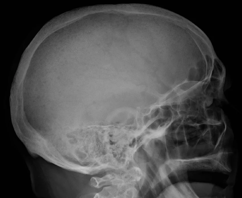
Long description
Lateral skull radiograph in multiple myeloma showing multiple punched-out lytic lesions.

Figure 2Serum protein electrophoresis normal and paraprotein
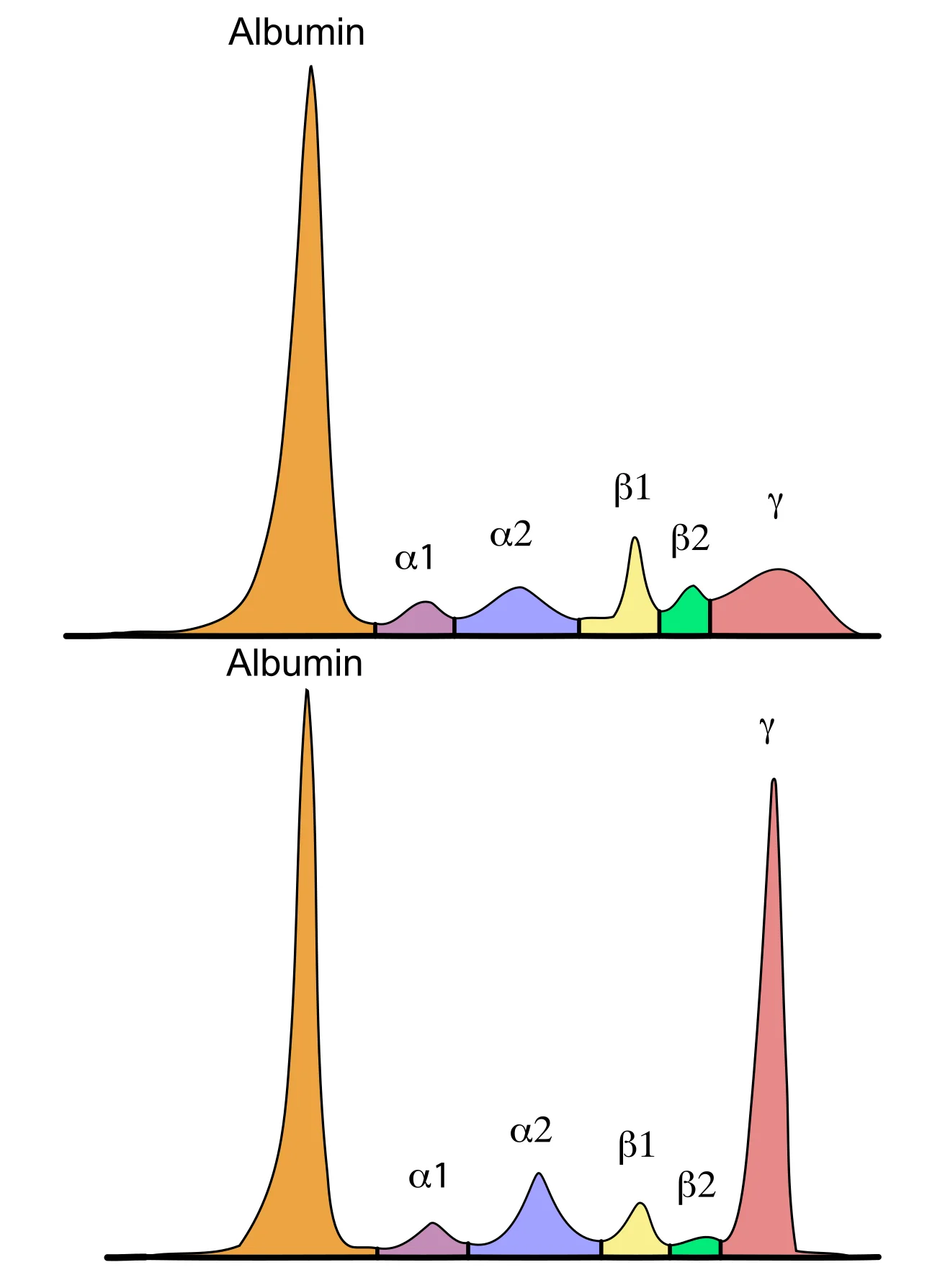
Long description
Serum protein electrophoresis: a normal pattern above and, below, a pattern with a tall narrow monoclonal (M) spike in the gamma region.

The diagnostic aim is to do three things in order: find the clone, define the damage, and stratify risk.
First-line and screening tests
- Full blood count with film: anaemia (often normocytic, low reticulocytes), and occasionally pancytopenia. The film may show rouleaux, the stacking of red cells like piles of coins caused by increased plasma proteins. Rouleaux is a clue, not a diagnosis.
- Serum creatinine and estimated glomerular filtration rate, serum calcium and albumin, and bone profile. Hypercalcaemia above 2.75 mmol/L is a myeloma-defining event.
- Serum protein electrophoresis (SPEP) and serum immunofixation electrophoresis (SIFE) to detect and type the monoclonal protein, together with the serum free light chain assay (sFLC) measuring kappa and lambda FLCs and their ratio. When all three are used together the sensitivity for a clonal plasma cell disorder is very high.[1] Immunofixation is the test that tells you which heavy chain (IgG, IgA, IgM, IgD, IgE) or light chain the clone is making.
- Urinalysis and a 24-hour urine collection for protein, with urine protein electrophoresis and urine immunofixation when myeloma is suspected, to detect Bence Jones protein (free light chains in the urine). Bence Jones protein is detected by heat precipitation or by the urine studies; it is a valuable confirmatory finding but the serum sFLC assay has largely replaced it for screening.
- Lactate dehydrogenase (LDH) and beta-2 microglobulin (β2M), which feed the ISS and R-ISS.
- Skeletal survey or, preferably, whole-body low-dose CT (WBCT) or FDG-PET/CT to look for osteolytic lesions. Whole-body MRI is most sensitive for early marrow infiltration and for spinal cord compression.
- Bone marrow aspirate and trephine biopsy, with immunophenotyping by flow cytometry (clonal plasma cells are usually CD138-positive and CD19/CD45-negative, with light chain restriction), and cytogenetics by interphase FISH on plasma cells for del(13q), del(17p), t(4;14), t(11;14), t(14;16), t(14;20), 1q gain or amplification and 1p deletion.[1]
What each result means
- An abnormal band on SPEP that types on immunofixation confirms a clonal plasma cell disorder, but not which one. The size of the monoclonal protein (in g/L) and the involved FLC level (in mg/L) measure tumour burden and guide both prognosis and response assessment.
- Marrow plasma cells below 10 percent usually means MGUS or another disorder. Ten percent or more, plus a myeloma-defining event, is active myeloma. If bone marrow has under 10 percent clonal plasma cells, then more than one bone lesion (and other criteria) is required to make the diagnosis.[1]
- A serum free light chain ratio of at least 100 with an involved FLC of at least 100 mg/L is itself a myeloma-defining event even before CRAB features appear.[1][2]
- Lytic, "punched-out" lesions on CT or PET/CT are typical. Purely osteoporotic or sclerotic changes should make you reconsider the diagnosis.
- A falling paraprotein and FLC during therapy is the best marker of response; normalization or near-normalization on serum and urine immunofixation is complete response (see response criteria in the literature).[1]
The IMWG 2014 update changed the disease definition to add validated biomarkers to the traditional CRAB features, so that patients with "smouldering" disease who are in fact destined to develop organ damage are identified and treated rather than watched.[2] A diagnosis of active multiple myeloma today requires clonal marrow plasma cells of at least 10 percent or a biopsy-proven plasmacytoma, plus at least one myeloma-defining event. Those events are the CRAB features and the SLiM biomarkers.[1][5]
The CRAB features, with their thresholds as most commonly applied, are:
- C (hypercalcaemia): serum calcium above 2.75 mmol/L (equivalent to 11.0 mg/dL).
- R (renal insufficiency): serum creatinine above 177 micromol/L (2.0 mg/dL) or a creatinine clearance below 40 mL/min.
- A (anaemia): haemoglobin below 10 g/dL, or 2 g/dL or more below the patient's own lower limit of normal.
- B (bone lesions): at least one osteolytic lesion 5 mm or larger on skeletal radiography, CT or FDG-PET/CT. An osteolytic lesion on conventional radiography counts only if it is lytic; generalised osteopenia alone does not.[1]
The SLiM biomarkers are:
- S (Sixty percent): 60 percent or more clonal plasma cells on bone marrow examination.
- Li (light chain): an involved-to-uninvolved serum free light chain ratio of 100 or more, with the involved free light chain concentration at 100 mg/L or more.
- M (MRI): more than one focal lesion on magnetic resonance imaging, each 5 mm or more.[1]
Each SLiM biomarker carries an approximately 80 percent risk of progression to symptomatic end-organ damage in the following two years, which is why they count as disease and not as "high-risk smouldering".[1] Note the distinction between a single focal lesion (which does not, on its own, meet the M criterion) and more than one, and between a lytic bone lesion and generalised demineralisation.
A related entity, monoclonal gammopathy of clinical or renal significance (MGCS/MGRS), now recognises that a small clone can damage the kidney or other organs without meeting the plasma cell threshold for myeloma; these patients are managed in the myeloma clinic but are not "multiple myeloma".
Staging and risk stratification
Staging in myeloma is risk stratification, not anatomical staging: there is no "stage I to IV" in the TNM sense, only groups that predict outcome and sometimes guide treatment intensity.
- ISS (International Staging System): uses only beta-2 microglobulin and serum albumin.
- Stage I: beta-2 microglobulin below 3.5 mg/L and albumin 3.5 g/dL or more.
- Stage II: beta-2 microglobulin below 3.5 mg/L and albumin below 3.5 g/dL, or beta-2 microglobulin 3.5 to 5.5 mg/L whatever the albumin.
- Stage III: beta-2 microglobulin above 5.5 mg/L.[3]
- R-ISS (Revised ISS): adds serum LDH (high or not) and high-risk FISH [del(17p), t(4;14), t(14;16)] to the ISS above.
- R-ISS I: ISS I features with normal LDH and no high-risk cytogenetics.
- R-ISS III: ISS III features together with high LDH or a high-risk cytogenetic abnormality.
- R-ISS II: everyone else.[3]
- Recent systems (R2-ISS and the Mayo Additive Staging System) refine this further, adding 1q gain or amplification and giving score-based stages, but the three-grade R-ISS is the most widely used in trials and in practice today.[3]
High-risk disease in practice means adverse FISH: t(4;14), t(14;16), t(14;20), del(17p), gain or amplification of 1q21, biallelic deletion of 1p32 and TP53 mutation, and combinations of these ("double-hit" or "triple-hit" when two or more are present). High-risk patients fare worse despite modern therapy, and in many centres they are given more intensive consolidation.
These are the situations that kill before treatment starts.
- Hypercalcaemia. Manage with intravenous fluids, loop diuretics only once volume is restored, and bisphosphonates (zoledronic acid 4 mg intravenously, or pamidronate); denosumab 120 mg subcutaneously is an alternative, including in renal impairment. Hypercalcaemia is a medical emergency because it worsens renal injury and because it self-amplifies through dehydration.[1][4]
- Spinal cord or cauda equina compression. Urgent MRI; high-dose dexamethasone (for example 40 mg daily is common in many protocols) and either radiotherapy or surgical decompression depending on structural stability and whether the lesion is due to retropulsed bone. Decompressive surgery comes first when the compression is from bone fragments in a structurally unstable spine.
- Acute renal failure. Rehydration, stop nephrotoxins, control calcium and uric acid, and start effective anti-myeloma therapy promptly (bortezomib-based). High-cutoff haemodialysis can clear free light chains quickly, but neither plasma exchange nor high-cutoff dialysis has consistently improved renal recovery in randomised trials and neither replaces chemotherapy.[5]
- Anaemia. Transfuse for symptoms; erythropoietin is used in some centres during therapy.
- Infection and sepsis. Neutropenic fever, or fever with hypogammaglobulinaemia, is treated with prompt broad-spectrum antibiotics after cultures. Antiviral prophylaxis with aciclovir is recommended during regimens that include proteasome inhibitors or anti-CD38 therapy, and antibacterial prophylaxis during the early months or active relapse. Intravenous immunoglobulin reduces the annual rate of all-grade infections by about 40 percent in patients with recurrent infections. Vaccination supports the whole strategy, but live vaccines are avoided during active treatment.[1]
- Pathological fracture and impending fracture. Orthopaedic stabilisation and radiotherapy as needed; systemic therapy controls the underlying disease.
Fitness, not age alone, decides this. Renal dysfunction and older age are not absolute barriers to transplant, but frailty and comorbidity must be weighed. Assessment of transplant eligibility should come early.
Induction uses three drugs: a proteasome inhibitor plus an immunomodulatory drug plus dexamethasone. The minimum should be VRd (bortezomib, lenalidomide, dexamethasone), VTd (bortezomib, thalidomide, dexamethasone), VCd (bortezomib, cyclophosphamide, dexamethasone) or KRd (carfilzomib, lenalidomide, dexamethasone); doublets are reserved for frail patients.[1] Quadruplets such as DVRd (daratumumab plus VRd), DVTd and Isa-VRd (isatuximab plus VRd) are now the enhanced-resource standard and show deeper responses, higher rates of minimal residual disease (MRD) negativity and better progression-free and overall survival, at the cost of access and toxicity. Induction typically runs for three to six cycles. Where melphalan or lenalidomide is part of the plan, stem cells are harvested after four to six cycles, before prolonged exposure, and VTD-PACE-type regimens are reserved for aggressive disease such as extramedullary plasmacytoma or plasma cell leukaemia.[1]
Induction is followed by high-dose melphalan (200 mg per square metre, reduced in renal impairment) and autologous stem cell transplant (ASCT) for eligible patients, then maintenance. Lenalidomide maintenance given for two to four years is the standard approach; two-drug maintenance (bortezomib plus lenalidomide, or daratumumab plus lenalidomide) is used in high-risk disease when resources allow; thalidomide maintenance can be considered in selected standard-risk patients who received thalidomide-based induction but is inferior in high-risk disease.[1] Tandem transplant may be considered for high-risk or ultra-high-risk myeloma. Consolidation with CAR-T cell therapy after first-line treatment is not recommended outside trials.
First-line therapy is individualised on frailty, myeloma risk, comorbidity and affordability. For fit patients VRd or DRd (daratumumab, lenalidomide, dexamethasone) is recommended, with quadruplets (DVRd, Isa-VRd) in fitter, under-80-year-olds, and DRd continuing until progression. For frail or very elderly patients, Rd or Vd, or dose-modified VTd, VCd, VMP (bortezomib, melphalan, prednisone) or MPT (melphalan, prednisone, thalidomide) may be safer. Induction is given for 8 to 12 cycles or until response plateaus.[1]
The MAIA trial established DRd as a standard for transplant-ineligible patients, with durable progression-free and overall survival benefit. Maintenance is lenalidomide after VRd, DRd or DVRd, with thalidomide maintenance only after thalidomide-based induction, and two-drug maintenance for high-risk disease; duration is individualised.[1]
Frailty assessment matters clinically. Tools such as the IMWG Frailty Score, which combines age, functional status and comorbidity, predict tolerance and survival better than age alone. Dose reductions, especially of dexamethasone, and careful renal monitoring are the practical interventions, not saying "no" on the basis of age.
Renal impairment at diagnosis (serum creatinine above 177 micromol/L or creatinine clearance below 40 mL/min) changes both prognosis and regimen choice. Bortezomib and daratumumab can be used without dose adjustment, even in dialysis-dependent patients, whereas lenalidomide, pomalidomide and ixazomib must be dose-adjusted to creatinine clearance.[1][5] General principles: rapid effective anti-myeloma therapy, hydration, avoidance of nephrotoxins (non-steroidal anti-inflammatory drugs, aminoglycosides, radiocontrast, renin-angiotensin blockers and loop diuretics where possible), treatment of hypercalcaemia, and monitoring of electrolytes and blood counts.[5]
Almost all patients eventually relapse, and relapsed myeloma is managed sequentially, adjusting for what has been used, how deep the response was and how long it lasted, fitness and the available drugs.
- Treatment at first relapse is usually a triplet containing at least two novel agents to which the patient is not refractory. If relapse occurs six months or more after stopping a drug class, that class can be reused. Autologous transplant is considered for transplant-naive patients or for those whose first transplant was followed by a sustained remission of at least three years.[1]
- Drug-class choices: combinations of daratumumab or isatuximab with bortezomib or carfilzomib or pomalidomide are the main options for lenalidomide- or bortezomib-refractory disease; elotuzumab-based combinations are another alternative.[1]
- After two to three prior lines, especially in triple-class refractory disease, the outlook without newer options is poor, with progression-free survival of only three to five months. Cell therapies and bispecific antibodies have transformed this setting: cilta-cel (a BCMA-directed CAR-T product) reduced progression or death compared with standard care in lenalidomide-refractory patients after one to three prior lines in the CARTITUDE-4 trial, and teclistamab (a bispecific antibody linking CD3 on T cells to BCMA on myeloma cells) combined with daratumumab outperformed conventional triplet therapy in the MajesTEC-3 trial. Elranatamab, talquetamab (a GPRC5D-targeting bispecific) and belantamab mafodotin are further options, subject to availability.[1]
- Late-line practice therefore favours participating in a clinical trial where possible, using bendamustine, cyclophosphamide or melphalan-based regimens where newer agents are not available, and treating transformation to plasma cell leukaemia, extramedullary disease or t(11;14)-driven disease specifically (for example with venetoclax-based combinations in t(11;14) myeloma).
Figure 3Left femur with myeloma
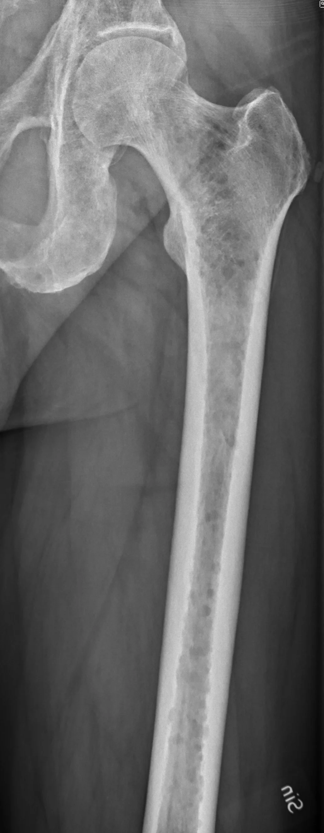
Long description
Radiograph of the left femur in multiple myeloma with scattered lytic lesions in the bone.

Bone-targeting agents are given to everyone receiving initial therapy, because skeletal-related events occur in up to 50 percent of patients within two years without prophylaxis, and up to 80 percent already have bone disease at diagnosis.[1][4] Bisphosphonates (zoledronic acid 4 mg intravenously, given every 4 or every 12 weeks, or pamidronate every 4 weeks) and denosumab (120 mg subcutaneously monthly) reduce skeletal-related events and bone pain. In the randomised trials denosumab is non-inferior to zoledronic acid for delaying the first event and causes less renal toxicity (fewer renal adverse events than zoledronic acid), but more hypocalcaemia, so calcium and vitamin D repletion are needed; zoledronic acid requires dose adjustment for creatinine clearance.[4] Breast and prostate cancer were studied with 20.6 to 26.4 months to first event in that class, whereas myeloma specifically showed 22.83 months with denosumab versus 23.98 with zoledronic acid, with progression-free survival favouring denosumab.[4] Duration of therapy is up to two years, after which a decision to continue or extend is made on individual clinical grounds.[1]
Radiotherapy is used for pain not controlled by systemic therapy, impending fractures or spinal cord compression; surgery is preferred for structural instability or a retropulsed bone fragment. Orthopaedic involvement should be sought early for weight-bearing instability. Dental assessment before starting bisphosphonates, and avoidance of extractions during treatment, reduces the risk of osteonecrosis of the jaw, which occurs in a minority of patients and rises with prolonged exposure.[4]
Traditionally SMM is observed, every three to six months, with the intensity guided by the 2/20/20 risk score: M-protein above 2 g/dL, marrow plasma cells above 20 percent and serum free light chain ratio above 20. With 0, 1 or at least 2 of these, the two-year risk of progression to active myeloma is about 6, 18 and 44 percent respectively.[5] Recent randomised trials (AQUILA with daratumumab, and studies of lenalidomide) support early intervention in carefully selected high-risk SMM, delaying progression to active disease, although whether overall survival improves is not yet established and overtreatment is a real risk. The Malaysian consensus supports lenalidomide or daratumumab monotherapy for high-risk SMM, added to close monitoring.[1]
Aside from the CRAB features themselves: skeletal-related events (fracture, spinal cord compression, pain requiring radiotherapy, surgery to bone), renal failure that can require dialysis in about 10 percent of patients, hyperviscosity syndrome, infection from hypogammaglobulinaemia, light chain deposition disease, amyloid light-chain amyloidosis, hypercalcaemia-associated nephropathy and, late in the disease, plasma cell leukaemia and extramedullary plasmacytoma. Peripheral neuropathy may be disease-related (amyloid, hyperviscosity) or treatment-related (bortezomib, thalidomide).
The disease is not curable in most patients, but is now controlled for years rather than months. Median overall survival in unselected patients is about 6 to 7 years and is longer in those who receive modern quadruplet therapy and transplant. The strongest prognostic factors are the R-ISS features, high-risk cytogenetics (especially del(17p), t(4;14), t(14;16) and 1q gain), high bone marrow plasma cell burden, elevated LDH, high beta-2 microglobulin and renal failure. Minimal residual disease negativity, now measurable by next-generation flow or next-generation sequencing to one cell in 100,000, is a powerful predictor of outcome and is increasingly used to refine duration of therapy.[1] Hypercalcaemia itself worsens prognosis: in one series survival was 26 against 48 months for patients with and without hypercalcaemia of malignancy.[4]
There is no population screening for myeloma. The places to intervene are the precursors: identifying MGUS (often accidentally, but increasingly as case-finding in unexplained symptoms), monitoring MGUS risk-stratified by paraprotein size and isotype and by free light chain ratio, and monitoring high-risk SMM. Early diagnosis still comes mainly from a low index of suspicion: the doctor who checks a paraprotein in an older person with unexplained back pain, anaemia, raised creatinine or recurrent infection.
- Older and frail patients: fitness, not age, decides treatment intensity. Formal geriatric and frailty assessment, comorbidity review and shared decision-making. The number of patients who receive only supportive care rises steeply above 80 years.
- Pregnancy: myeloma in pregnancy is rare. Management needs joint haematology, obstetric and neonatology planning; alkylating agents and proteasome inhibitors are used in selected second- and third-trimester cases. Breastfeeding during therapy is generally not advised.
- Children and young adults: myeloma is essentially a disease of adults; a case in a young person needs a critical review of the diagnosis and genetic counselling.
- HIV and other immunosuppression: renal mechanisms and infection risk are accentuated; coordinate with the infectious diseases team.
- Renal impairment: treatment should aim to recover renal function, because patients who become independent of dialysis do better; anti-myeloma therapy starts promptly, and nephrology input is early.
The Malaysian consensus explicitly builds its recommendations around a tiered picture, and the same reasoning applies globally in settings where sFLC assays, FISH, MRI, transplant and novel drugs are scarce.
- Diagnose with what is available. Full blood count, blood film, serum protein electrophoresis, immunofixation and a 24-hour urine for Bence Jones protein, plus a plain skeletal series, already diagnose and stage most patients. Serum free light chain testing and FISH are valuable but not always essential; a clinical diagnosis and risk assessment can be made without them.[1][5]
- Image pragmatically. Low-dose whole-body CT or PET/CT is more sensitive than conventional skeletal survey, and MRI of the spine when cord compression is suspected; where only a skeletal survey is available, remember that lesions may not show until 30 to 50 percent of bone is destroyed, so repeat imaging during treatment.[4][1]
- Use affordable induction. Where bortezomib, lenalidomide and anti-CD38 antibodies are unavailable or unaffordable, thalidomide, cyclophosphamide, melphalan and prednisone-based regimens (for example MPT, VMP or VCd, or VTd) remain effective. Oral thalidomide plus a steroid is the backbone in many such settings. Melphalan-containing induction is avoided only until stem cells are harvested, when transplant is available.[1]
- Transplant where capacity exists. A single transplant unit in a region, with melphalan 200 mg per square metre and stem cell harvest after four to six cycles, converts an incurable disease into durable control; resource sharing and referral networks bridge geography. Where transplant is not available, maximise first remission with prolonged induction and maintenance.
- Protect kidneys and bone with cheap measures. Hydration (aiming for a urine output of 100 to 150 mL per hour in acute situations), stopping nephrotoxins, treating hypercalcaemia with fluids, bisphosphonates and loop diuretics once volume is restored, calcium and phosphate replacement when denosumab is used, and dental screening before bisphosphonates. These inexpensive steps, coordinated by pharmacists in the Malaysian model, have a disproportionate effect on outcome.[5]
- Prevent infection with simple measures: co-trimoxazole, antiviral prophylaxis during proteasome inhibitor or anti-CD38 therapy, pneumococcal and influenza vaccination, and prompt intravenous antibiotics at first fever. Intravenous immunoglobulin is less accessible but oral antibiotics plus early assessment save lives.
- Pain and structure: radiotherapy for localised pain, orthopaedic stabilisation for fractures, spinal braces for collapse, and early palliative care.
- Access: the central inequality in myeloma is not the science, which is broadly the same worldwide, but the availability of novel drugs, diagnostics and stem cell transplant. Acknowledging this in every treatment plan, and enrolling patients in trials and registry studies where possible, is part of honest care.
- Multiple myeloma is a clonal plasma cell malignancy of bone marrow, producing a monoclonal protein and/or free light chains; it is incurable in most patients but controllable for years.
- The full spectrum is MGUS (under 10 percent plasma cells, no damage) to smouldering myeloma (10 to 60 percent) to active myeloma, defined by the CRAB or SLiM criteria.
- CRAB: hypercalcaemia above 2.75 mmol/L, renal creatinine above 177 micromol/L or creatinine clearance below 40 mL/min, anaemia below 10 g/dL or 2 g/dL below normal, and at least one osteolytic lesion. Hypercalcaemia occurs in about 30 percent of patients; renal impairment at diagnosis in 20 to 40 percent; bone disease in over 80 percent.
- SLiM biomarkers (about 80 percent progression risk): 60 percent or more marrow plasma cells, serum free light chain ratio 100 or more with involved FLC 100 mg/L or more, or more than one focal lesion of 5 mm or more on MRI.
- The monoclonal protein is found in serum or urine in about 97 percent of cases; SPEP plus immunofixation plus serum free light chain testing together detect almost all cases; Bence Jones protein appears in urine.
- Presentation is bone pain and pathological fracture, fatigue and breathlessness, recurrent infection, renal failure, hypercalcaemia, hyperviscosity and cord compression.
- Pathophysiology: clone crowds out normal marrow; free light chains obstruct distal tubules as casts with Tamm-Horsfall protein; RANKL up-regulation with osteoprotegerin loss and Dickkopf-1-driven osteoblast suppression give purely lytic bone disease.
- Diagnosis needs clonal marrow plasma cells of at least 10 percent plus a myeloma-defining event, with immunophenotyping and FISH for prognosis; imaging requires CT, PET/CT or MRI rather than (or as well as) plain films.
- ISS uses beta-2 microglobulin and albumin (stage I: below 3.5 mg/L and albumin 3.5 g/dL or more; stage III: above 5.5 mg/L); R-ISS adds LDH and del(17p), t(4;14), t(14;16).
- First-line for fit patients is a proteasome inhibitor plus an immunomodulator plus steroid (VRd, VTd, VCd, KRd), then autologous transplant and lenalidomide maintenance for two to four years; quadruplets with anti-CD38 antibody are now the enhanced standard.
- For transplant-ineligible patients, VRd or DRd, with maintenance; frail patients need dose modification, not exclusion from treatment.
- Bortezomib and daratumumab need no renal dose adjustment; lenalidomide, pomalidomide and ixazomib do.
- Bone disease: bisphosphonates up to two years (zoledronic acid 4 mg every 4 to 12 weeks, pamidronate monthly) or denosumab 120 mg monthly, with vitamin D and calcium; consider every-12-week zoledronic acid once stable.
- Relapse: sequential triplets, second transplant if suitable, and CAR-T/bispecifics for triple-class refractory disease; cilta-cel and teclistamab have the strongest recent data.
- Every patient needs infection prevention (antiviral prophylaxis, vaccination, immunoglobulin for recurrent infections) and early involvement of pain, orthopaedic and palliative care.
- Low-resource care can achieve the same diagnosis with cheaper tests, use oral/thalidomide or alkylator regimens where novel drugs are unavailable, share transplant capacity and concentrate on hydration, nephrotoxin avoidance, calcium and infection control.
Answer each one in your head first, then open the answer.
A 62-year-old man has eight weeks of thoracic back pain, fatigue, haemoglobin 86 g/L and creatinine 210 micromol/L. SPEP shows an IgG kappa paraprotein at 42 g/L and the serum free light chain kappa:lambda ratio is 180 with kappa 320 mg/L. Bone marrow shows 35 percent plasma cells. Whole-body CT shows several lytic vertebral and rib lesions. What is the diagnosis, and which IMWG criteria are met?
Show answerHide answer
Multiple myeloma. The marrow shows at least 10 percent clonal plasma cells and there are at least two CRAB features (anaemia with haemoglobin under 10 g/dL, renal creatinine over 177 micromol/L, and over one bone lesion). The light chain criterion is also met: the serum free light chain ratio is at least 100 with the involved free light chain at least 100 mg/L. The disease is therefore active myeloma even before considering the hypercalcaemia.
Why is the free light chain ratio above 100, with involved FLC above 100 mg/L, treated as 'disease' rather than 'high-risk smouldering myeloma'?
Show answerHide answer
Because data showed that such a ratio is associated with near-inevitable progression to CRAB features within about two years. Watchful waiting in those patients was therefore considered harmful, and the 2014 IMWG consensus reclassified them as active myeloma requiring treatment.
A patient with known myeloma presents with new paraparesis and a sensory level at T6, with constipation. Blood pressure is normal. What is the immediate management and why is the first drug given before imaging?
Show answerHide answer
Treat as spinal cord compression until proven otherwise: high-dose dexamethasone immediately (to reduce oedema), urgent MRI of the whole spine, then radiotherapy or surgical decompression depending on the structural findings. Dexamethasone is given first because it rapidly reduces cord oedema and pain while imaging is being arranged; delaying it for imaging risks permanent neurological deficit.
Which maintenance strategy follows VRd induction plus autologous stem cell transplant in a standard-risk patient, and for how long?
Show answerHide answer
Lenalidomide maintenance for two to four years. Thalidomide is an alternative in selected standard-risk patients who received thalidomide-based induction but is inferior in high-risk disease. Two-drug maintenance (bortezomib plus lenalidomide, or daratumumab plus lenalidomide) is reserved for high-risk or ultra-high-risk myeloma.
In a unit with no PET-CT, no FISH panel and no anti-CD38 antibody: which parts of the diagnostic and treatment pathway still have to be done, and which can safely be simplified?
Show answerHide answer
Diagnosis: serum and urine protein studies, marrow with morphology and immunophenotype, and at least a skeletal survey (repeated if needed); FISH and PET-CT improve accuracy but do not change the diagnosis. Staging can be done with the ISS alone using beta-2 microglobulin and albumin. Treatment: an affordable triplet such as VCd or MPT or VMP, with hydration, nephrotoxin avoidance, calcium control and bisphosphonate prophylaxis; thalidomide-based maintenance; and early referral for transplant if a unit is reachable. The keys that must not be dropped are measuring the paraprotein and FLC, hydrating, controlling calcium and infection prevention.
Why does bortezomib (rather than lenalidomide) become the default induction agent when a patient presents with acute renal failure?
Show answerHide answer
Bortezomib is cleared hepatically and can be given at full dose without adjustment in renal impairment, including dialysis, and it rapidly reduces the free light chain burden that is causing the renal injury. Lenalidomide is renally cleared and needs dose adjustment. Many renal impairment protocols therefore build induction around bortezomib, high-dose dexamethasone and an agent such as cyclophosphamide.
- Anaemia approach and iron deficiency
- Acute kidney injury
- Hypercalcaemia
- Osteoporosis
- Bisphosphonates
- Pain control and palliative care
- Transfusion reactions and safe transfusion
Evidence: sources/multiple-myeloma/
- Gan G, Wong L, Abdul Rahman M, et al. Malaysian Expert Consensus Guidelines for the Diagnosis and Management of Multiple Myeloma: A Modified Delphi Approach. Malays J Med Sci. 2026;33(4):7-36. doi:10.21315/mjms-03-2026-153. Accessed .
- Rajkumar SV, Dimopoulos MA, Palumbo A, et al. International Myeloma Working Group updated criteria for the diagnosis of multiple myeloma. Lancet Oncol. 2014;15(12):e538-e548. doi:10.1016/S1470-2045(14)70442-5. Accessed .
- Baysal M, Julian K, Sborov D, et al. The past, present, and future of myeloma staging and risk prognostication. Oncologist. 2025;30(10):oyaf299. doi:10.1093/oncolo/oyaf299. Accessed .
- Bernstein ZS, Kim EB, Raje N. Bone disease in multiple myeloma: biologic and clinical implications. Cells. 2022;11(15):2308. doi:10.3390/cells11152308. Accessed .
- Hussain A, Moniot JL, Sumaya M, Hughes DM. Multiple myeloma and renal health: navigating selected clinical complexities. Cureus. 2026;18(9):e115585. doi:10.7759/cureus.115585. Accessed .
Figure credits
Educational content for medical students and doctors. It is not a substitute for clinical judgement or local guidelines.