Hodgkin lymphoma
Classical Hodgkin lymphoma: pathology, presentation, staging, PET-adapted treatment, prognosis, late effects, and low-resource care.
- Hodgkin lymphoma is a B-cell neoplasm named for its HRS cells: giant, multinucleated cells that express CD30 in all cases and CD15 in most. [2]
- Classical disease (classic Hodgkin lymphoma, cHL) makes up about 90 to 95 percent of cases and is split into four histological subtypes; a smaller "nodular lymphocyte-predominant" entity (NLPHL) is biologically different. [2]
- It has a bimodal age curve: one peak in young adults aged 15 to 35 years, and a second after 60 years. [1]
- It usually declares itself as painless cervical or supraclavicular lymphadenopathy; mediastinal involvement occurs in about 60 percent of patients and B symptoms in about 40 percent.
- PET-CT is the staging investigation of choice, and treatment is risk-adapted and PET-guided, using chemotherapy (ABVD or newer regimens), radiotherapy, anti-CD30 antibody-drug conjugates, and checkpoint inhibitors.
- Because it is highly curable, long-term survivorship care matters: survivors have a two- to four-fold increased risk of second cancers and cardiovascular disease.
By the end you should be able to:
- Recognize the pathology of Hodgkin lymphoma, name its subtypes, and connect histology to EBV status and prognosis.
- Explain the bimodal age distribution, the EBV association, and the presentation in ordinary clinical terms.
- Use the Ann Arbor and Lugano staging systems and explain why PET-CT has changed staging and treatment decisions.
- Choose first-line management for early favourable, early unfavourable, and advanced disease, including when to add radiotherapy or antibody-drug conjugates.
- Recognise the principles of interim PET and how it guides escalation or de-escalation.
- List the important late effects and the simple monitoring that follows treatment.
- Adapt staging and treatment where PET-CT, pathology, and modern drugs are not available.
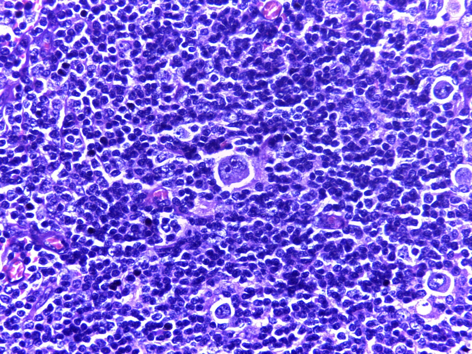
Hodgkin lymphoma (HL) is a neoplasm of B cells, but its defining feature is that the tumour mass is almost entirely reactive immune cells. Only a tiny fraction of the cells actually belong to the neoplasm: the HRS cells. These cells are large, often binucleated "owl's eye" cells with prominent eosinophilic nucleoli. They have lost much of the B-cell programme (they do not express the B-cell receptor) yet they keep the B-cell transcription factor PAX5 weakly, and they strongly express CD30 in every case and CD15 in most. [1][2]
This unusual structure has clinical consequences. The patient's symptoms come partly from the tumour and partly from the inflammatory reaction around it: fever, night sweats and weight loss are driven by cytokines (the B symptoms), while pruritus can be an early but easily missed clue. Because the malignant cells are so sparse, diagnosis is a hunting exercise in the biopsy rather than a matter of looking for "a lot of cancer cells".
HL accounts for about 0.4 percent of all cancers and has an annual incidence of roughly 2.5 new cases per 100,000 people. [1] It is one of the most curable cancers: about 89 percent of patients now survive five years. [1] Most HL-related deaths still come from the minority of patients who do not respond, and increasingly from treatment late effects in survivors.
Historically "Hodgkin disease" was one diagnosis. Today we separate classical Hodgkin lymphoma (cHL), which makes up roughly 90 to 95 percent of cases, from nodular lymphocyte-predominant Hodgkin lymphoma (NLPHL), roughly 5 percent. [2][3]
cHL has four histological subtypes, defined by how HRS cells look, how much fibrosis is present, and how dense the background of immune cells is. [2][3]
| Subtype | Histology | EBV association | Typical patient and prognosis |
|---|---|---|---|
| Nodular sclerosis (NScHL) | Fibrous bands divide lymphoid tissue into nodules; lacunar-type HRS cells; eosinophils, plasma cells, neutrophils common in the background | Less commonly EBV-positive | Most common subtype, especially in adolescents and young adults; often mediastinal involvement; excellent response to standard therapy [2] |
| Mixed cellularity (MCcHL) | Diffuse infiltrate rich in classic HRS cells, eosinophils, histiocytes, plasma cells and small lymphocytes; no fibrous bands | Often EBV-positive | Common in children, older adults, immunocompromised patients [2] |
| Lymphocyte-rich (LRcHL) | Scattered classic HRS cells in a background of small B lymphocytes; fewer tumour cells, so it can be hard to recognise | Variable | Indolent-like presentation; may resemble NLPHL morphologically |
| Lymphocyte-depleted (LDcHL) | Numerous pleomorphic HRS cells with very little reactive background | Often EBV-positive | Rarest; more common in immunocompromised patients; worst prognosis among the four subtypes [2] |
NLPHL is a separate clinicopathological entity. It has no HRS cells; instead it has "popcorn" lymphocytic and histiocytic (L&H) cells that retain a functional B-cell programme (CD20-positive, CD30-negative). It tends to present as localised peripheral lymphadenopathy, runs an indolent course, and has a tendency to late relapse. [2][3] The WHO classification still lists it under the Hodgkin family, but the International Consensus Classification (ICC) renames it "nodular lymphocyte predominant B-cell lymphoma" because it behaves like an indolent B-cell lymphoma. [1][2] This renaming is not just academic: treatment of NLPHL is more conservative and does not automatically follow the ABVD-like regimens used in cHL. [2]
The HRS cell profile is the immunophenotype that pins the diagnosis: CD30 is uniformly expressed, CD15 is present in about 75 to 80 percent, and PAX5 is weakly positive in about 95 percent. [1] CD20 is positive in up to 20 percent of cases, and OCT2 and BOB1 are often weak or absent, which helps separate cHL from anaplastic large cell lymphoma and from NLPHL. [2] CD30 is also the drug target: the antibody-drug conjugate brentuximab vedotin binds CD30. [2]
cHL is rare: about 15 percent of newly diagnosed lymphomas. [3] It is the commonest malignancy in the 15-to-35 age band in many populations, and its median age at diagnosis is 39 years. [1] There is a second incidence peak after 60 years. [1] This bimodal curve is one of the most important epidemiological facts about the disease, and it splits the condition into two problems: young people who need curative-intensity treatment with long survivorship, and older people in whom comorbidity is the limiting factor.
Men are affected more often than women. [1] The incidence is 5- to 25-fold higher in people living with HIV and in other immunodeficient states, and in these groups the disease is almost always EBV-positive. [1] HL is not an AIDS-defining malignancy and is generally treated as in immunocompetent patients. [1]
EBV is causally associated with 25 to 40 percent of cHL cases. [1][2] It is more common in older patients, in children, in immunocompromised patients (including those with HIV, and after solid organ transplant), and in much of the low-resource world. [2] EBV-positive disease usually shows the viral genome in HRS cells, and the mixed cellularity and lymphocyte-depleted subtypes are the ones most often EBV-associated. [2] EBV status is biologically important (it is linked to microenvironment and possibly to outcome) but is not yet used to select therapy. [2]
The host side of the equation matters too. Siblings have a higher risk than parents, consistent with a strong influence of HLA and non-HLA genetic variants. [1] Autoimmune diseases (notably autoimmune haemolytic anaemia, systemic lupus erythematosus, rheumatoid arthritis, sarcoidosis and several others) carry a raised standardised incidence ratio for HL. [1] HL can also arise as a transformation from an underlying low-grade B-cell lymphoma, usually chronic lymphocytic leukaemia ("Hodgkin-like Richter transformation"). [1]
cHL starts as neoplastic transformation of a germinal centre B cell whose survival programme has gone wrong. The HRS cell is the descendant that should have died by apoptosis but survives because of constitutively active NF-kappaB signalling, and in EBV-positive cases, viral proteins. [1]
Three mechanisms explain what the patient actually experiences:
- Local mass plus inflammation. The tumour usually begins in a single nodal group and spreads in a contiguous, stepwise fashion to adjacent nodal regions. This "centrifugal" spread is why a cervical node and a mediastinal node are commonly involved together, and it is why unilateral disease is the rule early on.
- Cytokine excess. HRS cells live in a rich microenvironment of reactive T cells, eosinophils, macrophages and fibroblasts that they manipulate. The inflammatory reaction produces fever, night sweats, weight loss, anaemia of chronic disease and, sometimes, prominent pruritus. B symptoms occur in about 40 percent of patients: roughly 20 percent in localised disease and 70 percent in advanced disease. [1] Pruritus without obvious skin disease can be the earliest clue.
- Alcohol-induced pain. A small but classic feature of Hodgkin disease is nodal pain that starts within minutes of drinking alcohol, almost certainly a vascular reflex within the involved node. It is not pathognomonic, but when it is reported it should be taken seriously.
"Laccunar" cells (the variant seen in nodular sclerosis) sit in clear spaces produced by retraction artefact; these are not real vacuoles, just a preparation effect. [2]
Figure 1Hodgkin's lymphoma with RS cell 10x
Long description
Histology of Hodgkin lymphoma at low power: Reed-Sternberg cells with large nuclei and prominent nucleoli among small lymphocytes.

The biopsy is almost always an excision lymph node biopsy; fine-needle aspiration is not adequate. The pathologist must see the whole architecture and have material for immunohistochemistry. [3]
Grossly, the node is replaced by firm, pale tumour, often with fibrotic bands in the nodular sclerosis subtype. Microscopically, the four cHL subtypes described in the table above are distinguished by the number of HRS cells, the background cell population, and the presence or absence of fibrous bands. [2] HRS cells are 15 to 45 micrometres across, with bilobed or multinucleated nuclei and "inclusion-like" nucleoli that give the "owl's eye" appearance. [2] Variants include mononuclear Hodgkin cells, "mummified" apoptotic-appearing cells, and the lacunar cells seen in nodular sclerosis. [2]
EBV can be demonstrated in HRS cells by in-situ hybridisation for EBV-encoded RNA. When the disease is EBV-positive, the latent membrane protein 1 (LMP1) oncogene product contributes to the NF-kappaB-driven survival of HRS cells. [2]
Most patients present with painless superficial lymphadenopathy, most commonly in the cervical and supraclavicular regions. [1] Mediastinal involvement occurs in about 60 percent of patients, and 20 to 25 percent have mediastinal bulk disease, which can present with cough, breathlessness or, rarely, superior vena cava syndrome. [1]
The lymph nodes are characteristically rubbery and sometimes tender early on. They do not fluctuate in size with infection the way reactive nodes do, but they may wax and wane.
B symptoms deserve specific attention, because they change stage and prognosis:
- Fever
- Drenching night sweats
- Unintentional weight loss of at least 10 percent of body weight in the six months before diagnosis. [3]
Other symptoms that are common but not B symptoms:
- Fatigue
- Generalised pruritus without a rash ("pruritus sine materia")
- Alcohol-induced nodal pain
- Symptoms of bulky mediastinal disease (cough, dyspnoea, stridor)
A thorough survey is needed because the disease is often more widespread than the patient thinks:
- All nodal groups: cervical, supraclavicular, axillary, epitrochlear, inguinal, femoral.
- The "Waldeyer ring" (tonsils, base of the tongue, nasopharynx) and the spleen; both count as nodal disease rather than extranodal in the staging system. [3]
- Chest and abdomen for hepatosplenomegaly.
- A mediastinal mass on chest X-ray if there are respiratory symptoms.
- General condition: cachexia suggests advanced disease.
Systemic features on examination include fever, pallor from anaemia and, occasionally, severe excoriation from pruritus.
Advanced disease involves extranodal sites. Roughly 40 percent of patients present at an advanced stage, with lung (21 percent), bone (15 percent), liver (10 percent) and bone marrow (9 percent) the common extranodal sites. [1] Bone marrow involvement is now detected well by PET-CT, which has largely removed the need for bone marrow biopsy in most cases. [1]
The differential falls into three broad groups: infections, inflammatory or autoimmune illness, and other lymphoid malignancies.
| Category | Examples | How it differs from Hodgkin lymphoma |
|---|---|---|
| Infection | Infectious mononucleosis (EBV), HIV, tuberculosis, Bartonella | Atypical or reactive cells, positive serology or microbiology, no HRS cells with the cHL immunophenotype |
| Inflammatory or autoimmune | Systemic lupus erythematosus, rheumatoid arthritis, sarcoidosis, haemophagocytic lymphohistiocytosis | Lymphadenopathy usually smaller, waxing and waning; serological and clinical clues; biopsy shows reactive hyperplasia |
| Other lymphoid malignancy | Non-Hodgkin lymphoma, anaplastic large cell lymphoma, NLPHL, diffuse large B-cell lymphoma | Different immunophenotype (CD30/CD15 often different; OCT2/BOB1 patterns differ) and architecture |
| Other solid cancer | Metastatic carcinoma, germ-cell tumour | Usually solid epithelial morphology, age- and risk-appropriate context |
A key diagnostic hazard is the reactive lymph node that contains immunoblasts. An excision biopsy with full immunophenotyping (CD30, CD15, CD20, PAX5, OCT2/BOB1, EBV in-situ hybridisation) is the only reliable way to tell.
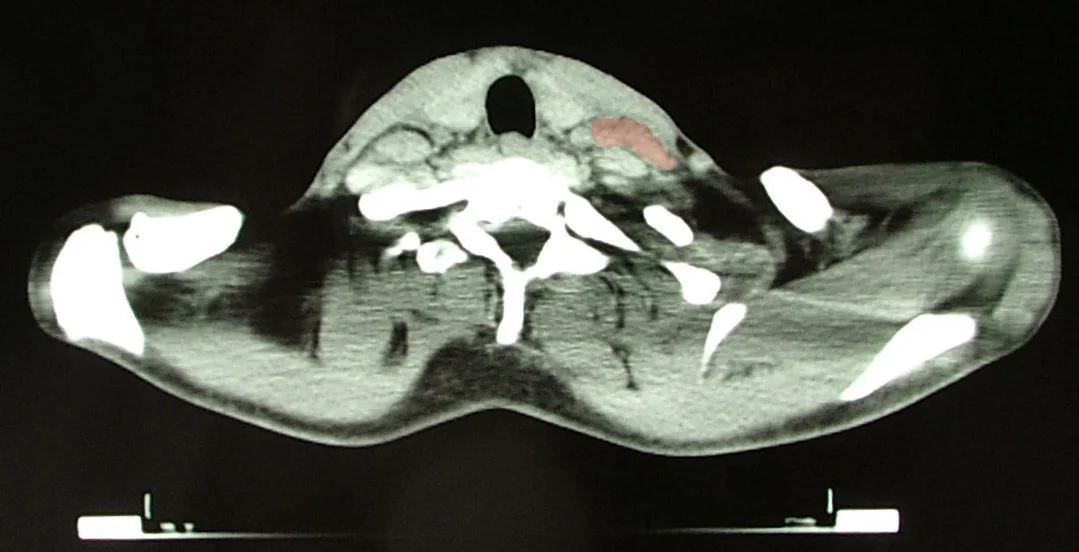
Figure 2Hodgkin Lymphoma CT2
Long description
Axial CT through the neck and upper chest in Hodgkin lymphoma, with the enlarged lymph node mass tinted.

- Excision lymph node biopsy for morphology and immunohistochemistry (diagnostic standard). [3]
- Full blood count, erythrocyte sedimentation rate, C-reactive protein, comprehensive metabolic panel, lactate dehydrogenase.
- HIV testing, tuberculosis screening where appropriate, EBV serology. [3]
- Chest radiograph, contrast-enhanced CT of neck/chest/abdomen/pelvis, and PET-CT.
- Echocardiogram in older patients or where anthracycline therapy is planned.
- Pulmonary function tests, including diffusion capacity, before bleomycin-containing regimens.
- Bone marrow biopsy is not required routinely, because PET-CT is more sensitive for marrow disease. [1]
- Fine-needle aspiration is never adequate for primary diagnosis.
- PET-CT stages the disease anatomically and metabolically. It stages correctly or upstages in up to 41 percent of patients, and downstages in up to 10 percent, compared with CT. [1]
- ESR and other inflammatory markers trend down with response and can reflect disease activity, but they are not specific.
The diagnosis rests on histology: a lymph node showing HRS cells (or variants) in the correct architectural setting, with the cHL immunophenotype (CD30 uniformly positive, CD15 in most, PAX5 weak, absent or weak OCT2/BOB1). [1][2] Clinical disease is then assigned to a stage.
Two systems are in common use. The Cotswolds-modified Ann Arbor classification remains the most widely used; the Lugano system modernises it by making PET-CT the mandatory first imaging study and removing the "X" suffix for bulky disease, while retaining "A" and "B". [3]
| Stage | Meaning |
|---|---|
| I | Disease limited to a single lymph node region or one extralymphatic organ (for example spleen, thymus or Waldeyer ring, which all count as nodal) |
| II | Two or more nodal groups on the same side of the diaphragm |
| III | Nodal regions or extralymphatic organs on both sides of the diaphragm |
| III1 | With or without splenic, hilar, coeliac or portal nodes |
| III2 | With para-aortic, iliac or mesenteric nodes |
| IV | Diffuse or disseminated disease of one or more extralymphatic organs, with or without nodal involvement |
| A | Absence of B symptoms |
| B | Presence of fever, night sweats and/or at least 10 percent weight loss in six months |
| E | Extranodal extension by direct local spread |
| X | Bulky disease: nodal mass greater than one third of the intrathoracic diameter on chest X-ray (a threshold still used in many guidelines; paediatric protocols use lower cut-offs) |
Bulky disease is usually defined as a mediastinal mass greater than one third of the intrathoracic diameter on a postero-anterior chest X-ray, or any mass 10 cm or more on CT at diagnosis. [3] Paediatric groups use 6 cm in the presence or absence of B symptoms. [3] These thresholds feed into risk group allocation, which in turn determines chemotherapy dose and whether radiotherapy is used.
The Lugano revision (2011, with continuing updates) builds on the same anatomy but makes three changes that matter at the bedside: [3]
- PET-CT is the staging modality of choice, which has improved accuracy and removed the need for routine bone marrow biopsy in most cases. [1][3]
- The "X" (bulky) suffix is dropped; bulk is handled in risk assessment instead. [3]
- Interim PET (after two cycles) is used as a treatment decision point: escalation for a positive scan, de-escalation for a negative one. [1][3]
Lugano stage 4 with bone marrow involvement still matters for risk, but it is now PET-defined rather than biopsy-defined in most patients. [5]
Response to treatment is measured with the Deauville five-point scale on PET-CT: scores 1 to 3 count as a complete metabolic response, 4 or 5 as residual metabolic activity. [3] Score 4 or 5 after treatment prompts further evaluation (sometimes biopsy) rather than assuming failure, because inflammation can be metabolically active.
Early-stage disease is split into favourable and unfavourable by the EORTC (European Organisation for Research and Treatment of Cancer) and GHSG (German Hodgkin Study Group) criteria. Advanced-stage risk is estimated with the International Prognostic Score, and newer continuous models (E-HIPI and A-HIPI) are beginning to replace the older discrete scores in trials. [1] Interim PET after two cycles is a strong, independent predictor of outcome. [1]
Approach to suspected Hodgkin lymphoma
- Painless lymphadenopathy, especially cervical/supraclavicular
- History and examinationB symptoms, alcohol-induced nodal pain, pruritus, mediastinal symptoms, HIV/TB exposure
- Excision lymph node biopsyMorphology plus immunophenotype (CD30, CD15, PAX5, CD20, OCT2/BOB1, EBV-ISH)
- Question: Is it classical Hodgkin lymphoma?
No
- Pursue the alternative (infection, other lymphoma, autoimmunity)
Yes
- Stage with PET-CT; compute bulk and B symptoms
- Assign risk group and interim PET after 2 cyclesEscalate if Deauville 4-5, de-escalate or omit bleomycin/radiation if 1-3
- Question: Early favourable (stage I-II, no bulk, A)?
Yes
- 2 ABVD then involved-site radiotherapy 20 Gy (or chemotherapy-only for selected patients)
Early unfavourable
- 4 ABVD then radiotherapy 30 Gy; consider bleomycin-free AVD or BV-AVD in advanced-risk disease
Advanced (stage III-IV, bulky IIB)
- 6 cycles ABVD or AVD; PET-guided escalation to escalated BEACOPP for PET2-positive disease; BV-AVD or checkpoint-based combinations in selected patients
- Document complete metabolic response on end-of-treatment PET-CT within 3 months
- Survivorship: thyroid function, echocardiography, second-cancer surveillance
All treatment is stage-based and, increasingly, response-adapted. Curability is high in most groups, so the clinical problem is usually not "how to cure the lymphoma" but "how to cure it with the least late toxicity". [1][3]
Current guidelines recommend two cycles of ABVD (doxorubicin, bleomycin, vinblastine, dacarbazine) followed by involved nodal or involved-site radiotherapy, about 20 Gray. [1][3] This combined-modality approach was validated by the GHSG HD10 trial: 10-year progression-free survival (PFS) and overall survival (OS) were about 87 percent and 94 percent in both the reduced and the standard arm, so dropping to two cycles plus 20 Gy did not hurt. [3]
Several trials then tested omitting radiotherapy in patients with a negative interim PET. In RAPID, H10 and HD16, omission did not achieve non-inferiority for PFS compared with combined modality, but overall survival was unchanged. [3] Many clinicians therefore accept a small loss of PFS in selected patients to protect them from late radiotherapy effects, while others stay with combined modality and accept higher local control. [3] Both are defensible.
Interim PET guides the decision: Deauville scores 1 to 3 count as a negative scan. [3] Score 4 or 5 leads to escalation (two more cycles of ABVD, or sometimes escalated BEACOPP) before radiotherapy. [3]
The standard recommendation is four cycles of ABVD followed by radiotherapy of about 30 Gy. [1][3] There is no benefit to adding escalated BEACOPP to ABVD in this setting without a PET reason. [3]
Six cycles of ABVD remain the international standard, with radiotherapy reserved for residual or initially bulky sites. [3] The radiotherapy field can now be limited by interim PET response. [1][3]
The main evidence-based variations are:
- RATHL (Response-Adapted Therapy in Advanced Hodgkin Lymphoma): two cycles of ABVD, then PET. If Deauville score is 1 to 3, patients received four more cycles of either ABVD or AVD (bleomycin omitted) with no difference in three-year PFS or OS, but markedly less pulmonary toxicity in the AVD arm. [3] If the scan is 4 to 5, treatment is escalated to escalated BEACOPP: three-year PFS and OS 71 percent and 85 percent. [3]
- EORTC/LYSA/GHSG guidelines: six cycles of escalated BEACOPP as an alternative for advanced disease, based on better tumour control; de-escalation to ABVD is possible after two cycles if PET is negative. [3]
- GHSG HD18 showed four cycles of escalated BEACOPP are enough for most patients; six to eight did not do better. [3]
- ECHELON-1 (brentuximab vedotin plus AVD versus ABVD): the BV arm improved three-year PFS (about 84 versus 74 percent) and caused less pulmonary toxicity, at the cost of more haematological toxicity and peripheral neuropathy. [3]
- Brentuximab-based modified regimens: BV plus a modified escalated BEACOPP (BrECADD) is being studied in GHSG HD21 as an attempt to reduce acute and long-term toxicity. [3]
- Checkpoint inhibitors: nivolumab and pembrolizumab are central to relapsed and refractory disease and are increasingly integrated into frontline combinations, although regimens still vary by country and guideline. [1][7]
Most patients who relapse can still be cured. Salvage high-dose therapy followed by autologous stem cell transplant remains the backbone. Brentuximab vedotin and checkpoint inhibitors are used before transplant, as consolidation after transplant, and as single agents. [1][3] For patients who relapse after autologous transplant, brentuximab, checkpoint inhibitors and allogeneic transplant offer real chances of cure; anti-CD30 CAR-T cells and the CD30/CD16A bispecific antibody AFM13 are emerging options. [1]
Older patients are about 20 percent of new diagnoses and carry the excess mortality. [1] Here the problem shifts from curing to not harming. Histology in the strict composite sense is unchanged, but complete cure is often precluded by comorbidity. Geriatric assessment tools identify patients who cannot safely receive full-dose chemotherapy. [1] Single-agent or low-toxicity combinations, dose reduction and, in very frail patients, palliative control are appropriate. [1] Checkpoint inhibitors and brentuximab may be preferable to conventional chemotherapy for patients with pre-existing lung or cardiac disease because of the different toxicity profiles, but each case needs individual assessment.
Both North American (COG) and European (EuroNet) groups base intensity on risk stratification (stage, bulk, B symptoms) and use response-adapted combined modality treatment, deliberately keeping radiotherapy volumes small to protect late growth and cardiac function. [3] The EuroNet-PHL C1 experience in a resource-limited country showed good outcomes with risk-adapted chemotherapy and selective radiotherapy, with complete remission in about 94 percent of children. [4]
Radiotherapy has not disappeared. It is a curative tool in early favourable and early unfavourable disease, and it remains essential for residual disease after chemotherapy. What has changed is the dose (20 to 30 Gray), the volume (involved site rather than extended fields) and the indication (guided by PET response). [1][3]
Common toxicities to anticipate: anaemia, neutropenia and febrile neutropenia during chemotherapy; nausea and alopecia; bleomycin-related pulmonary problems; anthracycline-related cardiac problems; vincristine-related neuropathy; and peripheral neuropathy from brentuximab (dose-related and mostly reversible). [3][2] Hospital admission for febrile neutropenia and granulocyte-colony stimulating factor support should follow local protocols.
- Disease-related complications. Superior vena cava syndrome from bulky mediastinum; haemolytic anaemia and thrombocytopenia from immune or marrow infiltration; infection in advanced disease.
- Treatment-related acute complications. Febrile neutropenia; bleomycin-induced pneumonitis; cardiotoxicity with anthracyclines; vinblastine neuropathy.
- Late complications. These dominate survivorship. Long-term observational studies show a two- to four-fold increased risk of second primary malignancies and cardiovascular disease, and these are the leading causes of late death. [1] Screening details are in follow-up below.
Overall, about 89 percent of patients survive at least five years. [1] Most HL-related deaths now occur in elderly patients (highest mortality at 75 to 84 years, median age at death 71 years) and in those who are refractory to modern therapy. [1]
Stage still matters: survival remains excellent for localised disease and somewhat lower for advanced disease, although the gap has narrowed with modern treatment. Other adverse features include older age, B symptoms, bulky disease, advanced stage and, in some newer models, molecular cluster and circulating tumour DNA. [1]
Risk has been stratified for decades by the EORTC (early disease, favourable versus unfavourable) and GHSG criteria and the International Prognostic Score (advanced disease). [1] PET status after two cycles of chemotherapy is a strong prognostic factor on top of these. [1]
For relapsed disease, outcome depends on chemosensitivity and time to relapse, but salvage transplant and modern immunotherapy now give most relapsing patients durable second remissions. [1][3]
There is no screening for Hodgkin lymphoma in the general population. What helps is awareness: a persistent unexplained painless lymph node in a young or middle-aged adult, especially with mediastinal bulk on chest X-ray, should prompt biopsy rather than antibiotics. Pruritus, alcohol-induced nodal pain and unexplained weight loss are atypical but useful triggers.
EBV vaccination strategies aimed at prevention are not yet in clinical use; the EBV association is a research and future-prevention target rather than a clinical tool today.
Pregnancy. HL is treatable in pregnancy. The placenta limits drug transfer, and single-agent or combined chemotherapy given in the second and third trimesters has produced favourable outcomes in series. ABVD-based treatment is generally usable from the second trimester, with staging and imaging modified to avoid radiation. Decisions should always be multidisciplinary (maternal-fetal medicine, haematology, oncology). [3]
Children and adolescents. Treatment is risk-adapted with deliberate reduction of radiotherapy volume and intensity to protect growth, cardiac function and second-cancer risk. [3] Adolescents do better when treated on paediatric-type protocols. [4]
Older adults (over 60). About 20 percent of new diagnoses fall here, and treatment-related mortality and cardiac and pulmonary toxicity dominate outcomes. Geriatric assessment, dose adjustment and, where appropriate, bleomycin-free or antibody-based regimens are the tools. [1]
HIV and immunocompromise. Incidence is much raised and the disease is almost always EBV-positive. [1] Antiretroviral therapy is continued, and treatment otherwise follows the immunocompetent pathway. [1]
Modern Hodgkin lymphoma care assumes three things that are scarce in many health systems: PET-CT for staging, accurate pathology with immunohistochemistry, and reliable supply of modern drugs. Each can be worked around, with honest trade-offs.
- Pathology first. Where immunohistochemistry is limited, an excision biopsy read by an experienced pathologist still separates Hodgkin from tuberculosis and most non-Hodgkin lymphomas, but NScHL can be subtle without CD30/CD15 staining. Where reagents are available, CD30 (and, if possible, CD15) should be prioritised. [3]
- Staging without PET-CT. Conventional CT of neck, chest, abdomen and pelvis still stages reasonably well, but it underestimates marrow involvement and extralymphatic disease, so more advanced disease will be missed. Where PET-CT is unavailable, bilateral bone marrow biopsies of adequate length (around 20 to 25 millimetres) can increase the chance of detecting marrow disease compared with a single biopsy, and should be considered. [5]
- Risk assignment without interim PET. In centres without PET machines, counting and measuring (stage, bulky disease, B symptoms, extranodal disease) remains the honest tool. ABVD given for the full course is a safe default where dose intensification guided by early PET cannot be done.
- Chemotherapy and radiotherapy. ABVD and AVD are inexpensive and effective, and both are on the WHO Model List of Essential Medicines. Radiotherapy is more access-dependent: where radiotherapy machines are unavailable, combined-modality plans must become chemotherapy-only plans, accepting a modest PFS loss that most patients still survive.
- Euronet paediatric experience from a resource-limited country showed about 94 percent complete remission with risk-adapted chemotherapy and selective radiotherapy. [4] Outcomes elsewhere will depend on delays in diagnosis, drug supply and follow-up, not on biology alone.
- Long-term surveillance. Where advanced imaging is unavailable, history and examination plus basic blood tests and, when indicated, plain chest X-ray remain the practical follow-up tools.
A note for where the patient lives: Hodgkin lymphoma in immunocompromised or HIV-positive patients remains almost always EBV-positive in many low-resource settings, and tuberculosis remains the most important masquerader. Treating the wrong diagnosis is commoner than failing to treat the right one.
- Hodgkin lymphoma is a B-cell neoplasm defined by sparse HRS cells sitting in a reactive infiltrate; CD30 is positive in all cases and CD15 in most.
- About 90 to 95 percent of cases are classical cHL, with four subtypes that differ in histology, EBV association and prognosis; the remainder (NLPHL) is a separate, more indolent disease.
- The age curve is bimodal: 15 to 35 years first, then a second peak after 60. Men are affected more often.
- EBV is causally implicated in 25 to 40 percent of classical cases, mostly mixed cellularity and lymphocyte-depleted disease, and the disease is almost always EBV-positive in HIV. [1]
- Patients present with painless cervical or supraclavicular node, mediastinal involvement in about 60 percent, B symptoms in about 40 percent, and sometimes pruritus or alcohol-induced nodal pain.
- Staging uses Ann Arbor (Cotswolds-modified) with A, B, E and X, and the Lugano revision makes PET-CT the staging investigation and removes the X suffix.
- PET-CT upstages up to 41 percent and downstages up to 10 percent of patients; it has largely removed the need for bone marrow biopsy.
- Early favourable disease: about two cycles of ABVD plus 20 Gray involved-site radiotherapy. Early unfavourable: four cycles plus 30 Gray. [3]
- Advanced disease: six cycles of ABVD (or AVD where pulmonary toxicity must be avoided), with PET-guided escalation to escalated BEACOPP in PET2-positive patients; brentuximab-based combinations where available.
- Interim PET (Deauville 1 to 3 versus 4 to 5) is the treatment rudder after two cycles.
- Survivors have a two- to four-fold increased risk of second cancers and cardiovascular disease; follow-up continues beyond five years with thyroid function testing after neck irradiation, echocardiography and second-cancer surveillance.
- Infertility is a real risk; patients who want future children should be counselled about fertility preservation before starting chemotherapy, because alkylating agents such as cyclophosphamide carry the highest risk. [6]
- Where modern staging and drugs are unavailable, ABVD/AVD and conventional CT, plus bilateral marrow biopsies, are honest, effective substitutes.
Answer each one in your head first, then open the answer.
A 24-year-old man notices a painless lump above his left collarbone. It does not hurt, he has lost 4 kg, and he sweats through his shirt at night. Chest X-ray shows a wide mediastinum. What is the most likely diagnosis and what features point to advanced rather than early disease?
Show answerHide answer
Hodgkin lymphoma, most likely nodular sclerosis in this age group given mediastinal involvement. Night sweats and weight loss are B symptoms, and a wide mediastinum suggests bulky/mediastinal disease; both count as unfavourable features that upstage risk even when the clinical Ann Arbor stage looks limited. [3]
Why can a patient with a mediastinal mass be 'stage II' and also 'unfavourable', and what does that change?
Show answerHide answer
Stage II means nodal disease on one side of the diaphragm only; 'unfavourable' is added by risk criteria such as bulky mediastinal disease and B symptoms, which are not part of Ann Arbor stage proper. It changes management from two to four cycles of ABVD and from about 20 Gray to about 30 Gray of radiotherapy.
What does an interim PET scan after two cycles of ABVD add, and how is the result used?
Show answerHide answer
It is a strong independent predictor of ultimate outcome. A Deauville score of 1 to 3 (negative) supports de-escalation, including using bleomycin-free AVD or omitting bleomycin in the RATHL design, or even omitting radiotherapy in selected trials. A score of 4 or 5 indicates residual metabolic activity and prompts escalation, for example to escalated BEACOPP before radiotherapy.
A patient inhales deeply and feels pain in a neck node within minutes of an alcoholic drink. Which disease is classical but uncommon for, and what should the clinician remember?
Show answerHide answer
Alcohol-induced pain in an involved node is a classic but uncommon manifestation of Hodgkin lymphoma. It is not pathognomonic, so it should trigger biopsy rather than be relied on as a diagnosis.
Which late effect matters most, and on what follow-up schedule?
Show answerHide answer
Second cancers and cardiovascular disease are the leading causes of late death, with a two- to four-fold raised risk. Follow-up in the first five years is every 3 to 6 months in the first two years, every 6 to 12 months to year three, then annually; beyond five years the focus shifts to late-effects monitoring, including annual thyroid function after neck irradiation and echocardiography for anthracycline-related dysfunction. [1]
A young woman with Hodgkin lymphoma is about to start ABVD and asks about children later. What do you tell her, honestly?
Show answerHide answer
ABVD contains an alkylating agent (dacarbazine) that can damage gonadal function, though less markedly than BEACOPP or BCNU-containing regimens. The best time to preserve fertility is before treatment (oocyte or embryo cryopreservation); after treatment, many women resume menstruation, but ovarian reserve predicts future pregnancy more than menstrual return. Refer to fertility services before chemotherapy starts. [6]
In a hospital without PET-CT, how would you stage a child with classical Hodgkin lymphoma, and how confident would you be?
Show answerHide answer
Use the same Ann Arbor clinical stages from physical examination, chest X-ray and CT of neck/chest/abdomen/pelvis, which miss marrow and subtle extranodal disease. Add bilateral bone marrow biopsies of adequate length to improve detection of marrow involvement. The result is adequate for treatment decisions using risk-adapted chemotherapy and selective radiotherapy, with slightly more disease likely to be underestimated.
How can you tell classical Hodgkin lymphoma from NLPHL on the stain?
Show answerHide answer
cHL has HRS cells that are CD30-positive (and CD15-positive in most cases), weakly PAX5-positive and usually lacking CD20; NLPHL has "popcorn" L&H cells that retain a strong B-cell programme and are CD20-positive and CD30-negative. The distinction matters because NLPHL is more indolent and may not be treated with ABVD-like regimens.
- Non-Hodgkin lymphoma
- Acute leukaemias
- Chronic lymphocytic leukaemia
- Febrile neutropenia and neutropenic sepsis
- Immune thrombocytopenia
- Transfusion and haemostasis
- Palliative care of cancer
- Fertility preservation before cancer treatment
(Only plainly listed; the topics themselves are reached as they become available.)
- D'Alò F, Schiaffini G, Mazzoni D, Alma E, Bellisario F, Viscovo M, Maiolo E, Bellesi S, Hohaus S. Classical Hodgkin Lymphoma Today. Mediterr J Hematol Infect Dis 2026;18(1):e2026055. doi:10.4084/MJHID.2026.055. Accessed .
- Carbone A, Alibrahim MN. Hodgkin Lymphoma Classification—From Historical Concepts to Current Refinements. Cancers (Basel) 2025;17(17):2929. doi:10.3390/cancers17172929. Accessed .
- Munir F, Hardit V, Sheikh IN, AlQahtani S, He J, Cuglievan B, Hosing C, Tewari P, Khazal S. Classical Hodgkin Lymphoma: From Past to Future—A Comprehensive Review of Pathophysiology and Therapeutic Advances. Int J Mol Sci 2023;24(12):10095. doi:10.3390/ijms241210095. Accessed .
- Bechara E, Eid T, El-Dhuwaib A, Tamim H, Noun D, Borghol R, Chakhachiro Z, Abboud MR, Muwakkit S. Outcome and Challenges in the Treatment of Pediatric Hodgkin Lymphoma With Euronet-PHL-C1 Protocol: Experience From a Resource-Limited Country. Cancer Med 2025;14(15):e71095. doi:10.1002/cam4.71095. Accessed .
- Kennedy SJ, Jonas MT, van Marle AC, du Plessis J. Should bilateral (versus unilateral) bone marrow biopsies be routine in paediatric classical Hodgkin Lymphoma where PET-CT is unavailable?. Afr J Lab Med 2026;15(1):3111. doi:10.4102/ajlm.v15i1.3111. Accessed .
- Quinn GP, Bayefsky M, Cherven B, Ghazal LV, Goldman KN, Francis Levin N, Nahata L, Najari B, Thakker S, Greenberg DR, Halpern JA, Vadaparampil ST. Update and new advances in fertility preservation and cancer. CA Cancer J Clin 2025;75(6):602-629. doi:10.3322/caac.70029. Accessed .
- Eyre TA, Cwynarski K, d'Amore F, de Leval L, Dreyling M, Eichenauer DA, Ferreri AJM, Giné E, Kersten MJ, Ladetto M, Specht L, Thieblemont C, Walewski J, Zucca E, Jerkeman M, ESMO Guidelines Committee. Lymphomas: ESMO Clinical Practice Guideline for diagnosis, treatment and follow-up. Ann Oncol 2025;36(11):1263-1284. doi:10.1016/j.annonc.2025.07.014. Accessed .
Figure credits
Educational content for medical students and doctors. It is not a substitute for clinical judgement or local guidelines.