Non-Hodgkin lymphoma
A heterogeneous group of lymphoid cancers: classification, painless lymphadenopathy, staging with PET-CT, and modern chemoimmunotherapy.
- Non-Hodgkin lymphoma (NHL) is not one disease. It is a family of more than 40 lymphomas that arise from mature (and occasionally precursor) lymphocytes, mostly B cells, with different biology, behaviour and treatment.
- The commonest presentation is painless lymphadenopathy, often with fever, night sweats, weight loss and (in aggressive disease) rapid growth of a mass. Many cases are found because a patient (or doctor) notices a lump.
- The diagnosis is made from a biopsy: a whole lymph node removed by excisional biopsy whenever possible. Blood tests and scans then stage the disease.
- Imaging is by PET-CT in most lymphomas; staging uses the Lugano (Ann Arbor) classification. Prognosis is estimated with the International Prognostic Index (IPI).
- Treatment splits into two broad groups. Indolent (slow-growing) lymphomas such as follicular lymphoma can be watched without treatment at first. Aggressive (fast-growing) lymphomas such as diffuse large B-cell lymphoma are treated with intent to cure, most often with R-CHOP or related chemoimmunotherapy.
- Newer options for relapsed or refractory disease include CAR-T cell therapy and bispecific antibodies, and autologous stem cell transplant remains standard salvage therapy.
By the end of this page you should be able to:
- Place the common NHL subtypes in the B-cell and T-cell categories, and pair each with its defining genetic lesion where one is recognised.
- Recognise the typical presentation of NHL and list the common extranodal sites.
- Explain why excisional (rather than needle) biopsy is preferred, and what immunophenotyping adds.
- Stage NHL with the Lugano classification and use PET-CT correctly.
- Distinguish indolent from aggressive lymphoma and describe the main treatment of each, including R-CHOP, watchful waiting, radiotherapy, CAR-T and bispecific antibodies. [10]
- Describe tumour lysis, central nervous system (CNS) prophylaxis and the approach to HIV-associated lymphoma.
- Outline the special problems of lymphoma care when resources are limited.
DefinitionNon-Hodgkin lymphoma
Non-Hodgkin lymphoma is a group of malignancies of lymphoid cells in which clonal proliferations of B, T or (rarely) natural killer (NK) lymphocytes replace normal lymphoid tissue. It is defined by excluding Hodgkin lymphoma. The cell of origin, degree of differentiation and genetic lesions determine subtype, behaviour and treatment.
Hodgkin lymphoma is a separate entity, with a different node architecture, Reed-Sternberg cells and a different therapeutic algorithm. This page covers the common non-Hodgkin (especially the B-cell) lymphomas; cutaneous T-cell lymphomas and Hodgkin lymphoma are treated separately.
The current World Health Organization classification divides non-Hodgkin lymphomas into B-cell neoplasms, T-cell/NK-cell neoplasms and precursor lymphoblastic neoplasms. T-cell (and NK-cell) lymphomas are the minority: they account for less than 15% of NHL, with B-cell lymphomas making up the large majority of cases.[2][5] The real-world distribution depends on region: in the developing world high-grade B-cell lymphomas are commoner and low-grade B-cell lymphomas less common than in the developed world.[9]
| Subtype | Defining feature(s) | Behaviour and usual sites | First-line outline |
|---|---|---|---|
| Diffuse large B-cell lymphoma (DLBCL) | CD20-positive large B cells, diffuse sheets; subtypes by gene expression (germinal centre vs activated B-cell). Dual MYC and BCL2 (with or without BCL6) rearrangements ("double hit") have particularly poor prognosis. | Aggressive. Nodal and extranodal. | Six cycles of R-CHOP (or RCHP-polatuzumab vedotin) with curative intent; radiotherapy consolidation in selected patients.[1][3] |
| Follicular lymphoma | Follicular growth of germinal-centre B cells; t(14;18)(q32;q21) juxtaposes the BCL2 gene with the immunoglobulin heavy-chain locus, giving BCL2 overexpression. Detected by FISH in about 85% of cases.[7] | Indolent. Nodal, with marrow involvement; median survival as long as 20 years.[2] | Watchful waiting in asymptomatic/low-tumour-burden disease; rituximab with or without chemotherapy when treatment is needed; radiotherapy for localised disease.[2][7] |
| Mantle cell lymphoma | CD5- and cyclin D1-positive B cells; t(11;14)(q13;q32) gives cyclin D1 overexpression. Accounts for about 5% of NHL.[4] | Usually aggressive (nodal, marrow, often gastrointestinal polyposis). A minority (about 20%) is indolent and can be watched. | Indolent: watchful waiting then lower-intensity therapy. Aggressive: chemoimmunotherapy (BR or R-CHOP/R-DHAP); BTK inhibitors; consolidation with CAR-T or allogeneic transplant in highest-risk/relapsed disease.[4] |
| Burkitt lymphoma | Monomorphic medium B cells; t(8;14) (or variants) deregulates MYC. Starry-sky histology (tingible-body macrophages). EBV is universal in the endemic form. | Highly aggressive, extranodal (jaw/facial bones in endemic, abdomen/genitourinary in sporadic). 20–30% lifetime risk of CNS involvement. | Short, dose-intensive multi-agent regimens with rituximab and mandatory CNS prophylaxis.[3][8] |
| Marginal zone (nodal, splenic) and MALT lymphoma | Extranodal disease, usually in stomach, lung, orbit, thyroid or salivary glands. Gastric MALT is strongly linked to Helicobacter pylori; antibiotic eradication resolves most localised gastric cases. | Indolent; disseminated disease behaves like other low-grade lymphomas. Fewer than 10% transform to a higher grade. | Eradicate H. pylori for gastric disease; radiotherapy, rituximab or chemotherapy for progressive/extranodal disease.[2] |
| T-cell and NK-cell lymphomas | Peripheral T-cell lymphoma not otherwise specified, angioimmunoblastic (T follicular helper origin), anaplastic large cell (ALK-positive or ALK-negative), extranodal NK/T-cell (nasal type), enteropathy-associated, hepatosplenic and others. | Almost all aggressive; worse prognosis than most B-cell lymphomas. 5-year survival about 30–40% in PTCL, better (70–80%) in ALK-positive anaplastic large cell lymphoma. | Usually CHOP-like chemoimmunotherapy first line; consolidation with autologous transplant in first remission is considered.[5] |
B-cell lymphomas make up about 85% of NHL, and NHL overall is more common in men and rises steeply with age.[2][5] In the United States an estimated 80,350 new cases and 19,390 deaths from NHL occurred in 2025.[2] Among the commoner subtypes, diffuse large B-cell lymphoma is the usual "aggressive" lymphoma; mantle cell lymphoma occurs in about 4,000 cases per year in the United States (about 5% of NHL) at a median age near 65 years.[4] Burkitt lymphoma is the commonest non-Hodgkin lymphoma of childhood.[3][8]
In the developing world the pattern differs: a review of 4,539 cases found 59.6% high-grade B-cell lymphomas versus 39.2% in the developed world, a lower frequency of low-grade B-cell lymphoma, a higher frequency of Burkitt lymphoma (2.2% vs 0.8%), and a significantly lower median age at diagnosis.[9] The median survival of follicular lymphoma can be as long as 20 years, but the disease is usually not curable in advanced stage; in contrast, more than 70% of patients with aggressive NHL can be cured with intensive combination chemotherapy.[2][3]
AIDS-related lymphoma is part of the definition of advanced HIV disease. Non-Hodgkin lymphoma accounts for 2–3% of newly diagnosed AIDS cases, and in the HAART era the incidence has fallen while outcomes have improved; the diagnosis of AIDS precedes the lymphoma in about half of patients.[6]
There is no single modifiable risk factor for most NHL. The main identifiable settings are:
- Immunodeficiency — HIV infection, congenital immunodeficiency and post-transplant immunosuppression. Post-transplant lymphoproliferative disorder occurs in 1–3% of solid-organ recipients and is usually EBV-driven.[3]
- Chronic infection and inflammation — Helicobacter pylori in gastric MALT lymphoma; Epstein-Barr virus in endemic Burkitt lymphoma and post-transplant lymphoma; HTLV-1 in adult T-cell leukaemia/lymphoma; hepatitis C virus in some marginal-zone lymphomas.
- Autoimmune disease (Sjögren syndrome, Hashimoto thyroiditis, systemic lupus) for marginal-zone lymphomas at the affected sites.
- Age, male sex and geography — endemic Burkitt lymphoma lies in the malaria belt.
- Prior chemotherapy or total-body irradiation — therapy-related second lymphoma and leukaemia years later.
Most cases remain idiopathic. Testing for HIV, hepatitis B and C, EBV or HTLV-1 is guided by the clinical picture and the subtype found.
Most non-Hodgkin lymphomas are B-cell tumours in which a single genetic lesion derails normal B-cell biology:
- Follicular lymphoma acquires t(14;18), putting the anti-apoptotic protein BCL2 under the immunoglobulin heavy-chain promoter. B cells survive when they should die; secondary mutations turn the indolent tumour aggressive.[7]
- Burkitt lymphoma acquires t(8;14) (or a variant), deregulating MYC, the driver of proliferation, with a Ki-67 close to 100% and extensive apoptosis (the "starry-sky" picture). EBV is ubiquitous in the endemic form and found in 30–50% of HIV-associated disease.[8]
- Mantle cell lymphoma acquires t(11;14), overexpressing cyclin D1, pushing cells through the cycle.[4]
- Diffuse large B-cell lymphoma is not one genetic entity: "double-hit" lymphomas (MYC plus BCL2 or BCL6 rearrangements) behave like Burkitt lymphoma, while germinal-centre vs activated B-cell cell-of-origin subtypes respond differently to targeted agents.[1][3]
AIDS-related lymphoma is thought to arise from the disordered immune milieu of HIV (B-cell hyperplasia plus EBV cofactors), with CD4 count reflecting the severity of underlying immunodeficiency.[6]
The consequences follow directly: a soft, rapidly growing mass can obstruct airway, bowel or biliary tree; a highly proliferative tumour can outgrow its blood supply and necrose; bulky disease destroys local structures. The same high proliferation explains tumour lysis syndrome.
- A painless lump in the neck, axilla or groin is the commonest presentation. Nodes may be single or multiple, rubbery, and persist for weeks.
- Constitutional upset — the "B symptoms" of unexplained fever, drenching night sweats or weight loss, which occur in advanced or aggressive disease.
- Mass effects — cough, dyspnoea or superior vena caval obstruction from a mediastinal mass; abdominal pain, bloating or early satiety from retroperitoneal or mesenteric nodes; testicular swelling; bone pain.
- Neurological symptoms — headache, visual change or focal deficits suggesting CNS disease.
- Extranodal organ symptoms — dyspepsia or haematemesis (stomach), diarrhoea and malabsorption (intestine), skin lesions.
- Generalised or regional lymphadenopathy: firm, non-tender nodes that may be matted or discrete. Cervical, supracalvicular and axillary nodes are inspected first.
- Hepatosplenomegaly and a palpable abdominal mass.
- Ascites or peritoneal signs if MALT disease perforates.
- Obstructive signs — SVC syndrome, bowel or biliary obstruction.
- Neurological deficits and meningism if the CNS is involved.
- Performance status and nutritional status.
- Follicular lymphoma is often silent, found incidentally, and runs a chronic relapsing course; many patients need no treatment for months to years.[7]
- Burkitt lymphoma explodes over days: large abdominal or jaw mass, very high LDH, and a propensity to spread to the CNS.[3][8]
- HIV-associated lymphoma usually presents with advanced, extranodal disease; bone marrow, liver, meninges and gastrointestinal tract are common sites.[6]
The mimics of NHL depend on the presenting site:
| Presentation | Common mimics | Feature that separates |
|---|---|---|
| Painless cervical node in an adult | Reactive hyperplasia, infectious mononucleosis, sarcoidosis, metastatic carcinoma, Hodgkin lymphoma | Persistence over weeks, rubbery firmness, lack of response to simple measures; biopsy distinguishes. |
| Mediastinal mass | Thymoma, germ-cell tumour, Hodgkin lymphoma, sarcoidosis | Biopsy and immunophenotype. |
| Abdominal mass/ascites | Crohn disease, tuberculosis, disseminated gastric cancer, ovarian cancer | CT/PET, endoscopy, biopsy. |
| Skin lesions | Cutaneous T-cell lymphoma, psoriasis, eczema, melanoma | Biopsy with immunophenotype. |
| Systemic (fever, weight loss, sweats) | Infection, tuberculosis, connective-tissue disease, sarcoid | Biopsy and marrow; raised LDH/C-reactive protein only suggests. |
The definitive separator is always tissue: a whole node or a core of adequate size, examined by a haematopathologist.
Figure 1B-NHL blood stain
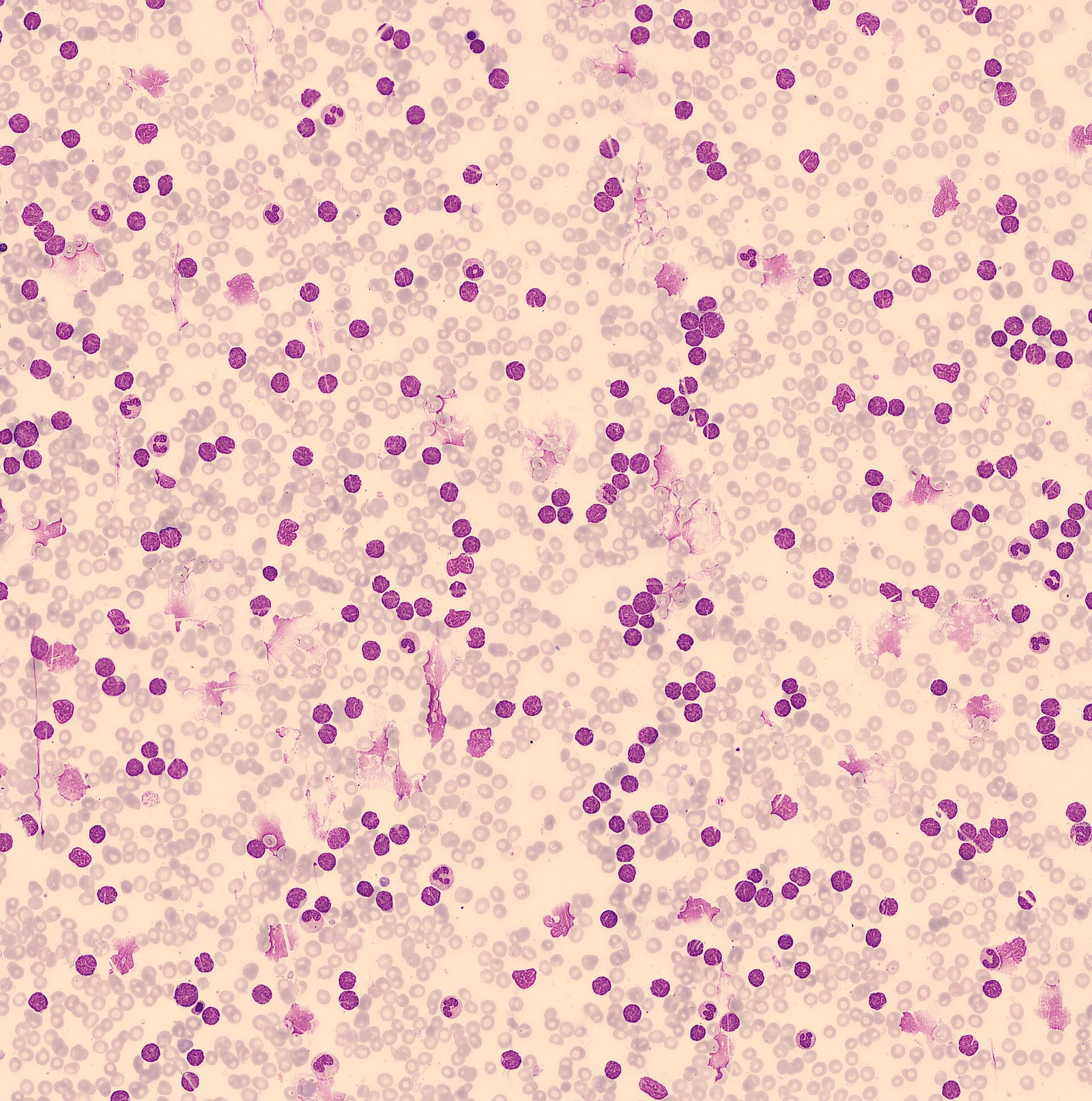
Long description
Low-power blood film in a B-cell non-Hodgkin lymphoma with a leukaemic phase, showing many circulating lymphoma cells among the red cells.

- Excisional biopsy of a whole lymph node is preferred; it gives architecture, full immunophenotype and material for cytogenetics and molecular tests. Core biopsy is acceptable when excision is impractical, risky or would cause delay; fine-needle aspiration is generally unhelpful and can delay the diagnosis.[1][2]
- Material is reviewed in a reference haematopathology laboratory, with CD marker panels, immunoglobulin/T-cell receptor gene rearrangement studies, cytogenetics (FISH for MYC, BCL2, BCL6 translocations) and, where relevant, gene-expression profiling.[1]
- All diagnoses are discussed at a multidisciplinary team meeting.
- PET-CT with [18F]fluorodeoxyglucose is the recommended staging modality for most lymphomas; contrast-enhanced CT is an alternative when PET is unavailable or as an additional study.[1][2]
- Stage I/II is single or regional nodal disease; stage III disease lies above and below the diaphragm; stage IV involves disseminated extranodal organs including bone marrow, liver, lungs or CSF. Hodgkin A/B designation is not used in NHL.[2]
- Bone marrow biopsy is not routinely needed for DLBCL or follicular lymphoma when PET-CT is available unless a co-existing marrow disorder is suspected or management would change.[1]
- CNS assessment (contrast MRI of brain/spine plus CSF cytology and flow cytometry) is done when CNS involvement is suspected or the CNS-IPI is high, especially in DLBCL with three or more extranodal sites.[1]
- Hepatitis B, hepatitis C and HIV serology are performed before treatment in all patients with lymphoma.[1][6]
- Baseline assessment: full blood count, renal and liver function, LDH, uric acid, ECG and cardiac assessment when anthracyclines are planned. Reproductive counselling and fertility preservation are offered (sperm storage, ovarian tissue or oocyte cryopreservation) before gonadotoxic therapy.
- Double-hit testing: FISH for MYC with immunoglobulin partner, and BCL2/BCL6 if MYC is rearranged, identifies high-risk disease that may change treatment.[1]
- Tumour lysis risk assessment before any treatment (LDH, uric acid, renal function), with a prophylaxis plan.[1]
- Geriatric and frailty assessment in older patients to decide whether full-dose or attenuated regimens are appropriate.[1]
Non-Hodgkin lymphoma has no single diagnostic threshold: it is a histological diagnosis made by an expert haematopathologist integrating morphology, immunophenotype and genetics. Staging is by the Lugano classification (the table above), and severity is estimated with:
- IPI for all patients (age, stage III/IV, performance status, LDH, number of extranodal sites). The NCCN-IPI gives finer separation in both low- and high-risk patients; stage-modified IPI (smIPI) is used for localised disease.[1][3][5]
- NCCN-IPI risk groups for aggressive (diffuse large cell) lymphoma: low (score 0–1) 5-year overall survival 96% with PFS 91%; low-intermediate (2–3) 82% and 74%; high-intermediate (4–5) 64% and 51%; high (more than 6) 33% and 30%.[3][5]
- CNS-IPI for the risk of CNS relapse.
Approach to suspected non-Hodgkin lymphoma
- Suspected NHL (painless node, B symptoms, extranodal mass)
- Assess urgency and siteAirway, bulky nodes, high LDH or tumour-lysis risk
- Question: Excisional biopsy possible without delay or excessive risk?
Yes
- Excisional (whole-node) biopsyPreferred: architecture plus full immunophenotype and cytogenetics
No
- Core needle biopsyAcceptable alternative when excision impractical, risky or delayed
- Expert haematopathology reviewMorphology, immunophenotype, genetics, FISH for MYC/BCL2/BCL6
- Staging PET-CT plus bloods, viral serology and CNS riskLugano stage; IPI and CNS-IPI
- Question: Indolent or aggressive biology?
Indolent (follicular, marginal zone/MALT, indolent MCL)
- Watchful waiting if asymptomatic and low tumour burden
- Rituximab, radiotherapy or chemo-immunotherapy when needed
Aggressive (DLBCL, Burkitt, aggressive MCL, T/NK cell)
- Curative chemo-immunotherapy (R-CHOP or RCHP-polatuzumab; Burkitt regimens)
- CNS prophylaxis if high risk; supportive care and infection prophylaxis
- Tumour lysis prophylaxis is started before therapy in any patient at risk: aggressive hydration, allopurinol or rasburicase, and monitoring of potassium, phosphate and uric acid. Burkitt lymphoma and bulky DLBCL carry the highest risk.[1]
- Antiemetics, G-CSF prophylaxis (primary prophylaxis recommended for all patients receiving curative chemoimmunotherapy) and infection prophylaxis (herpes zoster aciclovir/valaciclovir, Pneumocystis co-trimoxazole) are routine.[1]
- Hepatitis B prophylaxis with a nucleoside analogue (for example entecavir) when HBV DNA is detectable or even in resolved infection, with monitoring during and after immunotherapy. Patient positives for HCV are referred urgently to hepatology/infectious diseases.[1]
- EBV, HBV and HIV serology and referral for HIV co-care (patients on suppressive HAART with CD4 at or above 200 x 10^6/L can be treated on standard protocols).[1][6]
- Diffuse large B-cell lymphoma (noncontiguous stage II–IV, IPI 2–5): six cycles of RCHP-polatuzumab vedotin is preferred where available and affordable; R-CHOP remains standard, and six cycles of R-CHOP is the standard for stage III/IV IPI 1 disease. For ages 18–60 with early-stage non-bulky disease an abbreviated regimen (four cycles of R-CHOP plus two rituximab doses) is non-inferior.[1][3]
- Localised aggressive (stage I/II) DLBCL: four to six cycles of R-CHOP, with or without involved-site radiotherapy (about 30 Gy); in favourable FLYER-eligible patients four cycles of R-CHOP plus two rituximab doses avoids radiotherapy.[1][3]
- Follicular lymphoma: watchful waiting in asymptomatic, low-tumour-burden disease; when needed, rituximab alone, bendamustine-rituximab or (R-)CHOP, with radiotherapy for stage I/II; radiotherapy to involved sites at low dose (4 Gy) is used when tolerance is poor.[2][7]
- Marginal zone/MALT lymphoma: eradicate H. pylori (antibiotics resolve most localised gastric cases, sometimes taking 3–18 months); radiotherapy, rituximab, or systemic therapy for progressive or disseminated disease.[2]
- Mantle cell lymphoma: watchful waiting for indolent (non-nodal leukemic) disease; chemoimmunotherapy (bendamustine-rituximab or R-CHOP/R-DHAP), BTK inhibitors and rituximab maintenance for the aggressive form; targeted combinations for TP53-altered/blastic disease.[4]
- Burkitt lymphoma: short intensive multi-agent regimens (e.g. CODOX-M/IVAC or DA-EPOCH) with rituximab, with obligatory CNS prophylaxis. Patients with HIV benefit from dose-modified regimens with concurrent or sequential HAART.[3][6][8]
- T-cell lymphomas: CHOP-like regimens first line (with etoposide in younger patients); most need consolidation assessment. The 5-year survival of PTCL is about 30–40%, with better outcomes for ALK-positive anaplastic large cell lymphoma.[5]
- AIDS-related lymphoma: combination chemotherapy plus HAART (concurrent use is debated), with rituximab added for CD20-positive disease; dose-modified regimens for Burkitt lymphoma; intrathecal prophylaxis when CNS risk is high; and for resistant or relapsed disease, high-dose therapy with autologous stem cell rescue in selected patients whose HIV is well controlled.[6]
- Radiotherapy: involved-site radiation to about 30 Gy is used in early-stage aggressive disease and consolidation; extended or palliative fields in low-grade disease or symptomatic bulky sites. Even very low doses (4 Gy) can palliate indolent lymphoma.[2] [1]
- Surgery: usually only for diagnostic biopsy or for complications (perforation, obstruction, bleeding); gastric MALT rarely needs gastrectomy once H. pylori is eradicated.[2]
- Autologous stem cell transplant is standard salvage after relapse in fit patients (high-dose BEAM or similar then rescue), and as consolidation after remission-induction in some subtypes. It is not recommended as first-line consolidation in DLBCL. Allogeneic transplant is reserved for selected relapsed or high-risk (e.g. mantle cell) disease.[1][3]
- High-dose chemotherapy with autologous rescue has a treatment-related myelodysplasia/leukaemia rate of about 20% at 10 years in older series, an important counselling point.
- Response is assessed by PET-CT; complete metabolic response predicts good outcome, but PET has a real false-positive rate, so biopsy of a residual mass is warranted when management changes.[1]
- Most relapses of aggressive lymphoma occur within the first two years, so follow-up is most intense then; relapses of follicular and MALT lymphoma may be delayed by years to decades, and late toxicity (cardiac, second cancer, bone health, fertility) needs attention.[2]
- CNS prophylaxis: central nervous system relapse is highest in Burkitt lymphoma (20–30% lifetime), in young patients with testicular or breast DLBCL, and in high CNS-IPI disease. Prophylaxis is intrathecal (and high-dose systemic methotrexate where indicated) for Burkitt lymphoma and selected DLBCL; its routine use in mantle cell lymphoma has never been shown effective in a randomised trial.[1][3][4]
- HIV: suppress HIV viral load first or concurrently; patients with CD4 at or above 200 x 10^6/L and suppressed virus can be treated on standard protocols.[1][6]
- Pregnancy: treatment depends on subtype and trimester; HAART is essential for HIV-associated disease and standard regimens can often be deferred or modified to protect the fetus.
- Older patients: R-miniCHOP (cyclophosphamide 400 mg/m2, doxorubicin 25 mg/m2, vincristine 1 mg, with rituximab) is standard for patients aged 80 years and over; consider a steroid or vinvristine pre-phase when performance status is compromised by lymphoma burden; non-anthracycline regimens (RGCVP, RCEOP) when cardiac comorbidity forbids doxorubicin; and palliative, quality-of-life-focused approaches for the very frail.[1]
- T-cell lymphomas: ALK-positive anaplastic large cell lymphoma has a 5-year survival of 70–80%, but PTCL (not otherwise specified), angioimmunoblastic, enteropathy-associated and hepatosplenic types fare worse and are managed in specialist centres with early transplant evaluation.[5]
- Tumour lysis syndrome (Burkitt and bulky, rapidly growing DLBCL) — preventable, potentially lethal.
- CNS relapse (Burkitt, testicular/breast DLBCL) — prevent with prophylaxis as above.
- Infection — cytopenias plus immunosuppression; latent hepatitis B reactivation on rituximab/obinutuzumab; Pneumocystis; shingles.
- Late cardiac, pulmonary and renal toxicity from anthracyclines, bleomycin and cisplatin; second cancers, myelodysplasia and acute leukaemia 10–20+ years after therapy (21% second malignancy, 10% solid tumours, at 10-year follow-up in one autologous-transplant series).[2]
- Infertility after alkylating agents, and early menopause.
- Bispecific antibody and CAR-T therapy complications — cytokine release syndrome (fever, hypotension, neurotoxicity) require trained teams and, for CAR-T therapy, specialised centres with validated supportive-care protocols.[10]
- Aggressive NHL (DLBCL) is curable in more than 70% of patients with modern chemoimmunotherapy; the IPI separates risk. Even very high-risk disease can be cured with R-CHOP; advanced double-hit lymphoma remains difficult.[1][3]
- Indolent lymphomas run chronic courses with median survivals up to 20 years, but risk of transformation to DLBCL (about 3% per year in follicular lymphoma, up to 45% over a lifetime) and late relapse. Watchful waiting does not worsen survival.[2][7]
- Mantle cell lymphoma is not considered curable in the usual sense, but many patients achieve long functional remissions; prognosis is worse with blastoid or pleomorphic morphology, high Ki-67 (30% or more) or TP53 alteration.[4]
- T-cell lymphomas have the least favourable average outcomes and are usually managed in specialist centres.
- HIV-associated lymphoma in the HAART era has an overall survival comparable with that of the non-immunosuppressed population.[6]
- Response to HAART (HIV) and complete metabolic response on PET in DLBCL independently predict prolonged survival.[1][6]
- Older adults (80 years and over): R-miniCHOP is standard; geriatric assessment is useful even if not yet prospectively validated in lymphoma; pre-phase steroid or vincristine may improve tolerance.[1]
- Frail patients: palliative, quality-of-life-directed care (low-intensity regimens, rituximab, steroids, palliative radiotherapy) can be appropriate.
- Pregnancy: treatment depends on subtype and trimester; HAART is essential for HIV-associated disease and standard regimens can often be deferred or modified to protect the fetus.
- Children: Burkitt lymphoma is the relevant entity; curative chemotherapy with CNS prophylaxis gives excellent outcomes. Lymphoblastic lymphoma is treated on ALL-type protocols.
Chimeric antigen receptor (CAR) T-cell therapy (axicabtagene ciloleucel, tisagenlecleucel, lisocabtagene maraleucel) modifies the patient's own T cells to recognise CD19-positive B cells. Bispecific antibodies (epcoritamab, glofitamab, mosunetuzumab, odronextamab) are off-the-shelf T-cell-engaging constructs, usually CD20 x CD3. Both classes are now central to the treatment of relapsed or refractory B-cell lymphoma, with CAR-T therapy beating salvage chemotherapy plus transplant in early relapse in phase 3 trials of large B-cell lymphoma. They cause cytokine release syndrome and immune-effector-cell neurotoxicity, and need trained teams in accredited centres.[10]
Where diagnosis or treatment capacity is limited, the most useful practical steps are:
- Pathology is the bottleneck. A review of 4,848 cases in the developing world found a 7.5% misclassification rate (vs 2.2% in the developed world), with pathologists often lacking training in haematopathology or the ancillary tests needed to classify and stage lymphoma accurately.[9]
- The subtype mix differs: more high-grade B-cell disease, less follicular lymphoma, a younger median age at diagnosis and a higher proportion of Burkitt lymphoma, so assumptions imported from Western clinics may be wrong.[9]
- Excisional biopsy (or at least a good core, plus immunohistochemistry) should be pursued; avoid starting empirical chemotherapy on a clinician's impression.
- Stage with CT if PET-CT is unavailable; staging of lymphoma pre-dates PET and remains useful.
- Eradicate H. pylori for gastric MALT as a first, low-cost intervention; geriatric and infection-prophylaxis principles (G-CSF, anti-Pneumocystis prophylaxis, HBV prophylaxis) are cheap and high-yield.
- Transport, social support, and toxicity prevention matter as much as drugs: treatment abandonment, toxic death and delayed diagnosis are the main avoidable causes of poor outcomes in resource-constrained settings (especially in children with Burkitt lymphoma).
- Refer early for infusion-based treatments (PET-guided approaches, bendamustine-rituximab, anthracycline regimens) that need pharmacy and nursing support, central lines, and cardiac monitoring.
This page is written to the British (BSH) and NCI (PDQ) sources that are freely available. The North American (NCCN) and European (ESMO) guidelines are not free in full, but their direction can be summarised without over-claiming:
- All three (BSH, NCCN, ESMO) recommend PET-CT for staging and response, excisional biopsy before treatment, and rituximab-based chemoimmunotherapy as the backbone of first-line aggressive B-cell lymphoma.
- NCCN uses the NCCN-IPI and (in recent editions) allows polatuzumab vedotin (R-CHP) as an option for previously untreated DLBCL with IPI 2–5, while retaining R-CHOP where polatuzumab is unavailable or contraindicated.
- ESMO's 2025 lymphoma clinical practice guideline keeps the same broad architecture — watchful waiting in asymptomatic follicular lymphoma, rituximab combinations when treatment is needed, and CAR-T and bispecific antibodies in relapsed large B-cell lymphoma — with country-specific adaptations.
- CNS prophylaxis is the area where guidelines differ most: NCCN, ESMO and BSH all target high CNS-IPI patients but disagree on the exact risk score, the route (intrathecal vs high-dose systemic methotrexate) and the place of methotrexate in patients already receiving high-dose cytarabine. None recommends routine prophylaxis in mantle cell lymphoma, where it has never been shown to work.
- Non-Hodgkin lymphoma is a family of lymphoid cancers, not one disease; about 85% are B-cell derived, T-cell lymphomas make up less than 15%.
- The usual presentation is a persistent painless lymph node, sometimes with B symptoms or mass effects; extranodal sites are common in aggressive and HIV-related disease.
- Diagnosis requires a whole-node (or adequate core) biopsy reviewed by an expert haematopathologist; fine-needle aspiration is unhelpful.
- PET-CT is the staging tool of choice; staging is by Lugano (stage I–IV); IPI (including NCCN-IPI) estimates prognosis.
- Indolent (follicular, MALT/marginal zone): watchful waiting is safe at first; radiotherapy, rituximab or chemoimmunotherapy when needed.
- Aggressive (diffuse large B-cell, Burkitt): curative chemoimmunotherapy — R-CHOP or RCHP-polatuzumab in LBCL; intensive multi-agent regimens with mandatory CNS prophylaxis in Burkitt.
- Every patient is screened for hepatitis B, hepatitis C and HIV before treatment, and for CNS risk thereafter.
- HIV-associated lymphoma: HAART plus chemotherapy with rituximab; outcomes match the uninfected population in the HAART era.
- Bulky, high-risk disease is monitored for tumour lysis.
- CAR-T therapy and bispecific antibodies are now central to relapsed/refractory B-cell lymphoma and require specialist centres.
- In resource-limited settings, good pathology plus simple stage-directed therapy and toxicity prevention save the most lives.
Answer each one in your head first, then open the answer.
A 62-year-old man has a firm, painless, enlarging cervical node for 6 weeks, fatigue and 5 kg weight loss. What is the most useful first investigation and what must be avoided?
Show answerHide answer
An excisional (whole-node) biopsy, reviewed by an expert haematopathologist, with immunophenotype and cytogenetics. Fine-needle aspiration should be avoided because it may delay diagnosis and prevent full subclassification. Bloods including LDH, and hepatitis B/C and HIV serology, should be taken before treatment.
How is follicular lymphoma distinguished cytogenetically, and why does it matter clinically?
Show answerHide answer
t(14;18)(q32;q21) leads to BCL2 overexpression (found by FISH in about 85% of cases). It allows germinal-centre B cells to survive, producing an indolent nodal disease with a long survival, a 3% per year risk of transformation to diffuse large B-cell lymphoma, and good responses to anti-CD20 therapy and watchful waiting.
Which non-Hodgkin lymphoma is characterised by starry-sky histology and a 20–30% lifetime risk of CNS disease, and how is the CNS risk managed?
Show answerHide answer
Burkitt lymphoma (t(8;14) or variant, deranging MYC). CNS prophylaxis with intrathecal chemotherapy is a mandatory part of induction because of its very high proliferation and tendency to spread to the meninges and brain.
A 78-year-old, frailer patient has stage III diffuse large B-cell lymphoma. Describe the pragmatic modifications you would consider.
Show answerHide answer
Use R-miniCHOP (cyclophosphamide 400 mg/m2, doxorubicin 25 mg/m2, vincristine 1 mg, with rituximab) rather than full-dose R-CHOP; consider a steroid or vincristine pre-phase to improve performance status; give primary G-CSF and infection prophylaxis; screen cardiac function before anthracyclines; and avoid high-dose transplant-based consolidation.
Why is antibiotic eradication therapy the first, low-cost treatment for early gastric MALT lymphoma?
Show answerHide answer
Gastric MALT lymphoma is strongly associated with Helicobacter pylori; eradication resolves most localised gastric disease, with endoscopic resolution in about 50% by 3 months and more after 12–18 months. Deeper or progressive disease needs radiotherapy or systemic therapy. [2]
What does 'indolent vs aggressive' mean for treatment intent?
Show answerHide answer
Indolent lymphomas (follicular, MALT, indolent mantle cell) are often watched initially; immediate treatment does not improve survival in asymptomatic patients. Aggressive lymphomas (DLBCL, Burkitt, T/NK-cell) grow quickly but are potentially curable, so treatment with chemoimmunotherapy is given promptly and with curative intent.
Related topics (MedGrasp): Hodgkin lymphoma, chronic lymphocytic leukaemia, aplastic anaemia, immune thrombocytopenia, tumour lysis syndrome, central nervous system prophylaxis.
- Fox CP, Chaganti S, McIlroy G, Barrington SF, Burton C, Cwynarski K, Eyre TA, Illidge T, Kalakonda N, Kuhnl A, McKay P, Davies AJ. The management of newly diagnosed large B-cell lymphoma: a British Society for Haematology guideline. British Journal of Haematology 2024;204(4):1178–1192. Accessed .
- PDQ Adult Treatment Editorial Board. Indolent B-Cell Non-Hodgkin Lymphoma Treatment (PDQ): health professional version. National Cancer Institute PDQ Cancer Information Summaries, updated 14 May 2025. Accessed .
- PDQ Adult Treatment Editorial Board. Aggressive B-Cell Non-Hodgkin Lymphoma Treatment (PDQ): health professional version. National Cancer Institute PDQ Cancer Information Summaries, updated 12 May 2025. Accessed .
- PDQ Adult Treatment Editorial Board. Mantle Cell Lymphoma Treatment (PDQ): health professional version. National Cancer Institute PDQ Cancer Information Summaries, updated 12 May 2025. Accessed .
- PDQ Adult Treatment Editorial Board. Peripheral T-Cell Non-Hodgkin Lymphoma Treatment (PDQ): health professional version. National Cancer Institute PDQ Cancer Information Summaries, updated 13 May 2025. Accessed .
- PDQ Adult Treatment Editorial Board. AIDS-Related Lymphoma Treatment (PDQ): health professional version. National Cancer Institute PDQ Cancer Information Summaries, updated 18 September 2024. Accessed .
- Kridel R, Sehn LH, Gascoyne RD. Pathogenesis of follicular lymphoma. Journal of Clinical Investigation 2012;122(10):3424–3431. Accessed .
- Orem J, Mbidde EK, Lambert B, de Sanjose S, Weiderpass E. Burkitt's lymphoma in Africa, a review of the epidemiology and etiology. African Health Sciences 2007;7(3):166–175. Accessed .
- Perry AM, Diebold J, Nathwani BN, MacLennan KA, Müller-Hermelink HK, Bast M, Boilesen E, Armitage JO, Weisenburger DD. Non-Hodgkin lymphoma in the developing world: review of 4539 cases from the International Non-Hodgkin Lymphoma Classification Project. Haematologica 2016;101(10):1244–1250. Accessed .
- Kim J, Kim SJ. Advances in T-Cell-directed immunotherapy for adult mature B-cell lymphoma: a comprehensive review of CAR T-cell and bispecific antibody therapies. Cancer Research and Treatment 2025;57(4):905–922. Accessed .
Figure credits
Educational content for medical students and doctors. It is not a substitute for clinical judgement or local guidelines.