Acute sinusitis
Viral and bacterial acute sinusitis: how to tell them apart, diagnose by symptoms, treat, and avoid complications.
- Acute sinusitis (more precisely acute rhinosinusitis) is inflammation of the nose and the paranasal sinuses that comes on suddenly and lasts less than four weeks; viruses cause the large majority. [3]
- From that viral illness a secondary bacterial infection develops in a minority of people: the clues are symptoms that persist for at least 10 days without improvement, severe onset with high fever and purulent discharge for 3-4 days, or a "double worsening" after initial improvement [1][2].
- The bacterial pathogen is usually Streptococcus pneumoniae, Haemophilus influenzae or Moraxella catarrhalis.
- Diagnosis is made from the clinical picture; imaging is not needed routinely.
- Most viral cases need only symptomatic care, and even confirmed bacterial cases may first be managed by watchful waiting in uncomplicated adults; when an antibiotic is chosen, first-line treatment is amoxicillin with or without clavulanate for 5 to 10 days [1][2].
By the end of this page you should be able to:
- Define acute rhinosinusitis and separate acute viral from acute bacterial disease using the standard clinical criteria.
- Name the typical viral and bacterial organisms and say when a bacterial infection is most likely.
- Explain why routine imaging is avoided, and which clinical situations justify imaging.
- Treat acute rhinosinusitis: symptomatic care, watchful waiting, and first-line antibiotics with dose, route, duration and monitoring.
- Recognise and respond to the red flags and complications, including orbital cellulitis and intracranial spread.
- Outline how chronic and fungal rhinosinusitis differ from an acute episode.
- Adapt the approach when tests or first-line drugs are not available.
DefinitionAcute rhinosinusitis
Acute rhinosinusitis (ARS) is symptomatic inflammation of the nasal cavity and the paranasal sinuses lasting less than four weeks [3]. The word "sinusitis" is incomplete, because the nasal lining and the sinus lining are one continuous surface; most guidelines now use "rhinosinusitis". The term "acute sinusitis" remains common in everyday practice and in this page.
The condition matters because it is one of the commonest reasons people seek medical help, it accounts for a large share of antibiotic prescribing, and a small number of cases spread to the orbit or brain and become dangerous. A viral illness causes most episodes, so much of the practical skill is deciding who needs an antibiotic and who does not, rather than choosing a drug.
It helps to divide the illness along two axes: by cause, and by duration.
By cause.
- Acute viral rhinosinusitis (VRS): a viral upper respiratory infection with sinonasal inflammation.
- Acute bacterial rhinosinusitis (ABRS): bacterial superinfection on top of a viral illness, identified by the patterns below.
- Less common: fungal and other causes.
By duration. The same anatomical process is renamed by how long it has lasted [3].
- Acute rhinosinusitis: under four weeks [3].
- Subacute rhinosinusitis: more than four but less than twelve weeks [3].
- Chronic rhinosinusitis: at least twelve consecutive weeks [3][4].
- Recurrent acute rhinosinusitis: at least four separate acute episodes per year, each lasting 7 to 10 days, with no features of chronic disease between them [3].
Although they share a name, chronic rhinosinusitis is a different condition with a different mechanism (see "Chronic rhinosinusitis (outline)" below). This page stays with the acute illness, and only outlines the chronic one.
Acute rhinosinusitis is very common: one review estimates that about 31 million people in the United States are affected each year [3]. It is the commonest setting for acute sinusitis in healthy adults, and most episodes resolve without treatment. Among children brought to medical care with respiratory symptoms, roughly 6 to 7% have acute bacterial rhinosinusitis rather than a simple viral cold [6]. Complications are rare, but when they occur they are usually in otherwise healthy young children with acute ethmoiditis, or in adolescents with frontal sinusitis [6].
Viral triggers. Viral rhinosinusitis is the usual first step. Typical viruses include rhinovirus, influenza, parainfluenza, coronavirus, adenovirus and respiratory syncytial virus; rhinovirus is the commonest [3]. Viral mucus and inflammation obstruct the narrow drainage channels of the sinuses, especially the ostiomeatal complex, trap secretions and impair mucociliary clearance (the normal conveyor-belt movement of mucus out of the sinus), and this sets the stage for bacteria to grow.
Bacterial superinfection. In acute bacterial rhinosinusitis the main pathogens of the maxillary, ethmoid and frontal sinuses are Streptococcus pneumoniae, Haemophilus influenzae and Moraxella catarrhalis [3]. Additional organisms include other streptococci, Staphylococcus aureus and anaerobes [3]. Acute sphenoid sinusitis is more often due to Staphylococcus aureus [3]. Children share the same three main organisms [3].
Risk factors for bacterial disease. A primary or secondary bacterial infection is more likely with anatomical blockage, recent viral illness, allergic rhinitis, dental infection (especially of the upper teeth, which can seed the maxillary sinus), immunocompromise, cystic fibrosis, or poorly controlled diabetes. Recent antibiotic use, day-care attendance, age under 2 or over 65 years, recent hospitalisation, and immunocompromise are specifically used by the IDSA guideline as markers that justify high-dose amoxicillin-clavulanate (see "Management") [1].
The whole story is one of drainage and clearance.
- A viral or allergic insult inflames the sinus and nasal lining. The sinus drainage channels narrow and mucociliary clearance slows.
- Secretions are retained. The stagnant, warm, protein-rich fluid is an ideal breeding medium for bacteria.
- Bacterial overgrowth follows (in only a minority of viral colds), producing acute bacterial rhinosinusitis. The most common triad of organisms is Streptococcus pneumoniae, Haemophilus influenzae and Moraxella catarrhalis [3].
- If the inflammation continues for weeks, the bacterial flora shifts towards Staphylococcus aureus, coagulase-negative staphylococci, anaerobes and gram-negative organisms, and osteitis (inflammation of the sinus bone) can develop [3].
- In a few cases infection spreads through thin bone or valveless veins to the orbit or brain (see "Complications") [6].
Nothing in this process is helped by imaging in the early stages. The patient's history and examination carry the diagnosis.
Acute rhinosinusitis announces itself after a typical cold, or worsens during one. Common symptoms are nasal blockage or congestion, purulent (thick, coloured) nasal discharge, facial pain or pressure, and a reduced sense of smell. Post-nasal drip, cough (often worse at night), headache, ear fullness, dental pain and malodorous breath can accompany them. Uncomplicated viral sinusitis tends to resolve within about a week.
For the purpose of deciding whether the case is bacterial, three patterns matter. These are the criteria set out by the Infectious Diseases Society of America (IDSA, 2012) and adopted by the American Academy of Otolaryngology-Head and Neck Surgery (AAO-HNS) [1][2].
- Persistent symptoms. Symptoms compatible with acute rhinosinusitis that last for at least 10 days without any evidence of improvement. This is the commonest pattern in adults. [1]
- Severe onset. High fever of 39 degrees C (102 degrees F) or higher together with purulent nasal discharge or facial pain, lasting for at least 3 to 4 consecutive days at the beginning of the illness. In a viral cold, fever usually lasts only 24 to 48 hours and purulent discharge does not appear until around day 4 or 5 [1].
- Double worsening ("double-sickening"). Symptoms that improve after a typical viral upper respiratory infection lasting 5 to 6 days, and then suddenly worsen again, with a new fever, headache or increased nasal discharge. [1]
The AAO-HNS 2015 guideline adds the practical diagnostic package: purulent nasal drainage accompanied by nasal obstruction, facial pain-pressure-fullness, or both, lasting for at least 10 days beyond the onset of upper respiratory symptoms, or worsening within 10 days after initial improvement [2]. Daytime cough that worsens at night, with nasal discharge of any quality, is common in the persistent pattern, while fever, headache and facial pain are variable [1].
Viral and bacterial episodes cannot be separated cleanly, and even when bacterial infection is suspected the clinical criteria are imperfect. In a placebo-controlled trial of antibiotics in adults with symptoms for at least 7 days, only about 64% had positive bacterial cultures by sinus puncture [1]. The criteria are therefore a guide to a probability, not a laboratory diagnosis, and a clinician must reassess if the course is not typical.
Look for tenderness over the cheeks, forehead and between the eyes, and pain on percussion of the affected sinus. Purulent secretions can be seen in the middle meatus with a nasal speculum or endoscope. Check the teeth, because an infected upper tooth can cause one-sided maxillary sinusitis. Assess the eye and the nervous system when there is any hint of complication (see "Red flags"). In children the most common manifestations of bacterial infection are cough (about 80%), nasal discharge (about 76%) and fever (about 63%); headache, facial pain and swelling are rare, and malodorous breath is common in pre-school children [1].
The main distinctions are between viral, bacterial, and non-infectious causes of nasal and sinus symptoms, and between acute and chronic disease.
| Condition | Features that point to it |
|---|---|
| Viral upper respiratory infection | Improves within about a week; fever early and brief; no severe facial pain |
| Acute bacterial rhinosinusitis | Persistent 10 days, severe onset, or double worsening; may need antibiotic consideration |
| Allergic rhinitis | Clear watery discharge, sneezing, itchy eyes, seasonal pattern, no fever |
| Non-allergic rhinitis | Nasal stuffiness and drip without an allergic trigger (cold air, irritants) |
| Structural or obstructive disease | Deviated septum, polyps, tumour; often one-sided or recurrent; no acute infection |
| Odontogenic sinusitis | One-sided maxillary pain; infected upper tooth or recent dental work; anaerobic organisms |
| Foreign body (especially in children) | One-sided purulent discharge, often foul-smelling, with a normal contralateral side |
| Chronic rhinosinusitis | Symptoms for at least 12 weeks; see the outline below |
| Malignancy or granulomatous disease | Unilateral bleeding, crusting, cacosmia, mass, neurological or orbital findings |
Two simple rules help: chronic rhinosinusitis is usually bilateral, so unilateral symptoms should raise suspicion and prompt referral, and red-flag features such as bleeding, crusting, cacosmia, orbital symptoms, severe frontal headache, frontal swelling, or neurological or meningitic symptoms point to a diagnosis other than uncomplicated rhinosinusitis [4].
Figure 1CT of chronic sinusitis
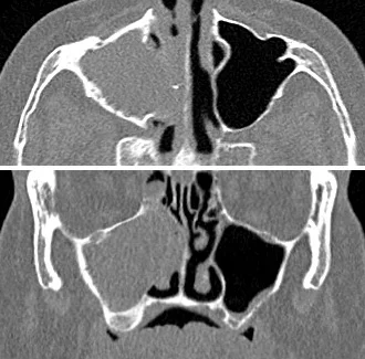
Long description
Axial and coronal CT slices of the sinuses with opacification and mucosal thickening of the maxillary sinus on one side and clear air on the other.

Routine imaging is not needed. Both IDSA and AAO-HNS state that radiographs or computed tomography should not be obtained for patients who meet the clinical criteria for acute rhinosinusitis, unless a complication or an alternative diagnosis is suspected [1][2]. The reasons are that imaging cannot separate viral from bacterial disease, mucosal thickening is common in simple viral colds, and unnecessary scanning exposes patients to radiation and cost. A normal scan also does not exclude infection.
When imaging is justified.
- Suspected complication: orbital cellulitis, cavernous sinus thrombosis, intracranial extension, or osteomyelitis (see "Red flags"). Cross-sectional imaging is mandatory in these situations [6].
- Suspected alternative diagnosis: tumour, structural lesion, or chronic rhinosinusitis.
- Suspected chronic rhinosinusitis, where nasal endoscopy or CT is needed to confirm objective sinus inflammation [2][4].
When cultures or aspiration are used. Sinus puncture or swab is not needed for ordinary acute disease. Cultures obtained by direct sinus aspiration are reserved for patients who have failed to respond to empiric therapy, to identify resistant organisms [1]. Nasopharyngeal swabs are unreliable for this purpose and are not recommended [1]. If therapy fails after reassessment, surgery may yield material for culture in the same way.
Other tests. Blood tests do not diagnose uncomplicated sinusitis. Measure glucose, renal function, blood counts and immune status only when invasive fungal disease or another underlying problem is suspected. Allergy testing is a consideration in chronic or recurrent disease [2].
Diagnostic criteria for acute bacterial rhinosinusitis combine two steps: evidence of acute sinusitis from symptoms and signs, and evidence that it is bacterial rather than viral based on onset, severity and time course (persistent 10 days, severe onset, or double worsening) [1][2]. Duration alone is not enough, because basing the diagnosis solely on 7 to 10 days of respiratory symptoms is inadequate to separate bacterial from viral disease [1].
Red flags that require urgent evaluation. Seek help immediately and consider imaging and hospital referral if any of the following appear:
- Swelling or redness around the eye, with or without displacement of the eye, pain on moving the eye, or any drop in vision.
- Cranial nerve palsies, severe or worsening headache, photophobia, seizures, or other neurological signs.
- Frontal headache with a tender swelling on the forehead.
- High fever with facial swelling, necrotic nasal mucosa, or a diabetic or immunocompromised patient.
- Rapidly deteriorating illness, lack of response to appropriate therapy, or relapse with worsening.
- One-sided symptoms or bleeding (which suggest an alternate diagnosis such as tumour).
These features overlap with the complications described below, and the threshold to image is deliberately low once they are present [6].
The goal is to relieve symptoms, to avoid unnecessary antibiotics, and to catch complications early.
Most cases need only supportive treatment. AAO-HNS 2015 allows analgesics, topical intranasal corticosteroids and saline irrigation as symptomatic relief for both viral and bacterial disease, and the same review notes that intranasal saline irrigation, antipyretics/analgesics and fluids form the backbone of care [2]. IDSA 2012 recommends intranasal saline irrigation with physiologic or hypertonic saline as an adjunct in adults, and intranasal corticosteroids as an adjunct to antibiotics, mainly in people with allergic rhinitis [1]. Neither topical nor oral decongestants or antihistamines are recommended as adjunctive treatment in acute bacterial disease [1]. Topical decongestants can help congestion for a few days but should not be used for long periods, because of rebound congestion.
Approach to acute rhinosinusitis in an adult (teaching algorithm)
- Symptoms of acute rhinosinusitisPurulent nasal drainage with nasal obstruction, facial pain-pressure-fullness, or both
- Assess duration and severityLook for persistent >=10 days, severe onset with high fever for 3-4 days, or double worsening after 5-6 days
- Question: Does the case meet ABRS criteria?
No: viral or non-infectious pattern
- Symptomatic careAnalgesics, saline irrigation, optional intranasal steroid
- Reassess if not improving in 7-10 days or if red flags appear
Yes: uncomplicated ABRS
- Question: Red flags or complication suspected?
Yes
- Urgent referral; contrast CT and treatment for the complication
No
- Offer watchful waiting OR initial antibioticsWatchful waiting only with guaranteed follow-up
- If treating, prescribe amoxicillin with or without clavulanate for 5-10 daysChoose high-dose for risk factors
- Reassess at 7 days (or sooner if worsening); escalate or change antibiotic if no improvement
AAO-HNS 2015 recommends that clinicians either offer watchful waiting without antibiotics or prescribe initial antibiotic therapy for adults with uncomplicated bacterial disease; watchful waiting is only appropriate when follow-up is assured, so that antibiotics can be started if the patient has not improved by seven days after diagnosis or worsens at any time [2]. Placebo-controlled evidence suggests that most uncomplicated cases recover within about two weeks without antibiotics, and antibiotics confer only a small benefit, with more gastrointestinal side effects [3]. Shared decision-making is therefore the right framing: in a healthy adult with mild illness and a clear route for follow-up, observation is reasonable; in a more ill patient, an antibiotic is justified.
When an antibiotic is chosen, IDSA 2012 recommends that empiric antimicrobial therapy be started as soon as the diagnosis of bacterial disease is established [1], and that a beta-lactam agent (amoxicillin-clavulanate) be used rather than a respiratory fluoroquinolone for initial therapy [1]. AAO-HNS 2015 recommends amoxicillin with or without clavulanate as first-line therapy for most adults [2]. IDSA 2012 goes further and recommends amoxicillin-clavulanate rather than amoxicillin alone for both children and adults (strong and moderate evidence in children, weak and low in adults), because of the prevalence of beta-lactamase-producing Haemophilus influenzae and other beta-lactamase-producing pathogens [1].
| Regimen | Role |
|---|---|
| Amoxicillin-clavulanate (standard dose) | First-line initial therapy for most adults and children [1][2] |
| Amoxicillin-clavulanate 2 g orally twice daily or 90 mg/kg/day orally twice daily ("high-dose") | For high endemic penicillin-nonsusceptible S. pneumoniae (10% or more), severe infection with systemic toxicity (fever 39 degrees C / 102 degrees F or higher), daycare attendance, age under 2 or over 65, recent hospitalisation, antibiotic use in the past month, or immunocompromise [1] |
| Doxycycline | Alternative initial regimen in adults [1]; also an alternative for penicillin-allergic adults [1] |
| Levofloxacin or moxifloxacin | Alternative for penicillin-allergic adults; clindamycin plus a third-generation oral cephalosporin in children with non-type I allergy [1] |
| Clarithromycin, azithromycin | Not recommended empirically because of high S. pneumoniae resistance [1] |
| Trimethoprim-sulfamethoxazole | Not recommended empirically because of high S. pneumoniae and H. influenzae resistance [1] |
| Second- and third-generation oral cephalosporins alone | No longer recommended as monotherapy; cefixime or cefpodoxime plus clindamycin is an alternative in selected children [1] |
Important cautions: routine cover for Staphylococcus aureus or MRSA is not recommended in initial empiric therapy, although S. aureus (including MRSA) remains a potential pathogen [1]. Culture-guided therapy is needed when first-line treatment fails.
IDSA 2012 recommends treatment for 5 to 7 days in adults with uncomplicated disease, and 10 to 14 days in children [1]. AAO-HNS 2015 recommends 5 to 10 days for most adults with antibacterial treatment [2]. That slightly wider range reflects the fact that the original guidance allows clinician judgement; if in doubt, follow the local guideline and extend toward the longer end for more severe cases. In children a longer course reflects slower resolution.
Treatment failure should be declared if symptoms worsen after 48 to 72 hours of initial empiric antibiotics or fail to improve after 3 to 5 days [1]. At that point IDSA and AAO-HNS agree that the clinician should reassess the patient to confirm the bacterial diagnosis, exclude other causes of illness and detect complications. If bacterial disease is confirmed and the patient was observed, start antibiotics; if an antibiotic was already used, change to an alternative agent guided by local resistance and, if possible, by culture from sinus aspiration [1][2]. Endoscopically guided middle meatal cultures may be an alternative in adults; nasopharyngeal cultures are unreliable [1]. Referral to an otolaryngologist, infectious disease specialist or allergist is appropriate for serious or immunocompromised patients, those still deteriorating, and those with recurrent episodes [1].
- Intranasal saline irrigation (physiologic or hypertonic) is recommended for adjunctive treatment in adults and is an option for bacterial disease [1][2].
- Intranasal corticosteroids are recommended as an adjunct to antibiotics in bacterial disease, mainly in people with allergic rhinitis [1][2].
- Analgesics and decongestants give symptomatic relief.
- Avoid antibiotics in purely viral disease; they do not speed recovery and can cause side effects and resistance [2].
Complications are uncommon but important. Orbital and intracranial spread, osteomyelitis and mucocele formation are the serious ones. Orbital complications are encountered most frequently, because the ethmoid sinus is separated from the orbit by a very thin sheet of bone (the lamina papyracea) [6]. Spread can also occur through valveless veins by bacterial thrombophlebitis, or through bone by osteomyelitis [6].
Chandler classification of orbital complications (least to most severe) [6].
- Preseptal cellulitis: eyelid swelling in front of the orbital septum, painless, normal eye movement and vision.
- Orbital cellulitis: orbital inflammation with proptosis, impaired eye movement and chemosis.
- Subperiosteal abscess: pus between bone and periorbita; proptosis and ophthalmoplegia, vision usually preserved.
- Orbital abscess: pus in the orbit; severe proptosis, ophthalmoplegia, often vision loss.
- Cavernous sinus thrombosis: bilateral eye findings, cranial nerve palsies; a medical and surgical emergency.
Chandler categories 1-3 are treated with intravenous antibiotics; if vision is lost or systemic disease progresses within 24 to 48 hours, functional endoscopic sinus surgery is added. Categories 4 and 5 are treated with surgery plus intravenous antibiotics; anticoagulation in cavernous sinus thrombosis is controversial because of the risk of intracranial haemorrhage [6].
Intracranial complications include cavernous sinus thrombosis, subdural and epidural empyema, cerebritis, brain abscess and meningitis [6]. They more often follow frontal sinusitis, which can erode or dehisce through the frontal bone, or ethmoid infection that spreads via valveless veins. They present with severe headache, photophobia, seizures or other focal neurological signs; if they occur, functional endoscopic sinus surgery is mandatory, with neurosurgical drainage when empyema or abscess is present [6].
Cavernous sinus thrombosis is particularly dangerous: classically ptosis, proptosis, chemosis and cranial nerve palsies; it arises from ethmoid or sphenoid sinus infection spread through valveless veins; and the dural venous system is valveless, so infection can propagate and lead to meningitis, subdural empyema or brain abscess [5][6].
If complex disease is suspected, cross-sectional imaging of the orbit and brain is mandatory [6], and treatment is hospital-based intravenous antibiotics with early surgical drainage.
Chronic rhinosinusitis is a different disease: an inflammatory rather than an infectious condition of the nose and sinuses, defined as symptomatic inflammation present for at least 12 consecutive weeks [4]. Diagnosis needs at least two of the core symptoms (nasal congestion or obstruction, anterior or posterior discharge, altered sense of smell, facial pain or pressure) plus an objective sign of inflammation on nasal endoscopy or CT [4]. Plain sinus radiographs lack sensitivity and should not be ordered [4]. Modifying conditions such as asthma, cystic fibrosis, immunocompromise and ciliary dyskinesia must be assessed, and red flags (unilateral symptoms, bleeding, crusting, cacosmia, orbital, severe frontal or neurological symptoms) should trigger early referral to rule out tumour or complicated disease [2][4].
Management is anti-inflammatory rather than antibacterial: long-term topical intranasal corticosteroids and saline irrigation are first-line, short courses of oral steroids help with polyps, and endoscopic sinus surgery is used when medical therapy fails (revision rates 10 to 30%) [2][4]. Systemic antibiotics have little evidence of benefit and should be reserved for acute exacerbations [4]. AAO-HNS 2015 explicitly recommends that topical or systemic antifungal therapy not be prescribed for chronic rhinosinusitis [2]. Viral and bacterial acute episodes can occur on top of a chronically inflamed baseline ("acute exacerbation of chronic rhinosinusitis"), in which case both the acute trigger and the underlying disease need treatment.
Fungal disease of the sinuses is a spectrum rather than one entity, and classification depends on tissue invasion and the host's immune state [3][5].
- Saprophytic fungal infestation: colonisation of crusts and secretions, often after surgery; managed with debridement and irrigation; excellent prognosis.
- Fungal ball (mycetoma): a matted extramucosal collection in an immunocompetent host, most often the maxillary sinus (about 94% of fungal balls); surgery clears it, and systemic antifungals are not appropriate [5].
- Allergic fungal rhinosinusitis: an allergic, non-invasive hypersensitivity disease in atopic patients, with eosinophilic mucin and nasal polyps (Bent and Kuhn criteria); treated with surgery plus steroids, with oral antifungals and immunotherapy reserved for refractory cases [5].
- Acute invasive fungal rhinosinusitis: a rare emergency in immunocompromised hosts (diabetes especially ketoacidosis, neutropenia, haematological malignancy, immunosuppression, iron overload), with high mortality (around 50 to 80%); it needs reversal of the predisposing condition, aggressive surgical debridement, and early systemic amphotericin B for Mucorales (voriconazole if Aspergillus is identified) [5].
- Chronic invasive fungal rhinosinusitis and chronic granulomatous invasive disease: slower courses in more immunocompetent hosts, managed with surgery and long-term antifungals [5].
The key clinical message is that fungal disease must be suspected in any immunocompromised, diabetic or profoundly unwell patient with sinus symptoms, black necrotic nasal mucosa, orbital or neurological signs, because delayed diagnosis is fatal.
Children. Children have the same main bacterial pathogens as adults [3]. Their presentations are dominated by cough, nasal discharge and fever rather than facial pain. IDSA recommends longer antibiotic courses in children (10 to 14 days) because of slower resolution [1]. Imaging is even more likely to be misleading in children, in whom mucosal thickening is common in uncomplicated illness [6].
Pregnancy. Uncomplicated acute sinusitis in pregnancy is managed supportively as usual, with the same red-flag precautions. Drug choices in pregnancy depend on local formulary guidance and gestational age; amoxicillin is generally regarded as acceptable; decongestants and many antihistamines need review. A pregnant patient with severe or complicated disease should be reviewed promptly by an obstetrician alongside the treating clinician.
Older and immunocompromised patients. More severe illness, and the high-dose amoxicillin-clavulanate criteria in IDSA 2012 include age over 65 and immunocompromise [1]. Any deterioration, atypical organism, or suspected complication warrants a lower threshold for investigation and referral.
Much of the guidance above can be delivered without imaging or specialist equipment, and low-resource adaptations include the following.
- Diagnosis is clinical. The bacterial-versus-viral criteria use only history and examination, so ABRS can be recognised without any scan or culture [1][2]. A simple nasal speculum is useful; even a pen-light inspection helps.
- No routine imaging. This is a quality improvement, not a compromise: avoiding unnecessary scans saves money and radiation while following the evidence [2].
- Generic antibiotics. Amoxicillin with or without clavulanate are on the WHO essential medicines list and widely available; where high-dose tablets are not, divided dosing can deliver an equivalent daily amount. Doxycycline is cheap and effective and is a useful oral alternative where penicillin allergy or supply problems exist. Macrolides, and expensive fluoroquinolones, are best avoided as first-line because of resistance and cost [1].
- Where first-line therapy fails. Reassess for complications such as orbital cellulitis, and for chronic rhinosinusitis or dental sources, before simply adding more antibiotics. Culture facilities are often scarce; sinus aspiration for culture is then particularly valuable when available [1].
- Low-resource management of complications. Orbital and intracranial complications can be recognised clinically by eye-movement restriction, proptosis, severe headache or focal neurological signs. Prompt referral, cross-sectional imaging when available, intravenous broad-spectrum antibiotics, and surgical drainage remain the standard of care; delays cost vision and lives.
- Fungal emergencies. Immunocompromised patients with black necrotic nasal mucosa or cranial nerve palsies need urgent referral to a centre capable of surgical debridement and intravenous amphotericin B.
- Patient education. Explaining viral aetiology and the harm of unnecessary antibiotics reduces demand and is one of the most cost-effective interventions available.
World Health Organization
WHO guidance emphasises rational antibiotic use for upper respiratory infection: antibiotics are reserved for bacterial infections confirmed by clinical criteria, and watching and waiting is appropriate for many cases. This page's treatment algorithm follows that principle, adapted from IDSA 2012 and AAO-HNS 2015.
- Acute rhinosinusitis is common, and viruses cause most of it.
- Bacterial superinfection is suspected on three patterns: symptoms for at least 10 days without improvement; severe onset with fever of 39 degrees C (102 degrees F) or higher plus purulent discharge for 3 to 4 days; or double worsening after initial improvement.
- The main bacterial organisms are Streptococcus pneumoniae, Haemophilus influenzae and Moraxella catarrhalis; acute sphenoid sinusitis more often involves Staphylococcus aureus.
- Diagnosis is clinical; routine imaging is not needed, and CT is reserved for complications or suspected alternate diagnoses.
- Uncomplicated viral disease needs symptomatic care only; decongestants and antihistamines are not recommended as adjuncts in bacterial disease.
- In uncomplicated bacterial disease in adults, either watchful waiting with clear follow-up or antibiotics is appropriate.
- First-line antibiotics, when used, are amoxicillin with or without clavulanate; IDSA 2012 recommends amoxicillin-clavulanate and favours high-dose regimens for high-risk patients.
- Treatment duration is 5 to 7 days in adults (AAO-HNS 5 to 10 days) and 10 to 14 days in children. [2]
- Reassess within 48 to 72 hours if worsening, or at 3 to 5 days if not improving; change the antibiotic if bacterial disease is confirmed and first treatment failed.
- Red flags include eye swelling and vision changes, neurological signs, black necrotic mucosa and severe immunocompromise; they need imaging and urgent specialist care.
- Complications (most often orbital) are rare but can be blinding or life-threatening; management is hospital-based, with iv antibiotics and surgical drainage.
- Chronic rhinosinusitis is an inflammatory disease of at least 12 weeks, treated with steroids and saline, not antibiotics.
- Fungal disease ranges from fungal ball to life-threatening acute invasive disease in immunocompromised patients.
Answer each one in your head first, then open the answer.
A 32-year-old has nasal congestion, yellow discharge and facial pressure for 12 days after a cold, with no improvement. What is the diagnosis and what is the first decision?
Show answerHide answer
This is acute bacterial rhinosinusitis by the persistent pattern (symptoms for at least 10 days without improvement) per IDSA 2012. The first decision is whether the patient is uncomplicated; if so, either watchful waiting with clear follow-up (reassess by day 7 after diagnosis) or a first-line antibiotic may be offered. If an antibiotic is chosen, amoxicillin with or without clavulanate for 5 to 10 days is appropriate. [1][2]
Why is routine imaging not ordered for this adult with classic acute sinusitis?
Show answerHide answer
Imaging cannot reliably distinguish viral from bacterial disease, mucosal thickening is common in uncomplicated infection, and it exposes the patient to radiation and cost without changing management. IDSA 2012 and AAO-HNS 2015 both recommend against imaging for patients who meet clinical criteria, unless a complication or an alternative diagnosis is suspected.
Which three organisms cause most acute bacterial rhinosinusitis, and which sinus more often has a different main pathogen?
Show answerHide answer
Streptococcus pneumoniae, Haemophilus influenzae and Moraxella catarrhalis. Acute sphenoid sinusitis more often has Staphylococcus aureus as one of its most common pathogens.
A patient with acute bacterial sinusitis started on amoxicillin-clavulanate still has worsening pain and new fever on day 3. What should you do?
Show answerHide answer
Reassess to confirm bacterial disease, exclude other causes and check for complications. If bacterial disease is confirmed and first treatment has failed, change to an alternative antibiotic guided by local resistance and culture if available; if red flags for orbital or intracranial spread appear, arrange urgent imaging and hospital care. Do not simply add decongestants or repeat the same drug.
Describe the difference between a fungal ball and acute invasive fungal rhinosinusitis.
Show answerHide answer
A fungal ball is an extramucosal matted collection in an immunocompetent patient (most often the maxillary sinus), managed by endoscopic surgery; antifungals are not appropriate. Acute invasive fungal rhinosinusitis occurs in immunocompromised or diabetic patients, can be rapidly fatal (mortality around 50 to 80%), needs surgical debridement plus early systemic amphotericin B (voriconazole if Aspergillus), and requires reversal of the predisposing condition.
A child aged 3 has 5 days of cough, nasal discharge and low-grade fever after a cold, with no red flags. Is this bacterial, and should antibiotics be prescribed?
Show answerHide answer
This does not yet meet criteria for bacterial disease (no persistent 10 days, no severe onset with high fever for 3 to 4 days, no double worsening). It is most likely viral and antibiotics are not indicated; treat symptomatically and reassess if the course changes.
Symptomatic relief and decongestants; chronic rhinosinusitis; allergic rhinitis and polyps; orbital cellulitis; cavernous sinus thrombosis; dental infections of the upper teeth; antibiotic stewardship in upper respiratory infection.
- Chow AW, Benninger MS, Brook I, Brozek JL, Goldstein EJC, Hicks LA, Pankey GA, Seleznick M, Volturo G, Wald ER, File TM Jr; Infectious Diseases Society of America. IDSA clinical practice guideline for acute bacterial rhinosinusitis in children and adults. Clin Infect Dis. 2012;54(8):e72-e112. doi:10.1093/cid/cis370. Accessed .
- Rosenfeld RM, Piccirillo JF, Chandrasekhar SS, Brook I, Ashok Kumar K, Kramper M, Orlandi RR, Palmer JN, Patel ZM, Peters A, Walsh SA, Corrigan MD. Clinical practice guideline (update): adult sinusitis. Otolaryngol Head Neck Surg. 2015;152(2 Suppl):S1-S39. doi:10.1177/0194599815572097. Accessed .
- Eloy JA, Govindaraj S. Microbiology and immunology of rhinosinusitis. In: Rhinosinusitis. Springer; 2008:1-12. Available via PMC7169636. Accessed .
- Chin CJ, Scott JR, Lee JM. Diagnosis and management of chronic rhinosinusitis. CMAJ. 2025;197(6):E148-E154. doi:10.1503/cmaj.241101. Accessed .
- Deutsch PG, Whittaker J, Prasad S. Invasive and non-invasive fungal rhinosinusitis: a review and update of the evidence. Medicina (Kaunas). 2019;55(7):319. doi:10.3390/medicina55070319. Accessed .
- Dankbaar JW, van Bemmel AJM, Pameijer FA. Imaging findings of the orbital and intracranial complications of acute bacterial rhinosinusitis. Insights Imaging. 2015;6(5):509-518. doi:10.1007/s13244-015-0424-y. Accessed .
Figure credits
Educational content for medical students and doctors. It is not a substitute for clinical judgement or local guidelines.