Anthrax
Bacillus anthracis infection in all its forms: cutaneous, inhalational, gastrointestinal, and injection disease, with diagnosis, CDC-guided treatment and.
- Anthrax is a zoonotic infection caused by the spore-forming bacterium Bacillus anthracis. Human disease usually follows contact with infected animals or their products; ingestion of contaminated meat; inhalation of spores; or, rarely, injection of drugs.
- Cutaneous anthrax is the most common form and is least lethal. Inhalation anthrax is the deadliest and can widen the mediastinum on chest imaging; gastrointestinal and injection forms are less common but serious.
- All forms are driven by the same two toxins (edema toxin and lethal toxin) acting through protective antigen, on top of an antiphagocytic capsule.
- Early antibiotics are lifesaving: single oral therapy for uncomplicated cutaneous disease; combination intravenous therapy with at least two bactericidal drugs plus a protein or RNA synthesis inhibitor and one antitoxin for systemic disease. [3]
- Anthrax is nationally notifiable. A suspected case should be notified to the local or state health department at once, and CDC can be consulted urgently by telephone.
By the end of this page you should be able to:
- Explain the structure and virulence of Bacillus anthracis, especially its spore, capsule and anthrax toxins.
- Recognise cutaneous, inhalational, gastrointestinal, injection and welder's anthrax, and explain why each form presents as it does.
- Plan the investigation of suspected anthrax and choose the correct specimen and imaging for the form of disease.
- Treat uncomplicated cutaneous anthrax, and build a combination regimen for systemic disease with the right agents, doses, routes and durations.
- Give evidence-based post-exposure prophylaxis after aerosol exposure, including when to add anthrax vaccine.
- Counsel patients and staff on transmission, infection control and when to notify public health.
DefinitionAnthrax
Anthrax is an acute infectious disease caused by Bacillus anthracis. It is a zoonosis: sheep, goats, cattle and other herbivores are the main hosts, and humans are infected only secondarily through contact with infected animals, contaminated animal products, ingestion of contaminated meat, inhalation of spores, or rarely injection of contaminated drugs.[1]
The disease is best understood as spores that enter the body by one of four routes, germinate locally, and then release toxins that cause oedema, necrosis, haemorrhage and shock.[3] The clinical picture depends on the route of entry, not on a different organism.
Bacillus anthracis is a large, gram-positive, spore-forming rod. It is encapsulated and facultatively anaerobic.[3] In unfavourable conditions it forms spores that can survive for decades in soil, wool, hair and animal hides; the spores germinate in an amino-acid- and glucose-rich environment such as blood or tissue.[3] Under the microscope the organism appears as gram-positive bacilli, often in chains.[3] Culture from blood, skin, pleural fluid, cerebrospinal fluid or stool remains the gold standard for diagnosis.[5]
Human anthrax is rare where livestock are vaccinated. In the United States, livestock vaccination and control have almost eliminated it, but wildlife and livestock anthrax still occur sporadically along a band from southwest Texas through Colorado, North and South Dakota and Montana.[1] Most human cases worldwide are cutaneous and follow occupational exposure to animals or animal products such as wool, hides, hair and meat.[3]
Risk factors and exposures include:
- Agricultural or veterinary contact with infected herbivores, carcasses or soil containing spores.[1][3]
- Tanning, wool sorting, butchery, or handling imported hides and animal hair, especially in enclosed or dusty environments.[3]
- Eating raw or undercooked meat from an anthrax-infected animal.[3]
- Laboratory work with concentrated Bacillus anthracis spores.[4]
- Injecting heroin or sharing needles, the route linked to the rare injection form seen in northern Europe.[3]
- Exposure during a deliberate release of spores: in 2001 letters containing Bacillus anthracis spores sent through the US Postal Service caused 11 inhalation and 11 cutaneous cases, of which five inhalation cases were fatal.[1]
Anthrax is considered a tier 1 select agent and a likely bioterrorism organism because spores are relatively easy to acquire, mass produce and disseminate as an aerosol.[1]
After entry, the spores are taken up by macrophages and carried to regional lymph nodes, where they germinate and multiply.[3] In inhalation disease the spores lodge in alveolar spaces, are carried by macrophages to the mediastinal lymph nodes, and cause a severe haemorrhagic mediastinitis rather than a routine pneumonia.[3]
Virulence rests on three elements.[3]
- An antiphagocytic poly-D-gamma-glutamic acid capsule, encoded on the pXO2 plasmid, which protects the bacterium from phagocytosis and immune clearance.[3]
- Two toxins made of three proteins: protective antigen (PA), lethal factor (LF) and oedema factor (EF). PA binds target cells and allows LF and EF to enter. Lethal toxin is PA plus LF; oedema toxin is PA plus EF.[1][3]
- Rapid bacterial replication, which can overwhelm host defences.[3]
The toxin effects explain the disease:
- Oedema toxin acts as an adenylate cyclase and causes massive local oedema, a hallmark of cutaneous lesions.[3]
- Lethal toxin causes a massive release of cytokines from macrophages and apoptosis of endothelial cells, which contributes to haemorrhage, sepsis and sudden death.[1][3]
- The capsule lets bacteria survive in blood and tissues, so bacteraemia develops in almost all fatal cases and meningitis is a common terminal complication.[3]
This toxin-based pathology is why systemic anthrax can deteriorate so quickly, and why antibiotics alone may not suffice once large amounts of toxin have been released.
Most patients present within one to seven days of exposure. In inhalation anthrax spores may remain dormant in the lungs for much longer, potentially up to 60 days or more after a very low inoculum.[3] [2]
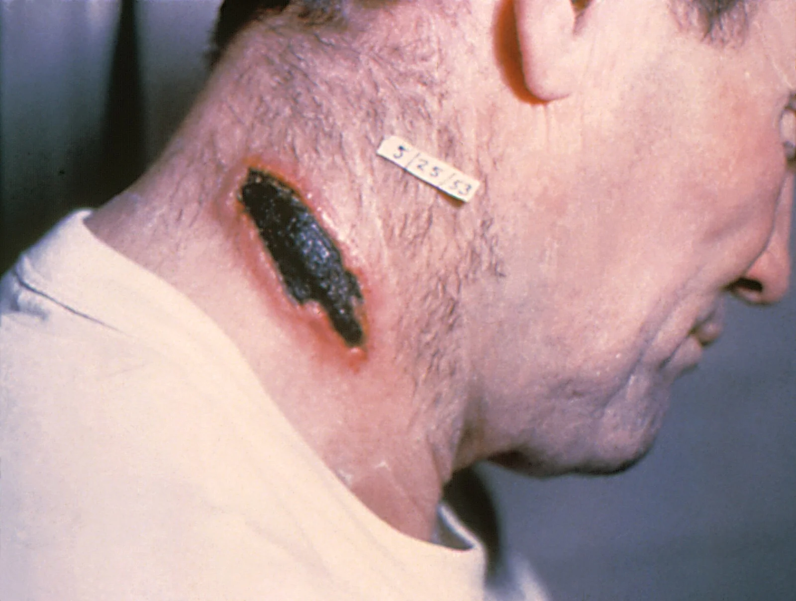
Figure 1Cutaneous anthrax lesion on the neck. PHIL 1934 lores
Long description
Clinical photograph (right lateral view of a patient’s neck) of a cutaneous anthrax lesion with characteristic coal-black coloration, caused by Bacillus anthracis.

This is the most common form and accounts for more than 95% of human cases.[1][2] Infection usually starts where spores enter through a cut or abrasion, most often on the head, neck, forearms or hands.[2] [3]
- Days one to twelve after exposure a painless, pruritic, reddish-brown papule appears.[3]
- The papule enlarges with a zone of brawny erythema and marked non-pitting oedema. Vesicles form, then central ulceration with serosanguineous exudate and a black, depressed, painless eschar (the so-called malignant pustule, though it is not a pustule).[3]
- Regional lymphadenopathy is common, with malaise, myalgia, headache, fever, nausea and vomiting.[3]
- Healing takes several weeks and the oedema resolves slowly.[3]
The eschar is characteristically painless, dry and black, surrounded by striking oedema; this combination is the clinical signature. Secondary spread occurs if the lesion is incised or manipulated.
Clinical pearlA useful clinical pointer
An eschar on an exposed site with surrounding brawny oedema and regional lymphadenopathy in a person with contact with animals or hides should make you suspect cutaneous anthrax even when the patient feels well. Early lesions can be mistaken for an infected insect bite or spider bite.[3]
This follows ingestion of inadequately cooked meat containing vegetative Bacillus anthracis, usually where mucosal disruption allows invasion.[3] The whole tract from mouth to caecum can be affected.[3] Latency is usually one to seven days.[3]
- Fever, nausea, vomiting, abdominal pain and bloody diarrhoea are common, with ascites in some patients.[3]
- Intestinal necrosis, mesenteric lymphadenitis and sepsis can follow.[3]
- Oropharyngeal anthrax produces oedematous tonsillar or pharyngeal ulcers with marked neck swelling, hoarseness, sore throat and dysphagia; airway obstruction is a risk.[3]
The deadliest form: often fatal without treatment. It is almost always an occupational disease from inhaling spores from contaminated hides or wool, or evidence of a deliberate aerosol release.[3] It must be considered an act of bioterrorism until proved otherwise whenever it is suspected.[3]
- Initial phase: a nonspecific influenza-like illness with fever, cough, chest discomfort and malaise, developing within about a week of exposure.[3]
- Severe phase: worsening fever, chest pain, severe respiratory distress, cyanosis, shock and coma within days.[3]
- Haemorrhagic necrotising mediastinal lymphadenitis spreads to adjacent structures; serosanguineous transudate, pulmonary oedema and bloody pleural effusion occur.[3]
- Typical bronchopneumonia is uncommon.[3]
- Haemorrhagic meningoencephalitis or features of gastrointestinal anthrax may develop.[3]
This is why the classic chest imaging shows a widened mediastinum and pleural effusions rather than lobar consolidation.
Rare and linked to heroin injection and needle sharing in northern Europe. It produces a severe deep soft-tissue infection with sepsis and systemic spread; there are no reported cases in people who do not inject drugs.[3]
A rare anthrax-like pneumonia in welders and metalworkers, caused by Bacillus cereus group organisms that carry anthrax toxin genes. It presents with high fever, cough, chest pain, rapid respiratory distress and hypoxia; systemic sepsis can occur.[3]
Red flagRed flags for severe or inhalational disease
| Condition | Why it can be confused | Pointer toward anthrax |
|---|---|---|
| Infected insect bite, spider bite or ecthyma | Early cutaneous papule or ulcer | Painless evolution to a black eschar with dramatic surrounding oedema and regional lymphadenopathy; relevant exposure history.[3] |
| Necrotising soft-tissue infection | Rapidly spreading erythema and necrosis | Anthrax eschar is dry, painless and depressed; patients often look less toxic early; no crepitus.[3] |
| Other occupational infections (orf, tularaemia, plague) | Ulcer with lymphadenopathy in animal-contact workers | Microscopy and culture distinguish; plague buboes, orf on fingers, tularaemia with tender nodes.[3] |
| Severe community pneumonia or mediastinitis | Chest pain, respiratory failure, mediastinal disease | Inhalation anthrax shows widened mediastinum and pleural effusions with little or no pneumonic infiltrate; prodromal influenza-like illness.[3] |
| Sepsis from any source | Fever, shock, bacteraemia | Exposure history; a cutaneous eschar; widened mediastinum; gram-positive bacilli in blood.[3] |
| Meningitis of other cause | Fever, headache, altered mental state | Anthrax meningitis often occurs secondary to systemic disease and may be haemorrhagic.[1][3] |
Every suspected case needs samples before treatment starts, as long as this does not delay therapy.[2][5] The usual set depends on the form:
- Cultures and Gram stain from clinically identified sites: cutaneous or mucosal lesions, blood, pleural fluid, cerebrospinal fluid, ascites and, when indicated, stool or rectal swabs.[3][5]
- A full-thickness skin biopsy of a papule or vesicle, including adjacent skin, before or shortly after antibiotics are started, for culture, PCR and histopathology.[5]
- Chest radiograph or CT in any patient with respiratory or systemic symptoms: expect widened mediastinum from haemorrhagic lymphadenitis and pleural effusions; pneumonic infiltrates are uncommon.[3][5]
- Lumbar puncture when meningeal signs or any change in mental status are present. If lumbar puncture cannot be done, contrast CT or MRI of the brain and spine is used to look for meningeal enhancement and haemorrhagic lesions.[3]
- Real-time PCR and immunohistochemistry can speed diagnosis.[3][5]
- Anthrax lethal factor toxin testing in plasma or serum is highly specific and rapid; plasma is preferred and samples may be collected from day zero to 18 days after suspected exposure or symptom onset.[5]
- Serology by ELISA can support the diagnosis later, confirmed by a four-fold rise in antibody titre between acute and convalescent samples, but this is not a point-of-care test.[3]
- Nasal swab testing for spores is not recommended after potential inhalation exposure because a negative result does not exclude exposure.[3]
A 10 ml blood sample in an EDTA or sodium citrate tube is required for PCR, and all specimens must be handled, packaged and shipped under the regulations for dangerous goods, after consultation with the state health department and CDC.[5]
Clinical classification of severity matters as much as the laboratory. In the CDC systemic data a patient was considered systemically ill if there was evidence of organ damage or fever or hypothermia, tachycardia, tachypnoea, hypotension, or leucocytosis or leucopenia.[1] Haemorrhagic meningitis is confirmed by CSF, which usually shows frank blood or xanthochromia.[3] Cytology, PCR and culture of CSF may be positive.
Sudden deterioration in systemic anthrax is usually due to meningitis, which is a predictor of death. CDC has derived and validated a simple screening tool: an adult is likely to have anthrax meningitis if two or more of severe headache, altered mental status, meningeal signs and other neurologic signs are present; unlikely to have it if none is present; and in an intermediate group, requiring brain imaging and lumbar puncture, if exactly one is present. The original tool had 86% sensitivity and 99% specificity.[1] A revised tool retained 86% sensitivity and 92% specificity but left only 2.5% of adults needing further testing.[1]
Predisposing clinical features flagged in the same data include fever above 38.0 degrees Celsius (above 100.4 degrees Fahrenheit), tachypnoea above 20 breaths per minute, tachycardia at or above 100 beats per minute, systolic blood pressure at or above 130 mmHg and diastolic pressure at or above 90 mmHg, meningeal signs, altered mental status, and evidence of coagulopathy.[1]
Approach to suspected anthrax
- Suspect anthrax from form and exposureCutaneous eschar with oedema; widened mediastinum after a viral prodrome; bloody diarrhoea with ascites after suspect meat; injection anthrax; unexplained sepsis after animal or hide exposure
- Ensure staff safety and keep lesion intactStandard precautions; no incision or drainage of the eschar; high-consequence pathogen, notify laboratory; state health department and CDC consultation
- Collect samples before antibiotics if feasibleBlood, lesion swab or biopsy, pleural or CSF as clinically indicated; do not delay treatment
- Question: Is the illness limited cutaneous disease?
Yes: localised cutaneous anthrax without systemic signs or meningitis concern
- Single oral antibiotic for 7 to 10 daysCiprofloxacin, doxycycline, levofloxacin or minocycline; continue 60 days if aerosol exposure possible
- Notify public health
No: inhalational, gastrointestinal, injection, severe cutaneous or systemic disease
- Start IV combination therapy promptlyTwo bactericidal drugs from different classes plus a protein or RNA synthesis inhibitor; add one antitoxin; consider meropenem if meningitis is suspected
- Imaging, lumbar puncture and critical care as neededChest radiograph or CT, pleural drainage, mechanical ventilation, vasopressors
- Continue oral monotherapy after clinical stabilityAt least 2 weeks IV first; total up to 60 days if aerosol exposure
The first principle is speed. Delayed diagnosis and treatment raise mortality sharply, so treatment must begin empirically as soon as anthrax is suspected, with antibiotic susceptibility results used to adjust the regimen.[3] The second principle is to match the antibiotic strategy to the form of disease, because toxin rather than simple infection drives severe illness. A third is source control and supportive care: continuous pleural drainage and mechanical ventilation can be life-saving in inhalation disease.[3] [1][2]
A single oral antibiotic for seven to ten days is adequate.[3] If there is a risk of inhalation exposure, therapy is continued for 60 days to complete post-exposure prophylaxis.[3] First-line empiric agents are ciprofloxacin, levofloxacin, minocycline and doxycycline.[3] Amoxicillin or penicillin V may be given if the isolate is known to be penicillin susceptible. Empiric adult oral doses used in CDC tables are ciprofloxacin 500 mg every 12 hours, doxycycline 100 mg every 12 hours, levofloxacin 750 mg every 24 hours and minocycline 200 mg once then 100 mg every 12 hours.[1] Cutaneous lesions are not drained or excised; mortality with treatment is uncommon, but the lesion will still progress through the eschar phase.[3]
Initial therapy requires three antibiotics: at least two from different classes with bactericidal activity, plus at least one protein or RNA synthesis inhibitor that may limit toxin production.[3] CDC recommends combination regimens with bactericidal agents preferred over protein synthesis inhibitors, plus one antitoxin as adjunctive therapy for noncutaneous systemic disease.[1]
A useful empiric combination is ciprofloxacin plus meropenem plus minocycline. CDC classifies regimens as follows.[1]
- Two bactericidal drugs from different classes plus a protein synthesis or RNA synthesis inhibitor.
- One bactericidal drug plus a protein synthesis inhibitor.
- Two bactericidal drugs from different classes.
- One bactericidal drug plus an RNA synthesis inhibitor (rifampin should not be used as monotherapy).
- Protein synthesis inhibitor plus RNA synthesis inhibitor.
- Two protein synthesis inhibitors.
- A single bactericidal drug.
- A single protein synthesis inhibitor.
First-line adult intravenous bactericidal options include meropenem 2 g every 8 hours, ciprofloxacin 400 mg every 8 hours, levofloxacin 500 mg every 12 hours, and penicillin G 4 million units every 4 hours or ampicillin 2 g every 4 hours for penicillin-susceptible strains; alternatives include imipenem/cilastatin 1 g every 6 hours and ampicillin/sulbactam 3 g every 6 hours.[1][3] Protein synthesis inhibitors include minocycline 200 mg once then 100 mg every 12 hours and doxycycline 200 mg once then 100 mg every 12 hours; alternatives include clindamycin, omadacycline and eravacycline.[1][3] Rifampin 600 mg every 12 hours is the RNA synthesis inhibitor.[3]
- If meningitis is suspected, meropenem is preferred because of central nervous system penetration; imipenem/cilastatin is an equivalent alternative. Intravenous therapy continues for at least 2 weeks or until the patient is clinically stable, whichever is longer.[1][3]
- Once combination intravenous therapy is completed, switch to a single oral antibiotic for 60 days to kill residual spores in the lungs.[3]
- For systemic anthrax from a non-aerosol event, prolonged post-exposure prophylaxis is not required; if aerosol exposure is possible, continue for the full 60 days from onset.[1]
- A single antitoxin is given as an adjunct: raxibacumab 40 mg/kg intravenously, obiltoxaximab 16 mg/kg intravenously, or anthrax immune globulin 420 units intravenously (840 units may be considered in severe cases), with premedication for hypersensitivity.[1]
- Raxibacumab and obiltoxaximab are also indicated, when available, as adjuncts to antibiotics in inhalation anthrax.[3]
Exam trapBeware the meningitis presentation
About one in four adults with cutaneous or gastrointestinal anthrax and one third with inhalation anthrax develop secondary meningitis in historical series. Confusion, severe headache or meningeal signs in a systemically ill patient should prompt brain imaging and lumbar puncture, with meropenem in the regimen.[1][3]
Continuous pleural fluid drainage by chest tube is critical in inhalation anthrax, and mechanical ventilation and vasopressors are often needed. Glucocorticoids may be useful for meningitis or severe mediastinal oedema, although they have not been studied adequately.[3] An antitoxin may be used if all recommended antibiotics are unavailable or inappropriate, including in cutaneous disease without meningitis.[1]
Anthrax is nationally notifiable and reportable in all US states and territories.[2] The health department should be notified immediately for any suspected or confirmed case; the health department then notifies CDC when the source is unknown, a bioterrorism exposure is suspected, or a severe naturally acquired case occurs.[3]
- Do not incise, drain or debride cutaneous lesions: this spreads organisms and worsens oedema.
- An exposed family member or healthcare worker does not need isolation: anthrax is not transmitted person to person in cutaneous, gastrointestinal or inhalation forms, although cutaneous lesions can rarely be transmitted by direct contact or fomites.[3]
- Laboratory specimens are biohazardous and must be collected, packaged and shipped only after consultation with the state health department and CDC; culture is done in the Laboratory Response Network.[5]
- Immediate guidance is available from CDC's Emergency Operations Center at 770-488-7100 (request the Bacterial Special Pathogens Branch on-call epidemiologist) for urgent clinical or public-health questions.[2]
Pregnancy and lactation. Infection in pregnant patients is associated with high maternal and foetal mortality, so pregnant patients are treated as vigorously as non-pregnant adults.[1][3] Combined with appropriate antibiotics, anthrax vaccine may be used under emergency arrangements for PEP. CDC includes doxycycline among first-line options in pregnancy because anthrax is severe and doxycycline has not been associated with the fetal problems seen with older tetracyclines; minocycline and other tetracyclines are avoided.[1][3] In practice MSD guidance prefers doxycycline, then ciprofloxacin, levofloxacin and minocycline, recognising that the usual restrictions on quinolones and tetracyclines in pregnancy are overridden by the risk of the disease.[3] [2]
Children. Usual cautions about fluoroquinolones and tetracyclines are again overridden: CDC considers doxycycline acceptable in children when tetracyclines are the preferred class, and the other first-line agents remain options, with the descending order doxycycline, ciprofloxacin, levofloxacin and minocycline.[1][3] Therapy should then be adjusted once susceptibility results and clinical recovery are known.[3]
Immunocompromised patients and older adults. These patients should receive 60 days of prophylaxis in any aerosol exposure, even if vaccine is given, and are more likely to need the full intravenous then oral course.[4]
Pre-exposure vaccination is for adults aged 18 to 65 years at high risk, such as laboratory and industrial workers handling spores, veterinarians and certain military personnel.[4] Anthrax Vaccine Adsorbed (AVA, BioThrax) is given intramuscularly as a priming series at 0, 1 and 6 months, with boosters at 12 and 18 months and annually thereafter. The 5-dose schedule of AVA is completed by these boosters.[3][4]
Post-exposure prophylaxis after a known or suspected aerosol exposure has two parts.[1][4] Antimicrobial prophylaxis and vaccination together, because animal data show that short antibiotic courses may be insufficient and vaccine meaningfully improves protection.[4]
- Healthy non-pregnant adults aged 18 to 65 years: a 60-day antibiotic course if used without vaccine; 42 days from the first vaccine dose or 2 weeks after the last vaccine dose if AVA is given on schedule; never more than 60 days.[1][4]
- Older adults (66 years or more) and immunocompromised people: 60 days.[1][4]
- Aerosol exposure in any age group: 60 days of antibiotics, with vaccine in children and adults as appropriate.[1][3]
- Non-aerosol exposures (cutaneous or ingestion): 7 days of antibiotics and no vaccine is needed.[1]
- AVA for PEP is 0.5 ml subcutaneously at 0, 2 and 4 weeks. AVA adsorbed, adjuvanted (Cyfendus) is an alternative 2-dose intramuscular series 2 weeks apart, approved for adults aged 18 to 65 years.[2][4]
- If an exposure occurs on a dose-sparing basis, 2 full doses 2 to 4 weeks apart or 3 half doses at 0, 2 and 4 weeks are acceptable in an emergency.[4]
- Antitoxins (raxibacumab or obiltoxaximab) are alternatives for PEP when antibiotics are not available or not appropriate.[4]
General prevention also involves vaccinating livestock in endemic areas, avoiding raw or undercooked meat from potentially infected animals, and occupational protection for handlers of hides, wool and hair. Cutaneous infection is usually not contagious, but people exposed to the material should be treated or observed appropriately.[3]
East Africa
In many East African settings anthrax is endemic in animals and spores contaminate soil. Local capacity may lack routine PCR, lethal-factor testing, blood cultures, antitoxins and anthrax vaccine. Diagnosis then rests on a careful exposure history, Gram stain and culture from lesion material or blood, and chest radiography where available. Treatment should still begin immediately with oral ciprofloxacin or doxycycline (or the best locally available equivalent) for limited cutaneous disease and with any locally available combination intravenous therapy for systemic illness. [1]
Practical points where advanced diagnostics are limited:
- Collect swabs, blood and any obtainable fluid before antibiotics start and keep them cold; send them to a reference laboratory, because diagnostic confirmation and susceptibility testing will guide longer-term therapy.[3][5]
- If imaging is unavailable, a patient with sudden respiratory deterioration after an influenza-like illness, widened mediastinum on any available film, or massive pleural effusion should be treated as inhalation anthrax until proved otherwise.[3]
- Drainage of pleural fluid and supportive care matter as much as antibiotics where intensive care is unavailable; refer early to a higher-level facility when possible.[3]
- Sputum Gram stain is unlikely to identify inhalation anthrax because airspace disease is frequently absent.[3]
- Vaccine, antitoxin and a full 60 day supply of antibiotics may need to be requested through national public-health or emergency channels rather than routine pharmacy procurement.
- Standard precautions are adequate for patient care; the danger is re-aerosolisation of spores from contaminated clothing, hides or soil, so contaminated materials should be bagged and discarded safely.
- Anthrax is a zoonotic, non-contagious infection caused by the spore-forming, capsule-protected bacterium Bacillus anthracis, acquired from animals, contaminated products, meat, spores in dust or air, or rarely by injection.
- Virulence comes from the capsule and from two toxins, lethal toxin and oedema toxin, which act through protective antigen.
- Cutaneous anthrax is the most common form, accounting for more than 95% of human cases; the lesion evolves from papule to vesicle to painless black eschar with marked oedema, with uncommon mortality when treated. [1]
- Inhalation anthrax is the deadliest form, presents with a flu-like prodrome followed by severe dyspnoea and shock, and shows widened mediastinum and pleural effusion rather than typical pneumonia.
- Gastrointestinal anthrax causes fever, vomiting, abdominal pain, bloody diarrhoea and ascites; injection and welder's anthrax are rare but may be lethal.
- Diagnosis is by Gram stain, culture, PCR, immunohistochemistry, lethal factor testing and, in appropriate cases, chest imaging; samples are ideally taken before antibiotics begin.
- Uncomplicated cutaneous disease is treated with a single oral antibiotic for seven to ten days. Systemic disease needs three antibiotics initially, two bactericidal from different classes plus a protein or RNA synthesis inhibitor, plus an antitoxin, with intravenous therapy for at least two weeks before oral step-down and total duration up to 60 days if aerosol exposure is possible. [1][3]
- Post-exposure prophylaxis after aerosol exposure combines antibiotics for up to 60 days with vaccine; healthy adults aged 18 to 65 can stop at 42 days if vaccinated on schedule. Non-aerosol exposures need 7 days of antibiotics. [1][4] [2][3]
- Anthrax is a notifiable disease in the United States; immediate notification goes to the local health department, which liaises with CDC. CDC accepts urgent consultation by telephone at all hours.
- Vaccines are reserved for high-risk workers before exposure and for exposed people after an event; modern mortality remains high for inhalational and meningeal disease despite treatment, so early recognition and treatment are the main determinants of survival.
Answer each one in your head first, then open the answer.
A shepherd develops a painless black eschar on his forearm with surrounding brawny oedema 5 days after skinning a goat. He is well, afebrile and has no pain. What is the most likely organism and the appropriate initial treatment?
Why does inhalation anthrax classically produce a widened mediastinum rather than lobar consolidation on chest imaging?
Show answerHide answer
Inhaled spores are carried by alveolar macrophages to the mediastinal lymph nodes, where the bacteria multiply and cause a haemorrhagic necrotising mediastinitis and bloody pleural effusions. Typical bronchopneumonia is uncommon because the process is nodal rather than airspace.
A 46-year-old man has fever, chest pain, stridor after a 3-day influenza-like illness and a widened mediastinum on CT. Which antibiotic strategy is appropriate and for how long?
Show answerHide answer
Start empiric intravenous combination therapy promptly with two bactericidal agents from different classes plus a protein or RNA synthesis inhibitor, add one antitoxin and include meropenem if meningitis is possible. Continue intravenously for at least 2 weeks or until clinically stable, then step down to oral monotherapy; give 60 days total if aerosol exposure is suspected. [3]
After a suspected aerosol release, an otherwise healthy 30-year-old woman is started on doxycycline and offered the 3-dose anthrax vaccine on schedule. How long should prophylaxis last?
Show answerHide answer
Antibiotic prophylaxis can stop 42 days after the first vaccine dose or 2 weeks after the last dose, whichever is later, and must not continue beyond 60 days. Without vaccine she would need the full 60 days. [4]
A patient with systemic anthrax becomes confused two days into treatment. What should you consider and how might the antibiotic plan change?
Show answerHide answer
Secondary anthrax meningitis is common in systemic disease. Assess brain imaging and lumbar puncture if safe, apply the meningitis prediction signs, and ensure the regimen contains meropenem because of its central nervous system penetration, with the antitoxin and supportive care continued.
What are the key virulence determinants of Bacillus anthracis and how does each contribute to disease?
Show answerHide answer
The poly-D-gamma-glutamic acid capsule resists phagocytosis, while lethal toxin causes cytokine release and vascular haemorrhage and oedema toxin causes massive local oedema. Protective antigen is the cell-binding moiety that lets both toxins enter cells.
Skin and soft-tissue infections; pneumonia; meningitis; sepsis evaluation; zoonotic infections; bioterrorism and mass-casualty preparedness; vaccination and chemoprophylaxis principles. All MedGrasp topics linked here are listed as plain text because topic linking is reserved for verified topics in that discipline.
- Bower WA, Yu Y, Person MK, et al. CDC Guidelines for the Prevention and Treatment of Anthrax, 2023. MMWR Recommendations and Reports 2023;72(6):1-47. doi:10.15585/mmwr.rr7206a1. Accessed .
- Centers for Disease Control and Prevention. Clinical Overview of Anthrax. CDC website (updated 2026). Accessed .
- Bush LM, Vazquez-Pertejo MT, peer reviewed by Tesini BL. Anthrax. MSD Manual Professional Edition (updated 2026). Accessed .
- Bower WA, Schiffer J, Atmar RL, et al. Use of Anthrax Vaccine in the United States: Recommendations of the Advisory Committee on Immunization Practices, 2019. MMWR Recommendations and Reports 2019;68(4):1-14. doi:10.15585/mmwr.rr6804a1. Accessed .
- Centers for Disease Control and Prevention. Laboratory Information for Anthrax Testing. CDC website (updated 2026). Accessed .
Figure credits
Educational content for medical students and doctors. It is not a substitute for clinical judgement or local guidelines.