Babesiosis and Rocky Mountain spotted fever
Babesiosis and Rocky Mountain spotted fever: tick-borne Babesia haemolytic anaemia and Rickettsia vasculitis, with diagnosis and treatment.
- Babesiosis is a malaria-like infection of red blood cells produced by the tick-borne parasite Babesia microti, seen mainly in the northeastern and upper midwestern United States; it is dangerous in asplenic and immunocompromised patients. [3][12]
- Rocky Mountain spotted fever (RMSF) is a rapidly progressive vasculitis caused by the intracellular bacterium Rickettsia rickettsii, spread by Dermacentor ticks, and is fatal within days if doxycycline is not given early. [7][4]
- Babesiosis presents with fever, chills, myalgias and haemolytic anaemia; the Maltese cross tetrad on a Giemsa-stained blood smear is unique to Babesia. [12]
- RMSF presents with sudden fever and headache, then a rash beginning at the wrists and ankles and spreading centrally, sometimes to the palms and soles, accompanied by thrombocytopenia and hyponatraemia. [6]
- Babesiosis is treated with atovaquone plus azithromycin, or clindamycin plus quinine, for at least 7 to 10 days, with exchange transfusion in severe disease. [1][12]
- RMSF is treated with doxycycline 100 mg every 12 hours for at least 5 to 7 days, started on clinical suspicion without waiting for laboratory confirmation. [4]
By the end of this page you should be able to:
- Name the agent, vector, reservoir and region for babesiosis and for RMSF, and explain why a tick bite in summer matters.
- Recognise babesiosis from haemolytic anaemia and the Maltese cross smear, and RMSF from fever, headache and the spreading macular rash. [6] [12]
- Order the correct confirmatory test for each, and say when serology is falsely negative. [5] [12]
- Prescribe the correct drug, dose, route, duration and adjustment for babesiosis and RMSF, with pregnancy, childhood and immunocompromise in mind. [1] [4]
- Judge severity in babesiosis and decide when exchange transfusion is indicated. [1]
- Distinguish RMSF, anaplasmosis and ehrlichiosis clinically and manage them in a setting with limited tests or drugs. [8] [9]
DefinitionBabesiosis
Babesiosis is an infection by intraerythrocytic protozoa of the genus Babesia, which are transmitted to humans by the bite of an infected Ixodes tick. In the United States the predominant species is Babesia microti. Disease ranges from silent infection to severe haemolytic anaemia and organ failure, most threatening to asplenic and immunocompromised patients. [2] [3]
DefinitionRocky Mountain spotted fever
RMSF is a tick-borne infection caused by the obligate intracellular bacterium Rickettsia rickettsii, a member of the spotted fever group rickettsiae. It infects the lining cells of blood vessels, producing a generalised vasculitis, fever, headache and rash. It is the most severe rickettsiosis in the United States. [7] [4] [6]
A useful way to think about them is that babesiosis destroys red cells, while RMSF damages vessels. Babesiosis therefore looks like malaria with haemolysis; RMSF looks like a febrile vasculitis. [12] [6]
Human babesiosis in the United States is most common in the Northeast and upper Midwest. Reported endemic areas include the coast and islands of southern New England and New Jersey, with foci in Wisconsin and Minnesota. [3] [12] Young nymphal blacklegged ticks are the usual stage that bites humans, in wooded and brushy places during the warmer months, so cases peak in late spring and summer and are often not recognised at the time because the tick is the size of a poppy seed. [3]
In 2020 there were 1827 reported cases from the 40 states where babesiosis was a reportable condition. [12] Most infections are probably asymptomatic, inferred from serologic surveys. [3][2] People residing in wooded areas with large deer populations carry the highest incidence. [12]
Elsewhere, different Babesia species infect humans through different ticks. In Europe the principal agent is Babesia divergens, classically in patients after splenectomy. [12]
Spotted fever rickettsiosis, the surveillance category that includes RMSF, is reported from every one of the lower 48 states. More than 60% of cases occur in North Carolina, Oklahoma, Arkansas, Tennessee and Missouri. [7] Although cases occur in every month, most occur during May to August. [7]
The vector is region-dependent. Dermacentor variabilis (American dog tick) predominates east of the Rocky Mountains, Dermacentor andersoni (Rocky Mountain wood tick) in the Rocky Mountain region, and Rhipicephalus sanguineus (brown dog tick) in parts of the southwestern United States and Mexico. [7] The brown dog tick lives on and near domestic dogs, in kennels, yards and houses, so in Arizona and northern Mexico transmission occurs year round with high incidence and high case fatality, particularly among children. [7]
Children represent less than 6% of spotted fever cases but 22% of spotted fever fatalities. [6]
United States
In the United States both babesiosis and RMSF are concentrated in particular regions, but the seasons and vectors differ: blacklegged nymphs for babesiosis in the Northeast and upper Midwest, and dog and wood ticks for RMSF across the country, including urban and peri-domestic settings in the Southwest. [3] [7]
Babesia microti cycles between the white-footed mouse, which is the principal reservoir, and Ixodes scapularis ticks. Larval ticks become infected while feeding on an infected rodent; after moulting into nymphs they transmit the parasite to another animal or to a human. Adult ticks usually feed on deer. [12] Many nymphs are coinfected with Borrelia burgdorferi, Anaplasma phagocytophilum, Borrelia miyamotoi or Powassan virus, so a patient may acquire more than one tick-borne infection from a single bite. [12] [10]
Rickettsia rickettsii passes to humans during the bite of an infected tick. The organism then enters the endothelial cells that line vessels, multiplies, and triggers widespread inflammation and leak. [6] Transmission through blood transfusion or organ transplantation is extremely rare or undocumented but is physiologically possible for RMSF. [7]
Babesiosis is often subclinical in healthy adults under 40 years. [12] It becomes severe or prolonged in:
- absence of a spleen or reduced splenic function; [2] [12]
- advanced age; [2]
- HIV infection and other immunocompromise; [2] [12]
- cancer, especially B-cell lymphoma; [2]
- immunosuppressive drugs, especially rituximab, corticosteroids and other causes of impaired immunity; [2]
- congestive heart failure; [2] and
- neonates exposed transplacentally, perinatally or by transfusion. [2]
For RMSF, delayed treatment, age under 10 years and glucose-6-phosphate dehydrogenase (G6PD) deficiency are markers of severe or fatal disease. [6]
After an infected nymph bites, sporozoites enter red blood cells, mature, and divide asexually. Infected cells rupture, releasing organisms that invade other red cells, and the process produces haemolytic anaemia, jaundice, haemoglobinuria and elevated liver enzymes, creatinine and urea. [12] [2] Host defence depends on CD4 T-cell immunity and on innate macrophage and natural killer cell responses; humoral immunity plays a less well defined role. [12] Splenectomy can convert an earlier silent parasitaemia into active, severe disease, which is why the spleen matters both biologically and clinically. [12]
Rickettsia rickettsii infects the endothelial cells lining blood vessels, causing vasculitis. Continued infection allows localised bleeding and clotting in the brain and other organs. [6] The resulting increase in vascular permeability explains the hyponatraemia, oedema and multi-organ involvement, and the drop in platelet count is both a diagnostic clue and a marker of severity. Skin and soft tissue necrosis, amputations and death reflect advanced vascular damage. [6] There is no evidence that Rickettsia rickettsii causes persistent or chronic disease. [6]
Symptom latency is usually 1 to 2 weeks, although Babesia infection can be subclinical for months to years. [12] When illness occurs it resembles influenza at first, with nonspecific symptoms: malaise, fatigue, chills, fever, headache, myalgia and arthralgia. [12] In healthy people these usually resolve within about a week. [12] In others there may be hepatosplenomegaly, jaundice, haemolytic anaemia, mild neutropenia and thrombocytopenia. [12]
In acutely ill patients the findings on routine tests frequently include haemolytic anaemia and thrombocytopenia, with proteinuria, haemoglobinuria, and elevated liver enzymes, blood urea nitrogen and creatinine. [2] Severe cases can produce disseminated intravascular coagulation, haemodynamic instability, acute respiratory distress, myocardial infarction, renal failure, hepatic compromise, altered mental status and potentially death. [2] In asplenic, HIV-infected or otherwise immunosuppressed patients the illness can mimic Plasmodium falciparum malaria, with high fever, severe haemolysis, haemoglobinuria, jaundice and renal failure. [12]
Figure 1Rocky mountain spotted fever
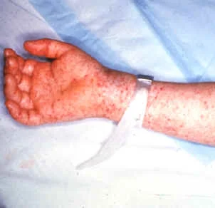
Long description
A 302 x 292 pixel clinical photograph of a hand rash caused by Rocky Mountain spotted fever, originally from the CDC.

Illness generally begins 3 to 12 days after the bite of an infected tick, with sudden onset of fever and headache. [6] Nausea, vomiting, anorexia, abdominal pain that can mimic appendicitis, myalgia and oedema of the eyelids and the dorsum of the hands are common in the first week. [6] Children are less likely to report headache but frequently have nausea, vomiting, loss of appetite, rash and abdominal pain. [6]
The rash appears 2 to 4 days after the onset of fever in a classic case, first as small, flat, pink macules on the wrists, forearms and ankles, then spreading to include the trunk and sometimes the palms of the hands and soles of the feet. [6] While 90% of people eventually develop a rash, fewer than half have it during the first 3 days, when most patients seek care, so RMSF must be suspected before the rash is present. [6] The petechial rash typically appears only on day 5 or 6 of illness and marks severe disease. [6]
General laboratory findings include thrombocytopenia and hyponatraemia, with elevated hepatic transaminases; they may not be abnormal early in illness. [6] Late disease, from day 5 onward, can include altered mental status, coma, cerebral oedema, pulmonary oedema and acute respiratory distress syndrome, skin and soft tissue necrosis with need for amputation or grafts, and multi-organ system damage. [6]
The main differential for both is malaria, especially Plasmodium falciparum, which also causes fever, haemolysis, thrombocytopenia and occasionally jaundice and renal impairment. [12] A blood smear looks for the parasites: Babesia trophozoites vary in shape and size and do not produce pigment, and the tetrad Maltese cross form, although not common, is unique to Babesia. [12] Other tick-borne illnesses (anaplasmosis, ehrlichiosis, Lyme disease) may coexist or cause a similar febrile illness, and RMSF must be separated from other spotted fevers such as Rickettsia parkeri rickettsiosis, Pacific Coast tick fever and rickettsialpox, and from meningococcaemia and other causes of fever with rash and thrombocytopenia. [6] [10]
| Infection | Agent | Vector | Rash and key labs | First-line treatment and duration |
|---|---|---|---|---|
| Babesiosis | Babesia microti (main US species) | Blacklegged or deer tick (Ixodes scapularis) | Often no rash; haemolytic anaemia, thrombocytopenia, elevated transaminases; Maltese cross on smear [3] [12] | Atovaquone 750 mg twice daily plus azithromycin 500 mg on day 1 then 250 mg daily, or clindamycin 600 mg three times daily plus quinine 650 mg three times daily, for at least 7 to 10 days [1] |
| RMSF | Rickettsia rickettsii | American dog tick, Rocky Mountain wood tick, brown dog tick (Dermacentor spp., Rhipicephalus sanguineus) | Macular rash wrists/ankles spreading to trunk, palms and soles; thrombocytopenia, hyponatraemia, transaminitis [6] [7] | Doxycycline 100 mg every 12 hours (children under 45 kg 2.2 mg per kg twice daily) for at least 3 days after fever subsides and clinical improvement; minimum 5 to 7 days [4] |
| Human granulocytic anaplasmosis | Anaplasma phagocytophilum | Blacklegged tick (Ixodes scapularis) | Rash uncommon; leucopenia, thrombocytopenia, elevated transaminases [10] | Doxycycline 100 mg every 12 hours; 10 to 14 days [8] |
| Human monocytic ehrlichiosis | Ehrlichia chaffeensis (most cases) | Lone star tick (Amblyomma americanum) | Rash in some patients; leucopenia, thrombocytopenia, transaminitis [10] | Doxycycline 100 mg twice daily; at least 5 to 7 days, until 72 hours after fever resolves [9] |
Diagnosis requires a high index of suspicion. For acutely ill patients the routine laboratory picture often includes haemolytic anaemia and thrombocytopenia, sometimes with proteinuria, haemoglobinuria and raised liver enzymes, blood urea nitrogen and creatinine. [2]
- Blood smear. Healthcare providers should explicitly request a manual (non-automated) review of the peripheral blood smear. In symptomatic acute infection Babesia parasites are typically detectable by light microscopy, but several smears may need to be examined because parasitaemia is sometimes low. Distinguishing Babesia from Plasmodium, especially P. falciparum, and from stain or platelet debris can be difficult. [2] [12] A tetrad Maltese cross is pathognomonic when seen, though not common. [12]
- Serology. Indirect fluorescent antibody testing with B. microti antigens can help in patients with low-level parasitaemia, but may be falsely negative early in infection and with other Babesia species. Antibodies persist for a year or more, so a positive antibody test should be confirmed by smear or PCR. [12]
- PCR. PCR detects and identifies the species, confirms infection at low parasitaemia, and separates Babesia from P. falciparum when smear findings are ambiguous. [12] [2]
- Coinfection testing. Because one tick may carry several pathogens, test for Borrelia, Anaplasma and other agents when the illness does not fit a single diagnosis or the region makes co-infection likely. [12] [10]
Clinical suspicion is the key step. Maintain suspicion for RMSF in nonspecific febrile illness and sepsis of unknown cause, especially in spring and summer after outdoor or dog exposure. [5]
- Serology. The standard test is IgG indirect fluorescent antibody against R. rickettsii. Antibody titres are frequently negative in the first week of illness, and one random titre is not diagnostic. Collect paired acute and convalescent sera 2 to 10 weeks apart to demonstrate a fourfold rise. Antibodies can remain elevated for months to years, and 10% or more of healthy people in some areas have elevated titres from past exposure, so timing matters. [5]
- PCR. PCR on whole blood, serum or plasma can help, especially in severe disease when rickettsiae have spread; a negative result does not exclude RMSF and should not delay treatment. Skin biopsy of a rash lesion can also be tested. [5]
- Other methods. Culture and immunohistochemistry are only available in specialised laboratories; routine hospital blood cultures do not detect R. rickettsii. [5]
- Routine tests. A complete blood count and chemistry panel show thrombocytopenia and hyponatraemia and may show elevated transaminases. [6]
There are no formal diagnostic criteria for either condition; both are diagnoses of clinical suspicion supported by epidemiology and laboratory evidence. [5] [12]
For babesiosis, severity is driven by parasitaemia and end-organ compromise. Severe disease is suggested by high-grade parasitaemia above 10% of red cells, severe haemolytic anaemia, or declining pulmonary, renal or hepatic function. [1] [12] Asplenic, HIV-infected and other immunocompromised patients warrant the closest monitoring because relapse can occur. [2]
For RMSF, severity is clinical: late illness with petechiae, altered mental status, respiratory compromise, bleeding or coagulopathy, multi-organ damage, skin necrosis, amputation or G6PD deficiency. [6] There is no validated scoring system in the sources used here. [6]
Diagnosis and treatment of babesiosis
- Patient with fever, chills, myalgia and haemolytic anaemia after tick exposureThink especially of asplenic, older or immunosuppressed patients; consider malaria
- Manual peripheral blood smear and, if available, PCRLook for Babesia trophozoites in red cells; a Maltese cross tetrad is diagnostic when seen
- Question: Is the patient symptomatic?
No, parasitaemia found and cleared or absent
- Usually do not treatTreat if parasitaemia persists for 1 month or longer
Yes, symptomatic babesiosis
- Preferred: atovaquone plus azithromycin (doses below)See the preferred regimen in the list below
- Alternative: clindamycin plus quinine (doses below)
- Continue at least 7 to 10 days; immunocompromised patients often need 6 or more weeks
- Hospitalised severe disease: use intravenous regimens, then oral to complete 7 to 10 days
- Severe disease with high parasitaemia or end-organ decline: add exchange transfusion and supportive care
- Preferred regimen. Atovaquone 750 mg orally twice daily (with food, preferably a fatty meal, to improve absorption) plus azithromycin 500 mg orally on the first day and 250 mg orally daily thereafter. [1]
- Alternative regimen. Clindamycin 600 mg orally three times daily plus quinine 650 mg orally three times daily. This combination is the usual choice when atovaquone is not tolerated or available, and it is generally the recommended regimen in pregnancy. [1]
- Severe or hospitalised disease. Atovaquone 750 mg orally twice daily plus azithromycin 500 mg intravenously daily, or clindamycin 600 mg intravenously four times daily plus quinine 650 mg orally three times daily. After symptoms subside, switch to oral doses and complete a total of 7 to 10 days. [1]
- Immunocompromise and pregnancy. Highly immunocompromised patients may need at least 6 consecutive weeks, with daily smears until parasitaemia is under 4%, then at least weekly, continuing until parasites are undetectable on smears for 2 consecutive weeks. [1] Atovaquone is pregnancy category C and there are limited data, so quinine plus clindamycin is generally used in symptomatic pregnancy unless those drugs are not available or tolerated. [1] Atovaquone has been used safely in children who weigh over 5 kg; azithromycin is pregnancy category B. [1]
- Higher azithromycin doses. A dose of 1000 mg followed by 500 mg daily has been used in immunocompromised patients and in more severe disease. [1]
- Exchange transfusion. Used for high-grade parasitaemia over 10%, or moderate to high-grade parasitaemia accompanied by severe haemolytic anaemia or declining pulmonary, renal or hepatic function; other supportive care includes blood transfusion, antipyretics, vasopressors, mechanical ventilation and dialysis. [1] [12]
- Paediatric and lactation notes. Atovaquone should be used with caution during breastfeeding when the infant weighs under 5 kg. [1] Senior review and local formulary advice apply in pregnancy and childhood. [1]
- Drug of choice. Doxycycline for all patients, including children of all ages and pregnant women, because other antibiotics increase the risk of severe illness and death. [4] In adults the dose is 100 mg every 12 hours; in children under 45 kg it is 2.2 mg per kg body weight twice daily. [4]
- Timing and duration. Start doxycycline when RMSF is suspected, ideally within the first 5 days of illness, and do not wait for laboratory confirmation. [4] Fever usually subsides within 24 to 48 hours, and treatment continues for at least 3 days after fever subsides with evidence of clinical improvement, with a minimum course of 5 to 7 days. [4]
- Alternatives. Chloramphenicol is the only alternative used, but patients treated with it have a higher death risk in epidemiologic studies, oral formulations are not available in the United States, and it carries risks including aplastic anaemia and grey baby syndrome. [4] In severe doxycycline allergy, inpatient rapid desensitisation may be considered. Sulfa-containing drugs may worsen the clinical course and increase the likelihood of death from RMSF; other broad-spectrum antibiotics are not effective. [4]
- Pregnancy and children. Short courses of doxycycline of 5 to 10 days have not been shown to stain permanent teeth or cause enamel hypoplasia, and both the CDC and the American Academy of Pediatrics recommend it for suspected rickettsial disease in children. [4] Suspected RMSF in pregnancy should be discussed with an infectious disease clinician. [4]
- Post-bite antibiotic prophylaxis is not recommended to prevent RMSF, anaplasmosis, ehrlichiosis or babesiosis. People who have been bitten should be advised to watch for symptoms and seek care if fever, rash, headache or other symptoms develop within 2 weeks. [4] [8] [9]
Babesiosis complications are the consequences of uncontrolled haemolysis and microvascular compromise: noncardiac pulmonary oedema, disseminated intravascular coagulation, haemodynamic instability, acute respiratory distress, myocardial infarction, renal failure, hepatic compromise, altered mental status and potentially death. [2] [12] Immunocompromised patients can relapse, and combinations such as atovaquone plus azithromycin plus clindamycin, or atovaquone plus azithromycin plus clindamycin plus quinine, have been used in selected cases on specialist advice. [1]
RMSF complications arise from vascular damage: petechiae, thrombocytopenia, hyponatraemia, multi-organ dysfunction, cerebral oedema and neurological deficits, and skin and soft tissue necrosis that may require amputation. The disease has no chronic or persistent form. [6]
Red flagA tick-borne febrile illness in an asplenic or immunocompromised patient
Babesiosis is often asymptomatic in healthy younger adults, resolves within a week in many others, and can leave PCR positivity and fatigue for weeks after treatment without indicating failure. [12] [1] Mortality is concentrated in older, asplenic, HIV-infected and immunosuppressed patients. [12] [2]
RMSF is fatal within days without early doxycycline, but patients who receive it early generally recover fully, and those without early vascular damage usually make a full recovery even after severe illness. [6] [4]
There is no vaccine for babesiosis, so prevention depends on avoiding ticks. [2] The practical measures are therefore tick avoidance and prompt removal: [11]
- know where ticks live (grassy, brushy or wooded areas, including gardens) and walk in the centre of trails; [11]
- use EPA-registered repellents containing DEET, picaridin, IR3535, OLE, PMD or 2-undecanone, with sunscreen applied first and repellent second; [11]
- treat clothing and gear with 0.5% permethrin and consider permethrin-treated clothing; [11]
- remove attached ticks promptly without crushing them; [12] [11]
- shower within 2 hours of coming indoors, do full body and gear checks, and tumble dry clothes on high heat for 10 minutes; [11]
- reduce ticks around the home by removing leaf litter, clearing tall grass and brush, creating a wood-chip or gravel barrier, and mowing regularly. [11]
People who have had babesiosis should not donate blood or organs until treatment is complete and parasitaemia is no longer detectable; screening of blood and organ donors is carried out in high-incidence areas of the United States. [12] For RMSF prevention rests on tick avoidance, early recognition and immediate treatment; no antibiotic is recommended after an asymptomatic bite. [4] [11]
- Pregnancy. For babesiosis, quinine plus clindamycin is generally recommended over atovaquone regimens in symptomatic pregnancy because more safety data are available, although quinine and clindamycin themselves have limited pregnancy data and require specialist judgement. [1] Atovaquone is pregnancy category C and azithromycin pregnancy category B. [1] Transplacental babesiosis can cause neonatal fever, anaemia and thrombocytopenia. [12] For RMSF, doxycycline is still recommended and pregnant patients should be counselled on benefits and risks, with specialist review where the disease appears mild. [4]
- Children. Dosing is by body weight for both diseases, and both CDC and the American Academy of Pediatrics recommend doxycycline for suspected rickettsial disease in children because short courses do not stain permanent teeth. [1] [4]
- Asplenia, HIV and cancer. Babesiosis in these patients is more severe, more prolonged and prone to relapse; treat for 6 or more weeks with close smear monitoring. [2] [1] Asplenic travellers to endemic areas need particular caution. [12]
- Older adults. Older age is a risk factor for severe babesiosis and RMSF carries higher case-fatality. [2] [6]
- G6PD deficiency. Patients with this deficiency have more severe RMSF. [6]
Babesiosis is concentrated in the United States, so outside endemic areas imported or transfusion-associated infection is rare but occurs. [3] [12] Where blood smears and PCR are unavailable, diagnosis depends on clinical suspicion, epidemiological exposure, and microscopy of a Giemsa-stained thin smear, which is cheap but operator-dependent; repeated smears increase yield. [2] [12] Where atovaquone is unavailable or unaffordable, quinine plus clindamycin is an effective alternative. [1] [12]
RMSF should be suspected in any patient with undifferentiated fever, severe headache and tick exposure in an endemic region; doxycycline is inexpensive, widely available and lifesaving, and should be given without waiting for confirmation. [4] [5] Where hospital capacity is limited, oral doxycycline and attention to fluids, electrolytes and tissue perfusion are the priorities; exchange transfusion for severe babesiosis, intensive support and specialist care may require referral. [1] [4] Removal of an attached tick should be done with steady traction using fine tweezers rather than irritants. [12]
NoteCo-infection and competing diagnoses
In low-resource settings a febrile, anaemic patient may simultaneously have malaria, babesiosis and a rickettsial infection. Where diagnostics are scarce, tailor empiric cover to local epidemiology and to clues such as haemolysis, rash, exposure and the season, and reassess when treatment fails. [10] [12]
- Babesiosis is a tick-borne Babesia microti infection of red cells in the northeastern and upper midwestern United States, diagnosed on a manual blood smear and PCR, and treated with atovaquone plus azithromycin or clindamycin plus quinine for 7 to 10 days. [3] [1]
- RMSF is a Rickettsia rickettsii infection of blood vessels spread by Dermacentor and brown dog ticks; it is the most severe rickettsiosis in the United States and fatal within days if not treated. [7] [4]
- Susceptibility to severe babesiosis is highest with asplenia, advanced age, HIV infection, B-cell malignancy, immunosuppressive therapy and congestive heart failure. [2]
- Severe babesiosis is marked by parasitaemia above 10%, severe haemolytic anaemia and pulmonary, renal or hepatic compromise; exchange transfusion is added to drug therapy. [1]
- The Maltese cross tetrad, seen occasionally on smear, is unique to Babesia but is not common; smear and PCR together confirm the diagnosis. [12]
- RMSF begins 3 to 12 days after the bite with sudden fever and headache; a rash begins at the wrists and ankles 2 to 4 days after fever onset and may spread to the palms and soles. [6]
- Fewer than half of RMSF patients have a rash in the first 3 days, so treat before a rash appears. [6]
- RMSF diagnosis rests on serology, with IgG indirect fluorescent antibody on paired sera 2 to 10 weeks apart, or PCR, but do not wait for results. [5]
- Doxycycline is first-line for RMSF, ehrlichiosis and anaplasmosis in adults and children, including pregnant women, at 100 mg every 12 hours in adults and 2.2 mg per kg twice daily in children under 45 kg. [4]
- Antibiotic prophylaxis after a tick bite is not recommended to prevent RMSF, anaplasmosis, ehrlichiosis or babesiosis. [4] [8] [9]
- Ongoing prevention relies on tick avoidance, repellents, permethrin on clothing, body checks and yard maintenance. [11]
- Co-infection from one tick is common, so severe or atypical disease may need broader evaluation. [10] [12]
Answer each one in your head first, then open the answer.
A 60-year-old man returns from Nantucket with fever, chills and haemolytic anaemia. He had a splenectomy 5 years ago. A single peripheral smear is negative. Which test is most useful now, and what empirical treatment should be started?
Show answerHide answer
Send PCR for Babesia and request a manual review of repeated smears, because parasitaemia can be low. Start atovaquone 750 mg orally twice daily with food plus azithromycin 500 mg orally on day 1 then 250 mg daily, and arrange close haematological monitoring because his splenectomy puts him at high risk of severe disease. [2] [1]
A 40-year-old woman in Tennessee has a tick bite two weeks ago and now has fever 39 degrees C, severe headache, nausea, platelets 80,000, sodium 128 and normal glucose. A faint macular rash is on her wrists and ankles. How should she be treated today?
Show answerHide answer
Start doxycycline 100 mg every 12 hours on suspicion of Rocky Mountain spotted fever today, without waiting for serology, which is often negative in the first week. Arrange a complete blood count and chemistry panel, advise that fever may settle in 24 to 48 hours, and continue treatment for at least 3 days after defervescence with a minimum of 5 to 7 days total. [4] [5]
Which blood smear finding separates babesiosis from malaria, and how common is it?
Show answerHide answer
Tetrad forms in a Maltese cross appear as four merozoites arranged in a cross; this configuration is unique to Babesia, but it is not common. Babesia trophozoites also vary in shape and size and do not produce pigment. In difficult cases use PCR. [12]
A pregnant woman in the second trimester has confirmed babesiosis with rising parasitaemia but is haemodynamically stable. Which drug combination is preferred, and what maternal and fetal cautions apply?
Show answerHide answer
Quinine 650 mg orally 3 times daily plus clindamycin 600 mg orally 3 times daily is generally preferred in pregnancy to atovaquone-based regimens. Atovaquone is pregnancy category C with limited data, so it is reserved for when quinine and clindamycin are not available or tolerated, with specialist advice. [1]
A 6-year-old child in an endemic area has suspected Rocky Mountain spotted fever. Her mother is worried that doxycycline will stain the child's permanent teeth. What is appropriate?
Show answerHide answer
Prescribe doxycycline at 2.2 mg per kg body weight orally twice daily. Short courses of 5 to 10 days have not been shown to stain permanent teeth or cause enamel hypoplasia, and doxycycline reduces the risk of severe illness and death. Chloramphenicol is the only alternative but carries higher mortality risk and availability problems. [4]
When is exchange transfusion added in babesiosis, and what is the usual drug duration in a highly immunocompromised patient?
Show answerHide answer
Exchange transfusion is indicated for high-grade parasitaemia over 10%, or moderate to high-grade parasitaemia with severe haemolytic anaemia or decline in pulmonary, renal or hepatic function. In highly immunocompromised patients the course is typically 6 or more weeks, continued until parasites are undetectable on smears for 2 consecutive weeks. [1]
Why is antibiotic prophylaxis not given after a tick bite to prevent Rocky Mountain spotted fever, ehrlichiosis, anaplasmosis or babesiosis?
Show answerHide answer
Babesiosis and Rocky Mountain spotted fever sit alongside other tick-borne and infectious diseases on MedGrasp, including Lyme disease, malaria, meningitis and tuberculosis. Cross-links between verified topics are added as their pages are confirmed.
- Centers for Disease Control and Prevention. Clinical Care of Babesiosis. Babesiosis: Clinical Care and Treatment for Health Care Providers (reviewed 13 Feb 2024). Accessed .
- Centers for Disease Control and Prevention. Clinical Overview of Babesiosis. Babesiosis: Clinical Overview for Health Care Providers. Accessed .
- Centers for Disease Control and Prevention. How Babesiosis Spreads. Babesiosis: How It Spreads. Accessed .
- Centers for Disease Control and Prevention. Clinical Care of Rocky Mountain Spotted Fever. Rocky Mountain Spotted Fever: Clinical Care and Treatment for Health Care Providers (updated 6 Mar 2025). Accessed .
- Centers for Disease Control and Prevention. Clinical and Laboratory Diagnosis for Rocky Mountain Spotted Fever. Rocky Mountain Spotted Fever: Clinical and Laboratory Diagnosis for Health Care Providers. Accessed .
- Centers for Disease Control and Prevention. Clinical Signs and Symptoms of Rocky Mountain Spotted Fever. Rocky Mountain Spotted Fever: Clinical Signs and Symptoms for Health Care Providers. Accessed .
- Centers for Disease Control and Prevention. Clinical Overview of Transmission and Epidemiology of Rocky Mountain Spotted Fever. Rocky Mountain Spotted Fever: Transmission and Epidemiology for Health Care Providers. Accessed .
- Centers for Disease Control and Prevention. Clinical Care of Anaplasmosis. Anaplasmosis: Clinical Care and Treatment for Health Care Providers (updated 30 Jan 2025). Accessed .
- Centers for Disease Control and Prevention. Clinical Care of Ehrlichiosis. Ehrlichiosis: Clinical Care and Treatment for Health Care Providers (updated 15 May 2024). Accessed .
- Centers for Disease Control and Prevention. Clinical Overview of Ehrlichiosis. Ehrlichiosis: Clinical Overview for Health Care Providers. Accessed .
- Centers for Disease Control and Prevention. Preventing Tick Bites. Ticks: Preventing Tick Bites (updated 28 Aug 2024). Accessed .
- Marie C, Petri WA Jr. Babesiosis. MSD Manual Professional Version, Infectious Diseases: Extraintestinal Protozoa (reviewed Sept 2025; last updated Sept 2026). Accessed .
Figure credits
Educational content for medical students and doctors. It is not a substitute for clinical judgement or local guidelines.