Brain abscess
A focal pus collection in the brain: sources, bugs, ring-enhancing imaging, why lumbar puncture is dangerous, and how to treat it.
- A brain abscess is a focal collection of pus inside the brain parenchyma, progressing from cerebritis to an encapsulated mass.
- It is rare but life-threatening. Men are affected two to three times more often than women.
- Infection reaches the brain by contiguous spread (25 to 50%, sinuses, ear, teeth), haematogenous spread (15 to 30%, endocarditis, lung) or direct inoculation (8 to 19%, trauma, surgery). Up to 40% are cryptogenic. [1]
- Fever is absent in 30 to 76% of cases. The classic picture is raised intracranial pressure plus focal signs. [1]
- Imaging shows a ring-enhancing lesion. Lumbar puncture is never indicated and can be dangerous.
- Treatment is drainage plus 4 to 8 weeks of intravenous antibiotics; the ESCMID guideline recommends a third-generation cephalosporin plus metronidazole.
- Primary antiepileptic prophylaxis is not routinely recommended, but epilepsy is common later. [2]
By the end of this page you should be able to:
- List the three routes of infection with their proportions and typical source lesions.
- Give the organisms for community, traumatic, cardiac and immunocompromised abscesses.
- Recognise the clinical presentation and explain why fever is often absent.
- Interpret CT or MRI ring enhancement and explain why lumbar puncture is contraindicated. [1]
- Plan surgical drainage, antimicrobial choice, duration, steroid use and seizure care.
- Distinguish a brain abscess from other ring-enhancing lesions.
- Adapt management to low-resource settings.
DefinitionBrain abscess
A focal, intraparenchymal infection that begins as localised cerebritis and becomes an encapsulated collection of pus. It is one of the intracranial suppurative disorders, alongside subdural empyema, epidural abscess and ventriculitis.
Solitary abscesses usually arise from contiguous spread; multiple scattered lesions point to haematogenous spread. A cryptogenic abscess (no source found, up to 40%) often proves to come from a silent right-to-left cardiac shunt such as a patent foramen ovale. [1]
Brain abscess is uncommon, but it is a major problem where oral hygiene, ear and sinus disease, and access to care are poor. Men are affected two to three times more often than women, the disease is most dangerous in the fourth decade and in young children, and paediatric cases account for roughly a quarter of hospital series. The share of intracranial masses that are abscesses is roughly 8% in developing countries versus 1 to 2% in the West. Mortality has fallen from about 50% to about 20%, and is under 10% in experienced centres. Epilepsy is reported in up to 80% of survivors in some series. A low Glasgow Coma Scale score at presentation marks a worse outcome. [1]
| Route | Share | Typical sources | Usual site |
|---|---|---|---|
| Contiguous | 25 to 50% | Sinusitis, otitis media, mastoiditis, dental infection | Frontal from sinus; temporal or cerebellum from ear |
| Haematogenous | 15 to 30% | Endocarditis, lung abscess, skin, pelvis or abdomen | Multiple lesions in middle cerebral artery territory |
| Direct | 8 to 19% | Penetrating head injury or neurosurgery | Along the track |
| Cryptogenic | up to 40% | Often a silent right-to-left cardiac shunt | Variable |
A review of imaging by source reports traumatic abscesses left-sided in 64.3% of cases; rhinogenic abscesses frontal in 83.8% and parietal in 7.1%; otogenic abscesses temporal 47.9% and cerebellar 45.5%; cardiac-source abscesses parietal 57.7% or frontal 34.6%.[1]
Risk factors include cyanotic congenital heart disease, advanced HIV disease, immunosuppressive drugs, diabetes, poor nutrition and chronic sinus or ear infection.
Streptococcus species, aerobic and anaerobic, cause about 70% of bacterial brain abscesses.[1]
- Community (sinus, ear, teeth): Streptococcus species, Bacteroides, Prevotella melaninogenica, Propionibacterium, Fusobacterium and Actinomyces, plus Gram-negative rods such as Morganella morganii and Pseudomonas.[1]
- Trauma or neurosurgery: Streptococcus viridans, the Streptococcus milleri (anginosus) group, Staphylococcus aureus, and Gram-negative rods (Klebsiella, Pseudomonas, Escherichia coli, Proteus).[1]
- Cardiac source: Peptostreptococcus and Streptococcus (viridans and microaerophilic species).[1]
- Immunocompromised host: the usual organisms plus Pseudomonas, Toxoplasma gondii, Listeria, Nocardia, Aspergillus, Cryptococcus and other fungi; reduced T-cell function invites Nocardia asteroides and Toxoplasma gondii, for which a sulphonamide or pyrimethamine-based therapy is most effective, while cryptococcal disease is treated with amphotericin plus 5-flucytosine.[1]
The Streptococcus milleri group is overrepresented in sinus and dental abscesses and tends to produce multiloculated pus. Polymicrobial growth occurs in more than a third of otogenic and metastatic abscesses.[1]
Infection reaches the brain along venous channels (contiguous spread, explaining the frontal lobe pattern in sinusitis) or through the arterial blood (haematogenous spread, near the grey-white junction). The capsule is thick and complete on the cortical side and thin on the ventricular side, which is why inward rupture produces sudden devastating ventriculitis while outward rupture may create a subdural empyema. Oedema is driven by inflammatory mediators as well as by water shifts. Hyphal fungi can invade and thrombose medium-sized cerebral arteries, so fungal abscess may present with infarction or haemorrhage.
Typical symptoms include fever (often absent, see below), headache, altered consciousness, nausea, vomiting, fits, poor balance, dysphagia and focal weakness.[1] Raised intracranial pressure gives a worsening headache (worst in the morning), projectile vomiting and drowsiness. Fits occur in 25 to 43% of patients at presentation.[1] Frontal lobe abscesses are "silent" and can smoulder for weeks. Occipital lesions can rupture into the ventricle or thrombose the transverse sinus, raising pressure and causing seizures.
Look for papilloedema, focal weakness or cranial nerve palsies, cerebellar signs and nuchal rigidity. Symptoms range from one day to eight weeks, averaging about 11 days.[1] Two traps matter most:
- Fever is absent in 30 to 76% of cases.[1] An afebrile patient with progressive headache and focal signs is not free of abscess.
- Antibiotics and analgesics can mask symptoms, allowing the abscess to mature, which is why late presentation is common in children and in areas with poor access to care.
In a large South African series (973 patients) trauma was the commonest cause (32.8%) and otorhinogenic (sinus or ear) infection accounted for 38.5% of an ear-disease cohort; most patients were young men.[1]
A 28-year-old man with a ventricular septal defect (a right-to-left shunt) presents after three weeks of frontal headache and increasing sleepiness, but never had fever. Examination shows left hemiparesis and papilloedema. MRI shows a single right cerebral abscess with a thin, uniform ring and marked oedema. Even without fever, the shunt and the imaging make the diagnosis, and this is one of the cryptogenic abscesses that turn out to be cardiac in origin.
Each lesion in the table below is separated mainly by the company it keeps (source, immunity, vascular history) rather than by a single radiological sign, which is why a thorough source search is as important as the scan.
| Lesion | Clues that favour it over bacterial abscess |
|---|---|
| Glioma or metastasis | Irregular, thick or nodular wall; disproportion of mass effect; no fever |
| Cerebral toxoplasmosis | Immunocompromised host; multiple basal ganglia lesions; eccentric target sign; responds to therapy |
| Tuberculoma | TB risk; basal cistern disease; conglomerate lesions; acid-fast bacilli |
| Fungal abscess | Profound immunocompromise; angioinvasion, vascular occlusion, haemorrhage; thin capsule |
| Resolving haematoma or infarct | Vascular history; blood products on MRI; rim fades over weeks |
| Radiation necrosis | Prior radiotherapy; delayed onset; sterile rim |
| Neurocysticercosis | Endemic area; scolices; multiple small lesions; calcification |
| Demyelinating plaque | Younger patient; incomplete rim; ovoid, periventricular |
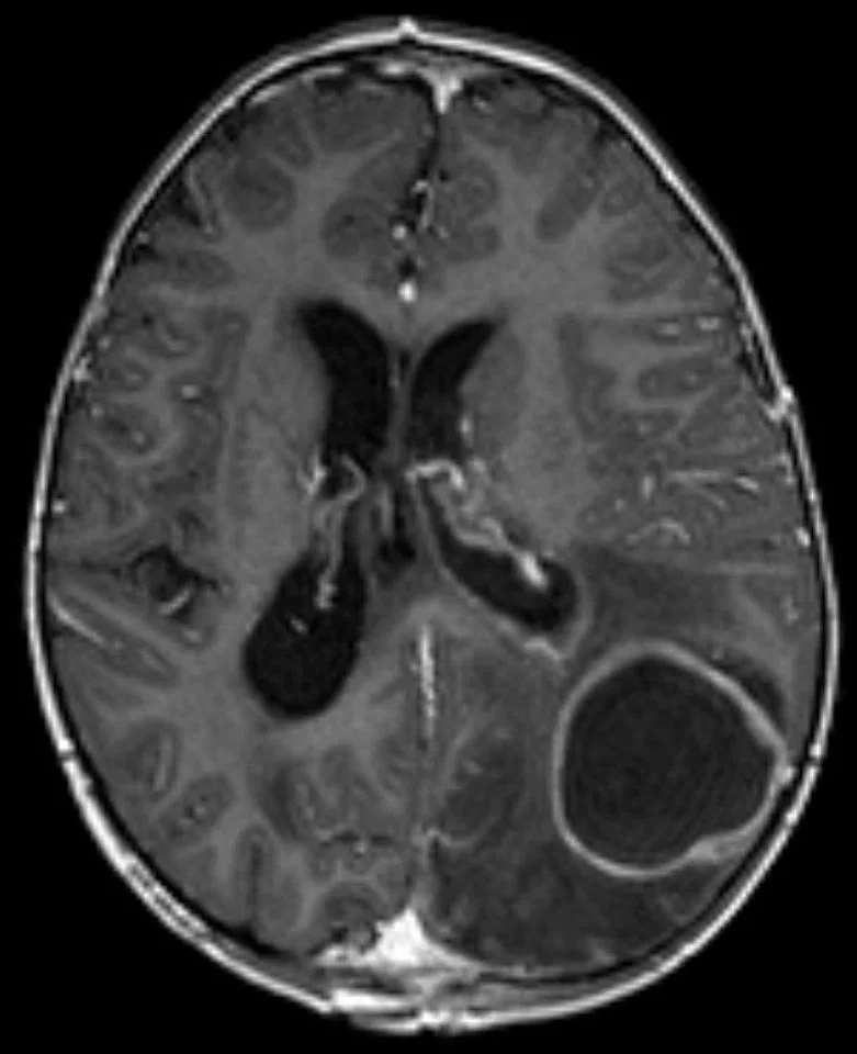
A brain abscess appears as a hypodense rounded collection with a contrast-enhancing ring and marked surrounding vasogenic oedema.[1] A complete uniform ring typifies the mature stage; nodularity or irregularity favours tumour or fungal infection.[1] DWI usually shows restricted diffusion in a pyogenic cavity (but not always), and MR spectroscopy can help separate abscess from necrotic tumour.
Approach to suspected brain abscess
- Suspect brain abscessRisk factors: sinus, ear or dental infection, trauma, surgery, cyanotic heart disease, immunocompromise
- Urgent brain imagingMRI preferred; CT with contrast if MRI unavailable. Do not perform a lumbar puncture
- Question: Ring-enhancing collection with surrounding oedema present?
No
- Reconsider the differentialTumour, toxoplasmosis, tuberculosis, stroke, demyelination
Yes
- Large abscess over 2.5 cm or mass effect present?Size, location, ventricular involvement, clinical state
- Question: Respiratory and neurological status permits intervention?
Yes, surgery available
- Drain: aspiration first, excision if neededObtain pus for culture and molecular tests
No or no surgical service
- Start empirical antibiotics and referThird-generation cephalosporin plus metronidazole; escalate where possible
- Send blood cultures and source investigationsCardiac echo, chest imaging, dental and sinus review
- Match antibiotics to host and sourceAdd vancomycin for trauma or surgery; add TMP-SMX and voriconazole in severe immunocompromise
- Severe perilesional oedema or herniation?Glucocorticoids for severe symptoms only
- Follow imaging and reassessRepeat scan at 2 to 3 weeks; complete 6 to 8 weeks and treat seizures as they occur
Figure 1Brain abscess - MRI T1 KM axial
Long description
An axial T1-weighted MRI after contrast showing a brain abscess with classic ring-shaped enhancement and marked perifocal edema.

MRI is the imaging investigation of choice; CT with contrast is used where MRI is unavailable or the patient is unstable.[2] A mature abscess shows a complete uniform ring, mass effect and surrounding (sometimes marked) vasogenic oedema, and in early cerebritis there is only patchy enhancement, so small abscesses can be missed on a single scan.
A brain abscess is the only central nervous system infection in which a lumbar puncture is never recommended and may be contraindicated.[1]
- It is unhelpful: cerebrospinal fluid shows a nonspecific pleocytosis with raised protein, grows no organism in 41.5% of cases, shows meningitis in 36.8% and is normal in the remainder. [1]
- It is dangerous: the abscess is a space-occupying mass, so intracranial pressure is often raised, and removing fluid below can create a pressure gradient that drives the brainstem downward (herniation). In one large series an LP had been performed in 19.8% of patients before specialist transfer; 13.5% of these deteriorated neurologically afterwards, with 7 deaths.[1]
Image the brain first, then drain the lesion for microbiology; do not touch the spine. Other tests are blood and source cultures, plus a formal search for the source (cardiac echo, chest imaging, dental and sinus review).
Successful care needs neurosurgery, infectious diseases, neurology, neuroradiology and microbiology working together.
Drainage is both therapeutic and diagnostic. Aspiration (burr hole or stereotactic) is the preferred first method and may be repeated. Excision is reserved for thick or multiloculated pus, cerebellar or brainstem lesions (risk of herniation), periventricular lesions (risk of ventricular rupture) and retained foreign bodies. Multiple abscesses are managed by aspirating the largest for diagnosis and draining the others if they cause mass effect. Non-operative treatment is an option for small encapsulated lesions under about 2 cm; failure (growing abscess or no change at 2 to 3 weeks) is an indication to drain. Bacterial abscesses over 2.5 cm generally require surgical drainage. Urgent evacuation is required for co-existing subdural empyema or cerebellar abscess. [1]
- Community-acquired disease in an immunocompetent adult: ESCMID recommends a third-generation cephalosporin (such as ceftriaxone) plus metronidazole; vancomycin is added after recent trauma or neurosurgery, or where Staphylococcus (including resistant strains) is suspected.[1][2]
- Severe immunocompromise: add trimethoprim-sulfamethoxazole and voriconazole.[2]
- Post-neurosurgical or post-traumatic disease: a carbapenem plus vancomycin or linezolid.[2]
Duration is 4 to 6 weeks intravenously for surgically drained abscesses and 6 to 8 weeks where the lesion is treated medically or lesions are multiple.[1] ESCMID settles on 6 to 8 weeks for aspirated or conservatively treated abscesses, with a shorter course possible after complete surgical excision, and does not routinely recommend oral consolidation after six weeks of intravenous therapy (no data support an early switch).[2] In immunocompromised patients antibiotics are given for 3 to 12 months.[1]
There is no randomised trial of corticosteroids in brain abscess. Current guidance supports them only for severe symptoms from perilesional oedema or impending herniation.[2] They should not be used routinely, because they blunt inflammation, reduce visibility on imaging and can worsen uncontrolled infection. Be cautious where tuberculosis is possible, since a ring-enhancing lesion may be tuberculous.
Seizures are part of the disease: 25 to 43% at presentation, with epilepsy in roughly 70% of survivors at 30 years.[1] Despite this, ESCMID recommends against primary prophylaxis, because the evidence of benefit is very weak.[2] Treat seizures when they occur. In post-operative patients, some discontinue antiepileptics after 2 seizure-free years with a normal electroencephalogram; others continue longer. Children who have seized are usually treated, with at least 3 months of prophylaxis if seizure-free.[1]
Repeat imaging, completion of targeted antibiotics, monitoring for recurrence (a risk for years), source control, seizure review and rehabilitation.
The clinical and radiological pictures do not always move together. In the cerebritis stage the brain shows patchy inflammation with little ring enhancement: the scan can look modest while the patient is deteriorating. As a capsule forms the collection becomes self-contained, ring enhancement becomes complete and uniform, and oedema usually peaks. In the late capsule stage the wall matures and enhancement may thin even before the cavity shrinks. That dissociation is why clinical response, inflammatory markers and repeated imaging together, rather than any single scan, should steer the duration of treatment. [1]
- Raised intracranial pressure, herniation and death (under 10% in modern series with early diagnosis).
- Intraventricular rupture, with ventriculitis and meningitis.
- Hydrocephalus.
- Seizures and epilepsy, both acutely and in the long term.
- Permanent focal neurological deficit.
- Cerebral venous sinus thrombophlebitis.
- Recurrence after incomplete drainage.
Outcome has improved dramatically: mortality from about 50% historically to about 20% now, under 10% in experienced centres. Neurological state at presentation matters more than lesion size. Early imaging, source control and prolonged antibiotics turn a fatal mass into a treatable infection.
Prevention is practical: good dental and oral hygiene, prompt treatment of sinus and ear infection, control of congenital and acquired shunts, structured review for survivors, and (in immunocompromised patients) good antiretroviral therapy, careful immunosuppression and glycaemic control.
- Immunocompromised patients: risk from Toxoplasma, Nocardia, Listeria, Pseudomonas and fungi; imaging often shows multiple lesions without a thick capsule. ESCMID adds trimethoprim-sulfamethoxazole and voriconazole and treatment is prolonged (3 to 12 months).[1][2]
- Children: roughly a quarter of cases; often late presentation; consider a cardiac right-to-left shunt or trauma.
- Pregnancy: imaging without gadolinium, standard drainage and safe antibiotics (metronidazole, third-generation cephalosporins), with obstetric input.
- Cyanotic congenital heart disease: single streptococcal abscess, often cortical, with need for cardiac and microbiological work-up and shunt discussion.
Most patients live in settings without MRI, stereotactic drainage or nearby neurosurgery.
- Image the brain before any lumbar puncture in suspected meningitis with focal signs or papilloedema. CT alone is adequate in the mature stage.
- Clinical suspicion is the key diagnostic tool: subacute headache with focal weakness, chronic ear or sinus disease, a heart murmur, or advanced HIV should be treated as brain abscess until imaging proves otherwise.
- Start empirical antibiotics immediately if imaging will be delayed and the patient is deteriorating: third-generation cephalosporin plus metronidazole, with vancomycin for trauma or recent surgery.
- Drain whatever is possible: burr-hole aspiration is far better than antibiotics alone for abscesses above about 2.5 cm. Refer early if no local neurosurgeon is available.
- In settings where fungi and tuberculosis are the commonest ring-enhancing lesions, empirical antifungal or anti-tuberculous therapy may need to be added or substituted on clinical and epidemiological grounds.
- Seizure care is usually oral phenobarbital, phenytoin or carbamazepine.
- Source control and rehabilitation are the steps most often skipped.
- A new or progressive focal neurological sign, papilloedema, or a falling conscious level in a patient presumed to have meningitis: image the brain before, not after, a lumbar puncture.
- Acute worsening with meningism after a recent abscess diagnosis or treatment: suspect intraventricular rupture, with ventriculitis or meningitis.
- Basal ganglia ring-enhancing lesions in advanced HIV: think toxoplasmosis, and note that cryptococcal and tuberculous disease also appear, sometimes all at once.
- A lesion whose enhancing rim is thick, irregular or nodular is more likely to be tumour or fungal disease than a bacterial abscess; biopsy before committing to long antibiotics.
- Systemic markers: a very low conscious level (Glasgow score below 12), advanced age, impaired immunity or diabetes signal much higher mortality and should prompt the most aggressive early drainage.
- Stopping antibiotics early because the patient looks well on the scan: the wall can shrink before the pus is sterile, and incomplete treatment invites recurrence.
Several areas of brain-abscess care remain poorly evidence-based, and current guidelines are built largely on observational data. There is no randomised trial of corticosteroids, no randomised trial of primary antiepileptic prophylaxis, no firm evidence on the ideal timing of oral consolidation, and no consensus on imaging intervals or the total duration of therapy for unusually slow responders. ESCMID gives most of these recommendations a conditional strength for exactly that reason. Clinicians should document their reasoning and seek specialist input rather than follow a single fixed protocol.
- A brain abscess is an encapsulated intraparenchymal pus collection that grows from cerebritis into a ring-enhancing mass.
- Infection arrives by contiguous (25 to 50%), haematogenous (15 to 30%) or direct (8 to 19%) routes; up to 40% are cryptogenic.
- Streptococci cause about 70% of bacterial abscesses; anaerobes dominate sinus, ear and dental sources. [1]
- Fever is absent in 30 to 76% of cases; frontal lesions are easily missed early.
- CT and MRI show a ring-enhancing lesion; nodularity or irregularity favours tumour or fungal infection.
- Lumbar puncture is never appropriate; image and drain.
- Aspiration is preferred; excision is reserved for thick, multiloculated, cerebellar, brainstem or periventricular lesions and foreign material; lesions under about 2 cm may be treated medically.
- Empirical antibiotics are a third-generation cephalosporin plus metronidazole; add trimethoprim-sulfamethoxazole and voriconazole in severe immunocompromise and a carbapenem plus vancomycin or linezolid after neurosurgery.
- Duration is 6 to 8 weeks intravenously; oral consolidation is not routinely recommended; immunocompromised patients need 3 to 12 months.
- Steroids are reserved for severe symptoms from perilesional oedema or impending herniation.
- Primary antiepileptic prophylaxis is not recommended routinely, but many survivors develop epilepsy.
Answer each one in your head first, then open the answer.
A person with chronic ear discharge develops subacute morning headache, vomiting and right-sided weakness. Fever need not be present. What is the likely diagnosis, and what must be avoided?
Show answerHide answer
Brain abscess by contiguous spread from otitis media. Urgent brain imaging comes first, followed by drainage and empirical antibiotics. A lumbar puncture is contraindicated because it can cause herniation and adds no useful diagnostic information.
A scan shows a well-defined low-density frontal cavity with a thin, uniform, complete ring enhancing after contrast and marked oedema in a patient with frontal sinusitis. Abscess or tumour?
Show answerHide answer
A mature brain abscess. The thin, uniform ring, intense surrounding vasogenic oedema and the sinus source fit a bacterial abscess. Tumour usually produces an irregular, thick or nodular rim with mass effect out of proportion and no febrile source.
A patient with advanced HIV has several basal ganglia ring-enhancing lesions. Which organism is most likely, and how does empirical therapy differ from immunocompetent disease?
Show answerHide answer
Cerebral toxoplasmosis is the leading diagnosis. For a suspected abscess in this host the empirical regimen adds trimethoprim-sulfamethoxazole and voriconazole to a third-generation cephalosporin plus metronidazole, and treatment runs from 3 to 12 months.
A patient with a new brain abscess is awake and has no oedema symptoms. Should antiepileptic prophylaxis be started routinely?
Show answerHide answer
No. Primary prophylaxis with antiepileptic drugs is not recommended routinely, because evidence that these drugs prevent late epilepsy is very weak. Treat seizures when they occur and counsel the patient about warning symptoms, as many survivors develop epilepsy.
Why is a lumbar puncture particularly dangerous in brain abscess, and what does the cerebrospinal fluid show?
Show answerHide answer
The abscess is a space-occupying mass with surrounding oedema, so intracranial pressure is raised; removing fluid below can drive the brainstem down (herniation). The fluid usually shows nonspecific pleocytosis and raised protein, grows no organism in 41.5% of cases, and may be normal.
A small encapsulated abscess lies in a deep, eloquent area and the patient is stable. Can it be treated without drainage?
Show answerHide answer
Yes: non-operative treatment is an option for small, encapsulated lesions under about 2 cm, with close imaging follow-up and drainage if there is growth or failure to respond. Larger abscesses generally require drainage.
What is the ESCMID-recommended empirical regimen and duration for community-acquired brain abscess in an immunocompetent adult?
Show answerHide answer
A third-generation cephalosporin (such as ceftriaxone) plus metronidazole, intravenously for 6 to 8 weeks. Oral consolidation is not routinely recommended. Add vancomycin where there has been trauma or neurosurgery, or where Staphylococcus aureus is suspected.
A patient has recovered from a brain abscess drained from the right frontal lobe. What must be done about the source and what are the long-term risks?
Show answerHide answer
Assess and treat the source: sinuses, teeth, ears and heart (endocarditis, right-to-left shunts). Recurrence can occur years later, and epilepsy affects about 70% of survivors, so follow-up continues.
Brain abscess usually starts outside the brain, which is why source control (sinuses, teeth, ears and heart) is as important as the antibiotics. Revise otitis media and mastoiditis, rhinosinusitis, dental infection and endocarditis for common sources; epilepsy and seizures, raised intracranial pressure and meningitis (where an LP is essential, in contrast to this condition) for surrounding clinical skills; and toxoplasmosis, HIV infection and other opportunistic infections for the immunocompromised patient. Low-resource and neurosurgical care also intersect with rehabilitation and long-term social care. These notes are educational and not a substitute for current local guidelines or for specialist neurosurgical and infectious diseases advice.
- Alvis Miranda H, Castellar-Leones SM, Elzain MA, Moscote-Salazar LR. Brain abscess: Current management. Journal of Neurosciences in Rural Practice 2013;4(Suppl 1):S67-S81. Accessed .
- Bodilsen J, D'Alessandris QG, Humphreys H, et al. European society of Clinical Microbiology and Infectious Diseases guidelines on diagnosis and treatment of brain abscess in children and adults. Clinical Microbiology and Infection 2024;30(1):66-89. doi:10.1016/j.cmi.2023.08.016. Accessed .
Figure credits
Educational content for medical students and doctors. It is not a substitute for clinical judgement or local guidelines.