Encephalitis
Encephalitis: causes, presentations, CSF and imaging, aciclovir dosing, autoimmune treatment, prognosis and low-resource care.
- Encephalitis is inflammation of the brain parenchyma itself, not just the lining around it. It shows as a new or worsening change in thinking, memory, behaviour or consciousness, often with fever, seizures or focal neurological deficits.
- In temperate regions the commonest infectious cause in adults is herpes simplex virus type 1 (HSV-1), which often affects the medial temporal lobes and can destroy tissue quickly.
- Diagnosis rests on clinical features plus cerebrospinal fluid (CSF), magnetic resonance imaging (MRI) and, when needed, an electroencephalogram (EEG). The CSF usually shows a lymphocytic pleocytosis with raised protein and normal glucose.
- Do not wait for a confirmed diagnosis to treat. If encephalitis is suspected, start intravenous aciclovir 10 mg/kg every 8 hours, ideally within 6 hours of arrival in hospital, and certainly within 24 hours. [2]
- Autoimmune encephalitis, including anti-N-methyl-D-aspartate receptor (anti-NMDA) encephalitis, is now at least as common as infectious encephalitis in many Western populations and must be considered from the start.
By the end of this page you should be able to:
- Define encephalitis and give the major-and-minor diagnostic criteria used to recognise it at the bedside.
- List the major infectious and autoimmune causes, including the settings in which opportunistic pathogens matter.
- Explain the typical CSF profile and the imaging and EEG findings, especially in HSV-1 encephalitis.
- Plan empirical treatment, including the dose, route and duration of intravenous aciclovir and the immediate management of raised intracranial pressure.
- Describe how autoimmune encephalitis is recognised and treated with first- and second-line immune therapy.
- Distinguish encephalitis from meningitis and outline care when imaging, PCR or drugs are limited.
DefinitionEncephalitis
Encephalitis is inflammation of the brain parenchyma, the functional tissue of the brain. In clinical practice the term usually means an acute or subacute illness with altered mental status plus evidence of inflammation in the CSF or on imaging. Low-grade, chronic inflammation (for example, in HIV-associated encephalopathy) is a different problem.
Inflammation is proved when at least two of the usual minor features accompany a subacute change in mental status: fever, a new seizure, a new focal neurological sign, a CSF white cell count of at least 5 cells per microlitre, or an acute imaging abnormality consistent with encephalitis. Meningitis, by contrast, is inflammation of the meninges around the brain and the brain itself may be spared. [2][3]
Encephalitis can be infectious or immune-mediated. Infectious causes are viruses most often, but bacteria, fungi and parasites can also inflame the brain (for example, Listeria, Toxoplasma and Cryptococcus in immunocompromised hosts). Immune-mediated encephalitis may follow infection or occur with tumours, and in many cases no immune trigger is found. A special case is post-infectious acute disseminated encephalomyelitis (ADEM), which produces demyelination rather than direct viral invasion and occurs mainly in children and young adults.
Reported incidence varies greatly by region, partly because studies use different definitions. European guidance cites a worldwide range of 0.5 to 16 cases per 100,000 people. [1] A recent UK review puts the global figure at 4 to 12.6 per 100,000. [2] In temperate regions the incidence is probably 3.5 to 7.5 per 100,000, and in the United States 10 to 20 per 100,000 for viral central nervous system infections overall. [3]
HSV-1 is the commonest cause of sporadic encephalitis among adults in Europe and in many other regions. [1] It affects about 1 to 3 people per million in Western Europe. [3] Other causes dominate in specific settings: Japanese encephalitis virus in parts of Asia, West Nile virus in Africa and the Americas, and rabies in dog-endemic regions of Africa and Asia.
Two epidemiological points matter for the bedside. First, only a minority of suspected cases turn out to have encephalitis at all, and the clinical picture is non-specific. [1] Second, the cause remains unexplained in a large fraction of cases, variously reported as 20 to 70 percent. [1] Historically an agent was found in roughly half of cases; more recent series and better molecular tests still leave a substantial gap. [3]
The table below groups the main infectious causes by the sort of patient, using European guidance. [1] Autoimmune causes are listed separately, because their treatment is different.
| Setting | Common causes |
|---|---|
| Immunocompetent, acute | Herpesviruses (HSV-1, HSV-2, varicella zoster virus), enteroviruses, paramyxoviruses (measles, mumps), influenza virus, adenovirus, parvovirus B19, LCMV, rubella virus, flaviviruses (West Nile, Japanese encephalitis, tick-borne encephalitis, dengue), alphaviruses, bunyaviruses, rhabdoviruses (rabies and other lyssaviruses), Nipah and other henipaviruses |
| Immunocompromised, acute | Herpesviruses (Epstein-Barr, cytomegalovirus, human herpesvirus 6 and 7, HSV-1/2, varicella zoster), Cryptococcus neoformans, Listeria monocytogenes, Toxoplasma gondii |
| Immunocompetent, subacute or chronic | Measles (subacute sclerosing panencephalitis), Mycobacterium tuberculosis, Treponema pallidum, Borrelia burgdorferi |
| Immunocompromised, subacute or chronic | CMV, HSV (especially HSV-2), HHV-6, enteroviruses, JC/BK virus (progressive multifocal leukoencephalopathy), HIV (dementia complex), Cryptococcus, Toxoplasma |
A UK review adds a useful rule of thumb: clinical features and risk factors are worth recording but should not be relied on alone to decide which organism is responsible. [2] Antibody tests are essential for arboviruses such as West Nile and Japanese encephalitis, but false positives and cross-reactions between flaviviruses are common. [1]
Autoimmune encephalitis is now a major part of the differential. It is the one other large group whose incidence approaches, or may exceed, that of infectious encephalitis in Western countries. [2] Anti-NMDA receptor encephalitis affects women more than men by about 4 to 1, and 37 percent have an ovarian teratoma. [2] Other autoimmune syndromes include leucine-rich glioma inactivated 1 (LGI-1) antibody encephalitis, with its characteristic faciobrachial dystonic seizures, and contactin-associated protein 2 (CASPR-2) antibody encephalitis. An HSV-1 encephalitis can later trigger an autoimmune encephalitis, usually weeks to months afterwards, which should be suspected when neurological symptoms worsen despite appropriate aciclovir. [3]
The symptoms of encephalitis come from three mechanisms, and understanding them explains the bedside picture.
- Direct viral injury. In HSV-1 encephalitis the virus replicates in neurons of the medial temporal lobes, the insula and the orbitofrontal cortex, causing cell death, oedema and sometimes haemorrhage. Déjà vu, olfactory hallucinations and an amnestic picture point to medial temporal involvement, and an acute onset over hours to days fits HSV-1. [2]
- Inflammation and oedema. Cytokines and recruited lymphocytes swell the brain. Raised intracranial pressure can then reduce consciousness, cause brainstem signs, or produce uncal herniation.
- Autoimmunity after infection. Molecular mimicry or release of neuronal antigens can create antibodies against NMDA receptors, LGI-1 and other targets weeks after HSV infection, producing a second neurological illness that looks like relapse but needs immune therapy rather than more aciclovir.
An acute or subacute onset of altered mental status lasting more than 24 hours is the defining historical feature. [2] The alteration may be subtle, a change in personality or behaviour, or obvious confusion, amnesia or coma. Because insight is often lost, a collateral history from a close contact is essential. [3]
European guidance lists the following syndrome for recognition. [1] Practically, suspect encephalitis when a patient has subacute altered mental status or psychiatric symptoms with no alternative cause, plus at least two of:
- fever of 38 degrees C or higher in the 72 hours before or after presentation
- new generalized or focal seizures not due to pre-existing epilepsy
- new focal neurological signs
- CSF white cell pleocytosis of at least 5 cells per microlitre
- an acute abnormality on neuroimaging consistent with encephalitis
German guidance adds an abnormal electroencephalogram consistent with encephalitis as a further supporting feature. [3]
Common additions are headache, photophobia, phonophobia, neck stiffness, nausea and vomiting, and movement disorders. Autoimmune cases often present with prominent psychiatric change, movement disorder or autonomic instability rather than fever. Brainstem encephalitis (rhombencephalitis) presents with cranial nerve palsies, ataxia or hemiparesis, sometimes with preserved consciousness; Listeria, Brucella, arthropod-borne viruses and enteroviruses are typical causes. [2]
Important clues in the history and examination include travel, animal or insect exposure, vaccination history, immunosuppression and pregnancy. A vesicular rash suggests varicella zoster virus, track marks suggest HIV, and early Parkinsonism or movement disorder raises arboviruses such as Japanese encephalitis and West Nile virus. [2]
Encephalitis has many mimics, and an infectious and an autoimmune cause should be investigated together rather than in sequence. [2]
| Feature | Encephalitis | Meningitis |
|---|---|---|
| Consciousness | Altered early and prominently | Usually preserved until late or severe disease |
| Seizures or focal deficits | Common | Uncommon except with complications |
| Headache and neck stiffness | May occur | Dominant, often severe |
| Fever | Often present, may be fluctuant | Often high and rigors common in bacterial forms |
| CSF: white cells | Lymphocytic, 10 to 200 per microlitre, sometimes up to 1000 early; may be normal early | Numerous cells, neutrophils in bacterial meningitis |
| CSF: glucose | Normal (viral) | Low in bacterial and tuberculous forms |
| CSF: protein | Mildly raised, 0.5 to 1.0 g/L (viral) | Raised [2] |
| Treatment | Aciclovir if suspected; antibiotics if Listeria suspected | Antibiotics; steroids in bacterial meningitis as indicated |
Other mimics to consider are septic encephalopathy, hypoglycaemia and other metabolic disturbances, stroke, malignancy, Creutzfeldt-Jakob disease, mitochondrial disorders, vasculitis and drug toxicity. Repeat imaging helps here: HSV encephalitis changes over days, whereas a tumour or glioma looks static. [2]
Lumbar puncture is the cornerstone of diagnosis and should be done as soon as possible unless contraindicated. [1] CT before a lumbar puncture should not delay treatment or the tap when there are no red flags, because over-reliance on imaging worsens outcomes in central nervous system infection. [1]
The CSF profile in viral encephalitis is typical but not absolute. [2]
| Measurement | Viral encephalitis | Non-viral or autoimmune encephalitis |
|---|---|---|
| Opening pressure | Normal or raised | Raised (bacterial and fungal) |
| White cell count | Lymphocytic, 10 to 200 per microlitre; rarely up to 1000 in the first 48 hours; may be normal early | Raised, predominantly neutrophils in bacterial infection; pleocytosis in most autoimmune cases [3] |
| CSF to blood glucose ratio | Normal | Low in bacterial, tuberculous and fungal infection |
| Protein | Mildly raised, typically 0.5 to 1.0 g/L | Raised |
| Xanthochromia | Possible in HSV-1 and varicella zoster | Not expected |
Specific testing matters more than the count. Send CSF, and sometimes blood, for molecular detection of HSV-1, HSV-2, varicella zoster virus and enteroviruses; add other tests guided by exposure and immune status. European guidance recommends against replacing pathogen-specific PCR with multiplex panels as first-line testing, because multiplex assays may be less sensitive. [1] Arboviruses are diagnosed best by antibody tests in serum and CSF, with the limits noted above. [1]
If the first lumbar puncture is done within the first 3 days of neurological illness and is non-diagnostic, repeat it 2 to 3 days later when encephalitis is still suspected. [1] A UK review suggests repeating a negative PCR after 48 hours while continuing aciclovir, because the CSF can be falsely negative very early or after treatment has begun. [2] It is exactly this logic that also allows aciclovir to be stopped: a negative HSV PCR combined with a normal white cell count and no temporal lobe abnormality on MRI, provided there is low clinical suspicion and more than 72 hours have passed since onset. [1]
In patients who remain without a diagnosis after thorough work-up, non-infectious causes, next-generation sequencing and, in selected cases, brain biopsy need to be considered. [1]
Figure 1Hsv encephalitis
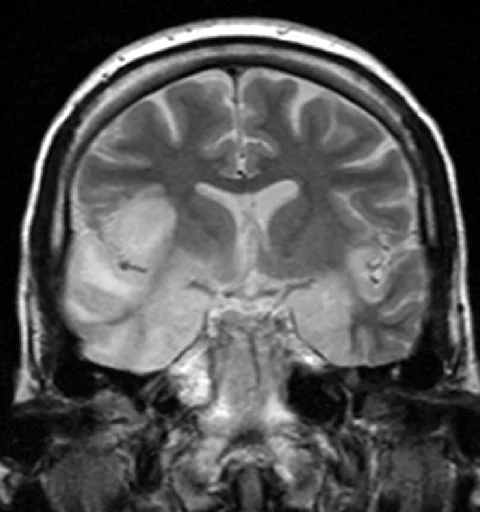
Long description
Coronal T2-weighted MR image showing high signal in the temporal lobes including the hippocampal formations and parahippocampal gyrae, insulae, and right inferior frontal gyrus in a patient whose biopsy was positive for HSV type I.

MRI is the preferred imaging modality and should be performed as soon as possible after admission, ideally within 24 hours and certainly within 48 hours. [1] CT is much less sensitive and is mainly useful to identify alternative diagnoses or raised intracranial pressure before a lumbar puncture. [1] Contrast-enhanced MRI is more useful than non-contrast imaging.
In HSV-1 encephalitis typical findings are T2 hyperintensity and restricted diffusion in the medial temporal lobes, often with frontobasal extension; these changes evolve over days. [3] Arboviruses such as Japanese encephalitis and West Nile virus more often produce basal ganglia and thalamic changes. Temporal lobe changes are the classic MRI signature of HSV-1, but their absence early in the illness does not exclude the diagnosis.
EEG is not needed in every case, but it should be performed when the diagnosis is uncertain, when psychiatric features dominate, or when subtle motor or non-convulsive seizures are suspected. [1] In HSV-1 encephalitis periodic lateralized epileptiform discharges are considered pathognomonic. [3] In anti-NMDA receptor encephalitis a pattern called extreme delta brush may appear. [3] EEG is also the key test for conditions that mimic encephalitis, such as Creutzfeldt-Jakob disease.
Blood tests support rather than diagnose. In acute viral central nervous system infection the total white cell count may be normal, high or low, but a relative lymphocytosis is common. Procalcitonin usually stays below 0.5 ng/mL in viral infection, compared with markedly raised levels in bacterial infection, and CRP is only modestly raised and rarely exceeds 50 mg/L. [3] HIV testing, toxoplasma and cryptococcal antigen testing in immunocompromised patients, syphilis serology, and screening for West Nile or Japanese encephalitis when exposure fits are guided by the clinical picture. [2]
Use the criteria above as a checklist at the bedside: one major criterion plus at least two minor criteria, with no more plausible alternative diagnosis. German guidance condenses this to a main criterion of altered consciousness, personality change or psychiatric symptoms for more than 24 hours, plus supporting findings of fever, seizures, new focal signs, CSF white cells of at least 5 per microlitre, typical imaging, or a consistent EEG. [3] [2]
A CSF white cell count below the usual threshold does not exclude encephalitis, particularly in very early disease or after antibiotics or aciclovir have been started. [1] Conversely, encephalitis is largely a clinical diagnosis; a raised cell count alone is not enough if the clinical picture does not fit.
Approach to suspected encephalitis
- Suspected encephalitisSubacute altered mental status over 24 hours plus at least two minor criteria
- Start IV aciclovir 10 mg/kg 8-hourlyIdeally within 6 hours, certainly within 24 hours of admission
- Lumbar puncture when safeCSF for viral PCR and basic studies; repeat if it is early and negative
- Question: Confirmed or likely cause?
HSV or VZV
- Continue aciclovir 14 days (21 if immunocompromised)Repeat CSF PCR if response is poor
Other organism
- Stop aciclovirSupportive care; specific treatment if available
Autoimmune syndrome
- First-line immune therapyHigh-dose IV methylprednisolone plus IVIG and/or plasma exchange
No diagnosis
- Repeat MRI and LP, screen for tumour, consider NGSBrain biopsy only when benefit outweighs risk
Treat before you know. Start empirical intravenous aciclovir as soon as encephalitis is suspected, ideally within 6 hours of presentation and certainly within 24 hours. [2] [1] Begin other antimicrobials alongside aciclovir when bacterial, Listeria or tuberculosis infection is possible, for example in immunocompromised patients or when the imaging pattern does not fit HSV. [1] Most patients need seizure control, fever control, careful fluid management and airway protection if consciousness is impaired. Patients with acute viral encephalitis should be managed in an intensive care or monitored setting. [3]
Aciclovir is the disease-modifying treatment for HSV and varicella zoster encephalitis. It is given intravenously at 10 mg/kg every 8 hours, with dose adjustment for renal impairment to avoid crystal nephropathy. [2] It should be continued for at least 14 days in immunocompetent patients and 21 days in immunocompromised patients. [2] Many clinicians obtain a repeat CSF PCR at the end of treatment, particularly if recovery is slow or the patient is immunocompromised, because viral DNA can persist and a long period of positivity correlates with worse outcome. [1]
If varicella zoster is confirmed or strongly suspected, guidance suggests considering a higher dose of 15 mg/kg every 8 hours, although the evidence for this in encephalitis is limited to in vitro data. [2]
Aciclovir is stopped when HSV is excluded: an alternative diagnosis is made, the PCR is positive for another virus, or, when clinical suspicion is low, there is a negative HSV PCR (after at least 72 hours of illness), a normal CSF white cell count, and no temporal lobe abnormality on MRI. [1] When suspicion remains high despite negative results, aciclovir may be continued with specialist and imaging review. [1]
Cerebral oedema is the main life-threatening complication. Supportive measures include head-of-bed elevation, osmotic diuretics, and, if these fail, decompressive hemicraniectomy in selected cases. [2] Corticosteroids can be used for significant brain swelling when intracranial pressure is raised. [2] Routine adjunctive dexamethasone is not recommended: a recent randomized trial found no clear benefit on outcome. [2]
Investigate and treat for infectious and autoimmune causes in parallel from the start. [2] If antibodies or a convincing syndrome are present, and viral PCR is negative, first-line immune therapy is standard. Current practice uses high-dose intravenous methylprednisolone, together with intravenous immunoglobulin and/or plasma exchange, chosen by availability and stability. [2]
For anti-NMDA receptor encephalitis many clinicians now use second-line agents early, such as rituximab (with cyclophosphamide an occasional alternative), to reduce relapse. Screen rapidly and remove any ovarian or testicular teratoma, which matters greatly for outcome. Tumour resection and immune therapy should run together. Other agents, such as tocilizumab or bortezomib, are reserved for refractory cases. [2]
LGI-1 antibody encephalitis often responds to first-line treatment or even corticosteroids alone. Intracellular antibody encephalitis (anti-Hu, anti-Ma) responds poorly to immune therapy and depends on treating the underlying cancer. Antibody-negative cases should have a trial of first-line therapy with objective evidence of improvement before escalation. [2]
The risk of relapse in NMDA- and LGI-1-antibody disease is 12 to 35 percent, and relapses need repeat courses of therapy. [2]
Enteroviruses, arboviruses such as West Nile and Japanese encephalitis, and most other viral causes have no reliable antiviral treatment, so care is supportive. Japanese encephalitis vaccination is available for travellers and residents of endemic regions. Mosquito and tick bite prevention reduces risk. Rabies is almost always fatal once symptoms appear, so exposure must be managed before illness, with wound washing and prompt rabies vaccination and immunoglobulin when indicated. [3]
Recovery should be mapped with neuropsychological, speech and language, occupational and psychiatric rehabilitation, because survivors frequently have memory, executive and personality change. Patients and carers benefit from early signposting to support organisations. NMDA-mediated autoimmune encephalitis triggered by HSV should be considered in the months after recovery. [3]
- Seizures and status epilepticus, sometimes refractory in autoimmune cases.
- Raised intracranial pressure and cerebral herniation.
- Severe amnesia, behavioural change and cognitive decline, especially after HSV-1 infection.
- Post-infectious autoimmune encephalitis weeks to months later.
- Thrombotic or cardiorespiratory complications of immobility.
- Iatrogenic renal injury from aciclovir if dosing ignores renal function.
- Death: without treatment, HSV encephalitis has a mortality rate up to 70 percent, and aciclovir reduces mortality to about 20 to 30 percent. [3] In a French cohort of patients with HSV encephalitis, 16.5 percent died overall and 20.8 percent of those needing intensive care died. [3]
Prognosis depends on the pathogen, the patient's age and general health, and above all on how quickly treatment is started. [3] Time to treatment is a key determinant of outcome in both infectious and autoimmune encephalitis. [2] About 10 percent of patients with HSV encephalitis are readmitted within six months. [3]
Survivors of HSV encephalitis often have significant residual deficits in memory and executive function. Autoimmune encephalitis has a better prognosis when recognised early and treated promptly, although relapse occurs in 12 to 35 percent of NMDA- and LGI-1-antibody cases. [2] Rabies, Japanese encephalitis and other untreatable arboviruses still carry high mortality where specific treatment is unavailable. [3]
There is no vaccine for HSV encephalitis. Prevention is therefore indirect: vaccination against measles, mumps and rubella, varicella vaccination where policy supports it, influenza vaccination for high-risk groups, and Japanese encephalitis vaccination for residents and travellers in endemic areas. Mosquito and tick bite precautions lower arboviral risk. HIV prevention and treatment reduce opportunistic causes. After recovery from HSV encephalitis, a low threshold for investigating autoimmune encephalitis is prudent.
- Children. Viral causes, especially enteroviruses, and ADEM are relatively more common than in adults. ADEM occurs mainly in children and young adults, and roughly half of cases have MOG antibodies. [2]
- Pregnancy and the immunocompromised. Opportunistic pathogens (Cryptococcus, Listeria, Toxoplasma, CMV, JC virus) dominate, with longer durations of aciclovir and specialist input needed when immunity is impaired. Toxoplasma in particular is covered in its own topic.
- Older adults. Morbidity and mortality are higher, and delayed diagnosis is more common. Listeria and VZV are proportionally more frequent.
Where CT, MRI, PCR and immune therapy are not available, the essentials still save lives:
- Recognise the syndrome clinically using the major and minor criteria, including the Glasgow Coma Scale and a reliable history.
- Do not wait for imaging before starting aciclovir if the clinical picture fits; imaging can follow when it is safe.
- Lumbar puncture with a basic cell count, glucose and protein is feasible in most district hospitals. Separate and refrigerate samples if transport for PCR is possible.
- Start aciclovir at 10 mg/kg intravenously every 8 hours when HSV or varicella zoster encephalitis is suspected, adjusting for renal function. Oral aciclovir is not a substitute for intravenous therapy in acute disease.
- Manage raised intracranial pressure with head elevation, modest fluid management and mannitol or hypertonic saline where available.
- Send paired serum and CSF for arboviral antibodies through reference laboratories when travel or residence fits.
- For suspected autoimmune encephalitis, first-line therapy depends on what is stocked: high-dose steroids plus immunoglobulin or plasma exchange where possible; check for teratoma when imaging allows.
- Plan early transfer to a centre with imaging and intensive care for deteriorating patients.
- Encephalitis is inflammation of the brain itself, presenting as subacute altered mental status plus fever, seizures, focal signs, CSF pleocytosis or compatible imaging.
- HSV-1 is the most common cause of sporadic encephalitis in adults in Europe, and it can devastate the medial temporal lobes.
- An aetiological cause is found in only about half of cases, and a substantial minority remain completely undiagnosed.
- CSF usually shows a lymphocytic pleocytosis of 10 to 200 cells per microlitre, mildly raised protein of 0.5 to 1.0 g/L and normal glucose.
- MRI, ideally within 24 hours, is the imaging modality of choice; EEG can be pathognomonic in HSV-1 and anti-NMDA disease.
- Start intravenous aciclovir 10 mg/kg every 8 hours on suspicion, ideally within 6 hours, and continue for 14 days in the immunocompetent or 21 days in the immunocompromised.
- Autoimmune encephalitis is at least as common as infection in many settings and should be treated from the outset when viral PCR is negative.
- Mortality in untreated HSV encephalitis is up to 70 percent, falling to roughly 20 to 30 percent with aciclovir.
- Prevention rests on childhood vaccination, travel medicine, vector control and HIV care.
Answer each one in your head first, then open the answer.
A 62-year-old man is brought in with two days of confusion and personality change. Temperature 38.2 degrees C. He has one tonic-clonic seizure. CSF shows 80 lymphocytes per microlitre and normal glucose. What is the most likely diagnosis and the immediate drug treatment?
Show answerHide answer
Suspected herpes simplex virus encephalitis. Start intravenous aciclovir 10 mg/kg every 8 hours without waiting for PCR, arrange MRI of the brain (ideally within 24 hours) and repeat CSF if results are negative and suspicion remains.
Explain why CSF glucose is normal and protein only mildly raised in viral encephalitis, but low glucose and markedly raised protein are expected in bacterial meningitis.
Show answerHide answer
Viral encephalitis inflames the brain parenchyma with relatively modest meningeal involvement, so glucose transfer is little affected and protein leaks only mildly. Bacterial meningitis is an intense purulent infection of the meninges that consumes glucose and breaks down the blood-CSF barrier, producing low glucose and very high protein.
A lumbar puncture done 18 hours after neurological onset is negative for HSV PCR. The patient is still febrile and confused. What should happen next?
Show answerHide answer
Continue intravenous aciclovir and repeat the lumbar puncture 48 hours later (or 2 to 3 days later if the initial puncture was early), because the first sample can be falsely negative very early or after treatment has started. Aciclovir should only be stopped later if a second PCR is negative alongside a normal white cell count and no temporal lobe abnormality on MRI.
A 24-year-old woman presents with one week of behavioural change, memory loss, orofacial dyskinesia and wide swings in heart rate. MRI is normal. What diagnosis should you consider first, what investigation follows, and what immediate management is needed?
Show answerHide answer
Anti-NMDA receptor encephalitis. Look for an ovarian teratoma on imaging and send serum and CSF for NMDA receptor antibodies. Start first-line immune therapy (high-dose intravenous methylprednisolone plus intravenous immunoglobulin or plasma exchange) in parallel with investigating for tumour, and screen rapidly for teratoma.
How would you tell HSV temporal-lobe encephalitis clinically and on MRI from an older left hemisphere stroke?
Show answerHide answer
HSV encephalitis evolves over days with fever, behavioural change and amnesia, and MRI shows bilateral or unilateral temporal lobe change including restricted diffusion and oedema that progresses on serial scans. Stroke is abrupt and vascular-territory, does not cause fever, and the imaging abnormality is fixed from the first scan.
In a district hospital without MRI or PCR, a previously healthy 45-year-old has fever, confusion, a seizure and CSF with 60 lymphocytes per microlitre. What are the three most important actions?
Show answerHide answer
Recognise the syndrome, start intravenous aciclovir 10 mg/kg every 8 hours (adjusting for renal function), and manage airway, seizures and intracranial pressure while arranging transfer to a centre with imaging and intensive care. A basic lumbar puncture alone has already given useful information.
Sepsis; meningitis; HIV infection; toxoplasmosis; cytomegalovirus infection; cryptococcal meningitis; malaria; mosquito-borne viruses; fever of unknown origin; stroke; seizures and status epilepticus; delirium.
- Granerod J, Sellner J, Stahl JP, Defres S, Mailles A, Schmutzhard E, Aurelius E, Whitfield T, Massacesi L, Titulaer MJ, Taba P, Michael BD, Bharucha T, Turtle L, Sargent B, Tamborska A, Cattrall J, Huang Y, Hodel EM, Lant S, Webb G, Shil RSK, Dhar S, van de Beek D, Studahl M, Brouwer MC, Meyding-Lamade U, Easton A, Singh B, Solomon T. Diagnosis and Management of Infectious Encephalitis in Adults: Guidelines From the European Academy of Neurology and European Society of Clinical Microbiology and Infectious Diseases. European Journal of Neurology 2026;33(10):e70647. doi:10.1111/ene.70647. Accessed .
- Auwal AM, Matthews R, Cook C, Sargent B, Easton A, Ray STJ, Ellul MA, Michael BD. Suspected encephalitis in adults. Practical Neurology 2026;26(5):e004299. doi:10.1136/pn-2024-004299. Accessed .
- Meyding-Lamade U, Craemer EM, Aydin K, Jacobi C, Wagner J, Pfausler B, Prange H, Schuchardt V, Mosthaf S, Mehling M, Klein M, Bassa B. S1 guidelines of the German Society of Neurology for Viral Meningoencephalitis. Neurological Research and Practice 2026;8(1):24. doi:10.1186/s42466-026-00487-3. Accessed .
Figure credits
Educational content for medical students and doctors. It is not a substitute for clinical judgement or local guidelines.