Bacterial and viral meningitis
Bacterial and viral meningitis: causes, red flags, CSF findings, empiric antibiotics, dexamethasone, chemoprophylaxis and vaccination.
- Meningitis is infection and inflammation of the meninges, the membranes that wrap the brain and spinal cord. It is a medical emergency: bacterial forms can kill within 24 hours.[4] [4]
- The classic triad is fever, headache and neck stiffness, but it is present in only about 41 to 51% of patients at presentation, so its absence never excludes the disease.
- Bacterial meningitis is distinguished from viral meningitis mainly by the cerebrospinal fluid (CSF): cloudy fluid, more than 1000 white cells per microlitre (mostly neutrophils), protein above 1 g/L, and glucose under 40% of the blood level; viral meningitis usually shows clear fluid, 5 to 1000 cells (mostly lymphocytes), protein 0.4 to 1 g/L and normal glucose. [2]
- When bacterial meningitis is suspected, do not wait for imaging or for laboratory proof: take blood cultures, start empiric antibiotics (usually a third-generation cephalosporin plus ampicillin, with vancomycin where pneumococcal resistance is high) and give dexamethasone, then perform a lumbar puncture, and take a CT first only when specific criteria are met. [5]
- Most people with bacterial meningitis survive if treatment starts early, but about 1 in 6 die and 1 in 5 survivors have lasting problems such as hearing loss or cognitive impairment. Vaccination, and antibiotic prophylaxis for close contacts, prevent cases. [2]
By the end of this page you should be able to:
- Explain the difference between bacterial, viral, tuberculous and fungal meningitis from their mechanisms and their cerebrospinal fluid profiles.
- List the common organisms by age group and risk group, and recognise the red flags that mean meningitis rather than a benign headache.
- Describe when a CT brain is needed before lumbar puncture, and what cerebrospinal fluid findings confirm or argue against bacterial infection.
- Choose the correct empiric antibiotics and doses by age and risk, and explain when to give and when to stop dexamethasone.
- Outline chemoprophylaxis for contacts and the vaccines that prevent meningitis.
- Apply the approach in low-resource settings, where imaging and rapid tests may not be available.
DefinitionMeningitis
Inflammation of the meninges: the thin membranes (the dura, arachnoid and pia) that cover the brain and spinal cord. When the inflammation is caused by infection, bacteria, viruses, mycobacteria (tuberculosis), fungi or parasites can be responsible; infection can also spread directly from a nearby focus such as otitis, sinusitis or a post-neurosurgical wound.
Meningitis is classified usefully by speed of onset and cause. Acute bacterial meningitis presents over hours to a day or two and is the most dangerous form. Viral meningitis (also called aseptic meningitis when the cause is not bacterial) is usually milder and self-limiting. Tuberculous and fungal meningitis run a subacute or chronic course over weeks. Because early treatment differs sharply by cause, the single most important task is to recognise the bacterial form and treat it before the organism is identified.
Meningitis also differs from encephalitis: meningitis affects the membranes, encephalitis the brain tissue itself. They can co-exist as meningoencephalitis, and bacterial meningitis can produce encephalopathy through raised pressure and inflamed vessels rather than through direct infection of brain tissue.
Meningitis affects an estimated 2.5 million people worldwide each year and kills over 200,000. [2] Bacterial meningitis accounts for about 1.6 million cases and 240,000 deaths. [4] About one in six people with bacterial meningitis die, and about one in five survivors is left with lasting disability. [4]
The burden is uneven. Sub-Saharan Africa's "meningitis belt", from Senegal to Ethiopia, carries the highest rates, with recurrent epidemics of meningococcal disease. [4] Incidence in children has fallen where conjugate vaccines against Haemophilus influenzae type b, Streptococcus pneumoniae and Neisseria meningitidis are used: to roughly 1 to 2 cases per 100,000 children. [6]
Age changes the organism and therefore the prognosis. Listeria monocytogenes favours older adults and immunocompromised people, who are more often critically ill. [2]
| Age / risk group | Common organisms |
|---|---|
| Neonates (under 4-6 weeks) | Group B streptococcus, Escherichia coli and other enterobacters, Listeria monocytogenes |
| Infants 6 weeks to adults under 50 years | Streptococcus pneumoniae, Neisseria meningitidis, Haemophilus influenzae |
| Adults over 50 years, or any immunocompromised patient | S. pneumoniae, N. meningitidis, H. influenzae, L. monocytogenes |
| Post-neurosurgery, trauma or with a CSF shunt | Staphylococcus aureus, Pseudomonas, Gram-negative enterobacters |
| Immunocompromised / HIV | Mycobacterium tuberculosis, Cryptococcus neoformans, Toxoplasma (in addition to bacterial causes) |
| Viral | Enteroviruses (coxsackie, echovirus), herpes simplex virus 1 and 2, varicella zoster, measles, mumps, influenza, HIV, arboviruses such as West Nile |
| Fungal | Cryptococcus neoformans or C. gattii (especially HIV), Coccidioides, Blastomyces, Candida, Histoplasma, Aspergillus |
Risk factors for acquiring bacterial meningitis include close contact in new groups (such as students in halls of residence), pilgrimages and travel to the meningitis belt, asplenia and complement deficiency, household members or nursery contacts of a case, and breaches of the dura from head injury, surgery or a CSF leak. [2] N. meningitidis is associated with septicaemia in about 70% of cases, producing a non-blanching purpuric rash and shock. [2]
Bacteria reach the meninges in three ways: through the bloodstream (from otitis, sinusitis, pneumonia, endocarditis or bacteraemia), by direct spread across a breached dura, or from a parameningeal focus. Once in the subarachnoid space they multiply and release cell-wall components that trigger an intense inflammatory cascade: cytokines (interleukin-1, tumour necrosis factor-alpha, interleukin-6), an influx of white cells, breakdown of the blood-brain barrier, cerebral oedema, raised intracranial pressure and sometimes vasculitis and thrombosis of cerebral vessels. [4]
This explains both the symptoms and the treatment:
- Fever and headache come from inflammation and raised intracranial pressure.
- Neck stiffness is reflex spasm of the neck muscles against an inflamed pain-sensitive meninges.
- Photophobia is inflammation of the cranial nerve sheaths and meninges around the optic pathways.
- Altered mental state results from oedema, ischaemia and toxins.
- Hearing loss (especially after pneumococcal and H. influenzae disease) comes from inflammation damaging the cochlea or auditory nerve.
- Hydrocephalus follows obstruction of CSF flow by thick exudate at the brain base, particularly in tuberculous meningitis.
Viral meningitis follows a milder version of the same process: viruses enter via the bloodstream or peripheral nerves, but viral replication is usually controlled quickly in immunocompetent people, producing lymphocytic rather than neutrophilic inflammation and little or no hypoglycorrhachia (low CSF glucose).
Tuberculous meningitis is a basilar, dense inflammation with exudate that chokes the cranial nerves and blocks CSF drainage, which is why hydrocephalus and cranial nerve palsies are common. Fungal meningitis, especially cryptococcal, is a chronic granulomatous or yeast-driven inflammation that is most dangerous when immunity is poor.
Patients present with fever, headache, neck stiffness (meningism), photophobia, nausea and vomiting, altered behaviour or consciousness, seizures and (in about 68% of meningococcal cases) a purpuric non-blanching rash. The classic triad of fever, neck stiffness and altered mental status is present in only about 41 to 51% of cases, and in immunocompromised people sometimes none of these is present. [5]
Other signs include cranial nerve palsies, focal deficits, papilloedema (a sign of raised intracranial pressure) and, in infants, a bulging fontanelle. Kernig's sign (pain on passive knee extension with the hip flexed) and Brudzinski's sign (hip and knee flexion triggered by neck flexion) are specific but not sensitive, so their absence rules nothing out, and studies show low sensitivity and specificity overall. [2] [5]
Reported frequencies in bacterial meningitis versus viral meningoencephalitis include fever (77 to 97% vs 72%), headache (58 to 87% vs 88%), neck stiffness (69 to 83% vs 36%), nausea and vomiting (45 to 74% vs 54%) and altered mental status (30 to 69% vs 68%). [2]
| Feature | Bacterial meningitis (%) | Viral meningoencephalitis (%) |
|---|---|---|
| Fever | 77-97 | 72 |
| Headache | 58-87 | 88 |
| Neck stiffness | 69-83 | 36 |
| Nausea and vomiting | 45-74 | 54 |
| Altered mental status | 30-69 | 68 |
| Rash | 20-52 | Not reported [2] |
Infants, older adults and immunocompromised patients often present subtly: low-grade fever, irritability, poor feeding, lethargy or a flu-like illness rather than meningism. [2] In neonates, signs may be only temperature instability, apnoea, irritability or a bulging fontanelle.
Red flagMeningitis red flags
Suspect meningitis, and treat empirically, with any two of: fever plus headache; neck stiffness; photophobia; vomiting; altered behaviour or consciousness; seizures; a non-blanching rash; or a new focal neurological deficit. A "clinical picture of bacterial meningitis" (fever or a focus such as otitis, sinusitis or pneumonia, plus headache and neck stiffness) is more reliable than the classic triad. [5]
The differential is wide: migraine, tension headache, viral upper respiratory infection, influenza, sepsis of another source, subarachnoid haemorrhage, stroke, brain tumour or metastasis, encephalitis, intracranial abscess, autoimmune encephalitis and drug-induced aseptic meningitis (for example with NSAIDs, penicillins, sulphonamides or intravenous immunoglobulin). [2]
Meningitis can be given a non-infectious name only after infection is excluded. "Aseptic meningitis" describes meningeal inflammation that is either non-bacterial or caused by organisms that do not grow on ordinary culture; about half of aseptic cases remain undiagnosed. Enteroviruses are the most common cause, followed by other viruses, fungi, TB, parasites, spirochaetes and autoimmune disease. [2]
Take two pairs of blood cultures before antibiotics, a full blood count, C-reactive protein and procalcitonin (procalcitonin has reported sensitivity 95% and specificity 97% for separating bacterial from viral meningitis in meta-analytic data). [5] Throat swabs for meningococcal PCR and viral PCR are useful when meningococcal disease is suspected. [2]
Lumbar puncture is the diagnostic test. It should be performed as soon as it can be done safely. CSF should be examined for opening pressure, appearance, cell count and differential, protein, glucose (with a paired blood glucose), lactate, Gram stain, culture, and PCR for meningococcus, pneumococcus, H. influenzae, L. monocytogenes, group B streptococcus, HSV 1 and 2, varicella zoster, enteroviruses and parechovirus. [2]
A CT brain before lumbar puncture is recommended when there are new focal neurological deficits, recent seizures (within days), severely impaired consciousness, signs of raised intracranial pressure (papilloedema) or suspected immunocompromise where indicated by the guideline used. [1] [5] It has also been recommended on history of central nervous system disease, anisocoria or abnormal eye movements, visual field defects, limb drift, age over 60 years or altered language. [1]
Guidelines differ in how many patients they flag: the IDSA 2004 criteria (Glasgow Coma Scale under 15, immunocompromised state or history of central nervous system disease, new seizures, focal deficits, papilloedema, cranial nerve abnormalities) flag about 64% of suspected cases, whereas ESCMID 2016 (Glasgow Coma Scale under 10, immunocompromised state, new seizures, aphasia, gaze palsy, limb paresis, ataxia) flags about 39%. [1] If imaging delays treatment, start antibiotics before the scan. [5]
Figure 1Neisseria meningitidis CSF Gram 1000
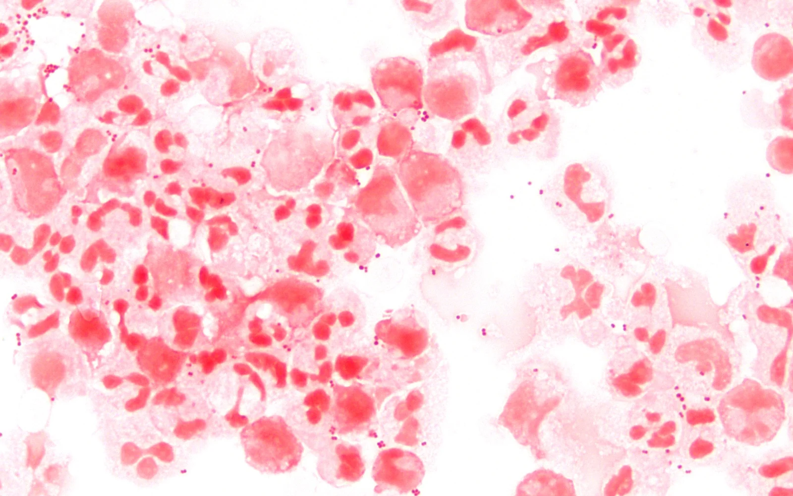
Long description
Photomicrograph of a Gram stain of spinal fluid at 1000x from which Neisseria meningitidis grew.

| Parameter | Normal | Bacterial | Viral | Tuberculous | Fungal |
|---|---|---|---|---|---|
| Opening pressure | 10-20 cmH2O | Raised | Normal or raised | Raised | Normal or raised [2] |
| Appearance | Clear | Cloudy | Clear | Cobweb-like or clear | Clear or cloudy |
| White cells | 0-5 per microlitre, lymphocytes | Over 1000 per microlitre, neutrophils | 5-1000 per microlitre, lymphocytes (PMNs early) | 10-500 per microlitre, lymphocytes (mixed early) | 0-500 per microlitre, lymphocytes |
| Protein | 0.15-0.45 g/L | Over 1 g/L | 0.4-1 g/L | 1-5 g/L | 0.4-2.5 g/L |
| Glucose (CSF:serum) | Over 60% | Under 40% | Usually normal | Under 30% | Under 40% |
| Lactate | Under 2 mmol/L | Over 3.5 mmol/L typically | Under 2 mmol/L | Over 2 mmol/L (often over 3.5) | Over 2 mmol/L [2][5] |
Two cautions matter clinically. First, one third of bacterial meningitis cases show under 1000 cells per microlitre in the first CSF sample, and 11% show under 100; but 96% have at least one abnormal CSF parameter (pleocytosis, raised protein, or low CSF:serum glucose ratio). [5] Second, antibiotic pretreatment reduces culture yield sharply (positivity falls after 4 hours and is often negative after 8), so taking CSF and blood before antibiotics, or as soon as possible after, preserves the diagnosis. [1]
The Gram stain is rapid and cheap and is positive in about 90% of pneumococcal and 70-90% of meningococcal cases, but only about 25-35% of Listeria and about 50% of H. influenzae cases. [5] PCR has reported sensitivities of 79-100% for S. pneumoniae, 91-100% for N. meningitidis and 67-100% for H. influenzae, with specificities of 95-100%. [5]
Bacterial meningitis is a clinical and laboratory diagnosis rather than a single score. A useful working definition for acute community-acquired cases is CSF pleocytosis with neutrophil predominance plus at least one of raised protein or low CSF glucose, supported by a positive Gram stain, culture or PCR. [5] Severity is tracked clinically (conscious level, seizures, focal deficits, need for ventilation) and by complications (hydrocephalus on imaging, stroke). There is no staging score in routine use.
- Start droplet precautions and isolate if meningococcal disease is possible. [5]
- Take two pairs of blood cultures (and a throat swab for meningococcal PCR). [5]
- Perform lumbar puncture unless contraindicated (see above), ideally within 1 hour of arrival; if delay is unavoidable, give antibiotics first. [5]
- Start empiric antibiotics within 1 hour of arrival (ESCMID 2016). [5]
- Give dexamethasone with or 15-20 minutes before the first antibiotic dose. [4]
| Situation | Antibiotics | Dose |
|---|---|---|
| Standard community-acquired (all adults) | Ceftriaxone plus ampicillin (add vancomycin where pneumococcal cephalosporin resistance is high) | Ceftriaxone 2 g IV every 12 h; ampicillin 2 g IV every 4 h; vancomycin 15-20 mg/kg IV every 8-12 h [3] |
| Age over 50 years, or Listeria suspected | As above (ampicillin covers Listeria) | Same doses |
| Immunocompromised, or Gram-negatives/Pseudomonas suspected | Replace cephalosporin with cefepime or meropenem; plus ampicillin; vancomycin as above | Meropenem 2 g IV every 8 h; cefepime dosing per local protocol [5] |
| Herpes simplex encephalitis possible | Add acyclovir | Aciclovir 10 mg/kg IV every 8 h |
| High resistance setting (alternative to vancomycin) | Rifampicin | 600 mg IV once daily [5] |
Notes: in penicillin allergy, meropenem (2 g IV every 8 h) is used instead of ampicillin. [5] Cefotaxime 2 g IV every 4-6 h is an acceptable alternative to ceftriaxone. [3] Empiric therapy is continued until the organism and its sensitivities are known; L. monocytogenes requires at least 21 days of treatment. [5]
In neonates, empiric cover is aimed at group B streptococcus, E. coli and Listeria: ampicillin plus gentamicin (or cefotaxime), with dosing by local neonatal guidelines. In infants and children over 3 months, empiric parenteral antibiotics are a third-generation cephalosporin (ceftriaxone or cefotaxime) plus vancomycin where indicated, again by local resistance patterns. Vancomycin (and gentamicin) dosing follows body weight and renal function. [5] Exact neonatal and paediatric dosing is weight-based and follows the same augmentin/ampicillin and cephalosporin principles as the adult regimens above; use the local formulary for milligram-per-kilogram doses and intervals.
Give dexamethasone to adults and to children over 3 months when bacterial meningitis is suspected:
- Adults: 10 mg IV every 6 hours for 4 days, started 15-20 minutes before or with the first antibiotic dose. [4]
- Children over 3 months: 0.15 mg/kg IV every 6 hours for 4 days (2-4 days in some regimens, started before or with antibiotics). [4]
- Neonates: routine dexamethasone is not recommended; available evidence (dexamethasone 0.25 mg/kg IV every 12 hours for 3 days) shows no consistent benefit. [4]
Dexamethasone reduces mortality in pneumococcal meningitis (from about 36% to about 30% in pooled studies) and reduces hearing loss and short-term neurological sequelae in high-income settings, especially in H. influenzae type b meningitis in children. [3] [6] It should be continued for 4 days if S. pneumoniae or H. influenzae is confirmed, and stopped if another organism is identified; it is possibly harmful in cryptococcal and possibly in Listeria meningitis, where observational studies show worse outcomes. [5] [6]
Two sequencing cautions: start dexamethasone before or with antibiotics (delayed dosing is much less effective), and because dexamethasone may reduce vancomycin penetration into CSF, replace vancomycin with rifampicin when dexamethasone is used if you are covering resistant pneumococci. [4] [5]
Tuberculous meningitis needs four-drug anti-tuberculous therapy plus adjunctive dexamethasone at 0.3-0.4 mg/kg per day IV for the first 2 weeks, tapered over 6-8 weeks and transitioned to oral prednisolone, which reduced 9-month mortality from 33% to 22% in a landmark trial. [4]
Cryptococcal meningitis, the commonest fungal form (especially in HIV), is treated with antifungal therapy (amphotericin B plus flucytosine, then fluconazole), and adjunctive corticosteroids have shown harm rather than benefit and should not be given. [6] Other fungal infections are treated with targeted antifungals on microbiology advice.
Patients with bacterial meningitis should be managed in a unit with neurological or intensive care expertise. Monitor for and treat hydrocephalus (external ventricular drain where indicated), raised intracranial pressure, seizures, hyponatraemia and circulatory failure. [5] Search for a focus of infection (otitis, sinusitis, mastoiditis, endocarditis, spinal infection) and treat it; ENT infection needs early surgical treatment, ideally within 24 hours. [5]
Approach to suspected meningitis in the emergency department
- Suspected meningitis: fever plus headache, neck stiffness, or altered consciousness
- Droplet precautions and isolation until N. meningitidis excluded
- Blood cultures x 2; throat swab; basic labs including procalcitonin
- Question: CT before lumbar puncture needed?
Yes: new focal deficit, recent seizure, GCS < 10-15, papilloedema, immunocompromise, CNS disease, cranial nerve abnormality
- Start antibiotics and dexamethasone now, before imaging
- CT brain; then lumbar puncture if safe
No: none of the criteria
- Lumbar puncture now; do not delay for imaging
- CSF: cell count, protein, glucose ratio, lactate, Gram stain, culture, PCR
- Start empiric antibiotics plus dexamethasone within 1 hour of arrival
- Tailor once organism identified; continue dexamethasone only for S. pneumoniae or H. influenzae
- Monitor for hydrocephalus, raised pressure, seizures; treat focus (ENT, endocarditis)
Complications of bacterial meningitis include hearing loss (a particular risk after pneumococcal and H. influenzae infection), hydrocephalus, seizures, stroke from cerebral vasculitis, brain or subdural abscess, venous sinus thrombosis, cerebral oedema with herniation, Waterhouse-Friderichsen syndrome in meningococcal disease, and cognitive and behavioural impairment. About 40% of survivors leave hospital with some disability. [2] Hearing loss is the most common sequela in children. [4]
Complications of viral meningitis are uncommon in immunocompetent adults. Tuberculous meningitis complications include hydrocephalus, cranial nerve palsies, stroke, and cognitive impairment, with high mortality even with treatment.
With modern treatment, mortality from community-acquired pneumococcal meningitis falls significantly when dexamethasone is used; in the Netherlands, mortality fell from about 30% to 20% after routine adjunctive dexamethasone was adopted. [6] Poor outcome is associated with older age, coma on admission, seizures, focal deficits, delayed treatment beyond 4-6 hours, and immunocompromise. [1] [5]
Vaccination has transformed the epidemiology of meningitis: the Hib vaccine, pneumococcal conjugate vaccines and meningococcal conjugate vaccines (including serogroup A in the African meningitis belt) reduce cases; BCG protects children against disseminated tuberculosis including TB meningitis. [2] [4] Vaccination is therefore the most effective long-term prevention.
Chemoprophylaxis for close contacts of a meningococcal case should be given within 24 hours of exposure to all people with droplet contact: ciprofloxacin 500 mg orally once, ceftriaxone 250 mg intramuscularly once, or rifampicin 600 mg intravenously twice daily for 2 days. [5] Close household contacts should be vaccinated according to the serogroup identified in the index patient. [5]
Prevention of invasive pneumococcal and Hib disease also relies on vaccination of infants and on antibiotic prophylaxis guidelines for high-risk groups (such as asplenia), which are set locally. Prevention of TB meningitis rests on BCG vaccination of infants and on detecting and treating latent tuberculosis in contacts.
- Neonates: cover group B streptococcus, E. coli and Listeria; ampicillin plus an aminoglycoside; do not give routine dexamethasone. [4] [5]
- Children over 3 months: third-generation cephalosporin (plus vancomycin where indicated) plus dexamethasone 0.15 mg/kg IV every 6 hours for 4 days. [4]
- Pregnancy: Listeria coverage matters (ampicillin), and many standard drugs (such as vancomycin and acyclovir) are used only when justified; specialist and obstetric input is required.
- Older adults and immunocompromised patients: always add ampicillin for Listeria; consider M. tuberculosis and Cryptococcus where HIV or immunosuppression is present. [2] [3]
- Post-neurosurgical or CSF shunt patients: treat as nosocomial meningitis with meropenem plus vancomycin (see doses above), and involve neurosurgery. [5]
In settings where imaging, CSF biochemistry and PCR are limited:
- Start droplet precautions and treat empirically on clinical suspicion, within 1 hour of arrival. [5]
- A lumbar puncture can be done without prior CT if there are no focal deficits, recent seizures or severely impaired consciousness, and no signs of raised pressure. [5]
- Even basic CSF examination (appearance, crude cell count, protein, and glucose compared with blood using a glucometer) separates bacterial from viral meningitis well enough to guide treatment. [2]
- If no resistant pneumococci are expected locally, ceftriaxone alone, or benzylpenicillin plus chloramphenicol where cephalosporins are unavailable, are used by local protocol; add ampicillin if Listeria is plausible.
- Dexamethasone remains useful in bacterial meningitis where good medical care is available; evidence in low-resource settings is weaker, and the benefit is smaller when presentation is delayed. [3] [6]
- Vaccination (Hib, pneumococcal conjugate, meningococcal A conjugate in the belt, BCG) is the single most cost-effective intervention, and should be prioritised. [2] [4]
NoteLow-resource settings note
Guideline recommendations based on high-income data (IDSA, ESCMID, NICE) may not generalise to every setting; adapt antibiotics to local resistance and availability, and follow national meningitis protocols.
- Meningitis is inflammation of the meninges; bacterial forms are rapidly lethal emergencies.
- The classic triad is present in only 41-51% of cases: never exclude meningitis because it is absent.
- N. meningitidis causes about 70% of its infections with septicaemia and a non-blanching purpuric rash.
- Age dictates the organism: neonates get group B strep, E. coli and Listeria; older children and young adults get pneumococcus, meningococcus and Hib; over-50s and immunocompromised get the same plus Listeria.
- CSF is the key to the diagnosis. Bacterial: cloudy, over 1000 cells per microlitre (neutrophils), protein over 1 g/L, glucose under 40% of blood. Viral: clear, 5-1000 cells (lymphocytes), protein 0.4-1 g/L, glucose usually normal.
- Do not delay treatment: blood cultures, lumbar puncture and empiric antibiotics plus dexamethasone, all within 1 hour of arrival when bacterial disease is likely.
- Empiric antibiotics are a third-generation cephalosporin plus ampicillin, with vancomycin (or rifampicin) where pneumococcal resistance is high; add aciclovir when viral encephalitis is plausible.
- CT is needed before lumbar puncture with new focal deficits, recent seizures, coma, papilloedema or raised-pressure features; otherwise LP should come first.
- Dexamethasone (10 mg IV every 6 hours for 4 days in adults; 0.15 mg/kg IV every 6 hours for 4 days in children) is given with or before antibiotics and continued only for pneumococcal or Hib meningitis. [4]
- Steroids are harmful in cryptococcal meningitis and probably in Listeria meningitis; monitor and stop when the organism argues against them.
- Close contacts of meningococcal cases need chemoprophylaxis within 24 hours: ciprofloxacin 500 mg orally once, ceftriaxone 250 mg IM once, or rifampicin 600 mg IV twice daily for 2 days.
- Vaccination (Hib, pneumococcal, meningococcal, BCG) is the most effective prevention.
- Tuberculous meningitis receives anti-TB therapy plus dexamethasone 0.3-0.4 mg/kg per day IV for 2 weeks, tapered over 6-8 weeks.
- About 40% of bacterial meningitis survivors have long-term disability, particularly hearing loss; early treatment is the key to prevention. [2]
Answer each one in your head first, then open the answer.
A 62-year-old man with chronic lymphocytic leukaemia presents with 18 hours of fever, headache and neck stiffness. What empirical antibiotics should you start, pending culture, and why is ampicillin included?
Show answerHide answer
Start ceftriaxone 2 g IV every 12 hours plus ampicillin 2 g IV every 4 hours (plus vancomycin if local pneumococcal resistance is high, unless dexamethasone is also given, in which case use rifampicin). Ampicillin is included because Listeria monocytogenes is common in patients over 50 and in the immunocompromised, and cephalosporins do not reliably cover Listeria. Add aciclovir 10 mg/kg IV every 8 hours if herpes encephalitis is plausible. [3]
Lumbar puncture in bacterial meningitis most typically shows which cells, and at what counts, compared with viral meningitis?
Show answerHide answer
Bacterial: cloudy CSF with usually over 1000 white cells per microlitre, predominantly neutrophils, protein over 1 g/L and glucose under 40% of the blood level. Viral: clear fluid, 5 to 1000 cells per microlitre, predominantly lymphocytes, protein 0.4 to 1 g/L, glucose usually normal. Under 40% glucose in bacterial versus usually normal in viral is often the most useful single difference.
A 25-year-old student has fever, headache and a Glasgow Coma Scale of 13. No focal deficits or seizures. Should you CT before lumbar puncture, and what treatment must not be delayed?
Show answerHide answer
CT is not required in this case (no focal deficit, no recent seizure, no coma, no papilloedema, no cranial nerve lesion). Proceed to lumbar puncture; if imaging were delayed for any reason, start empiric antibiotics and dexamethasone first. Antibiotics should be started within 1 hour of arrival. [5]
For which organisms is adjunctive dexamethasone clearly beneficial, and which mandate that you stop it?
Show answerHide answer
It clearly benefits pneumococcal meningitis (mortality reduction, reduced hearing loss) and reduces hearing loss in Haemophilus influenzae type b meningitis. Continue it for 4 days if either organism is confirmed. Stop it if another organism is identified; it has been associated with worse outcomes in Listeria and cryptococcal meningitis.
What are the standard chemoprophylaxis options for an adult close contact of a case of meningococcal meningitis, and within what time window must they be given?
Show answerHide answer
Options: ciprofloxacin 500 mg orally once, ceftriaxone 250 mg intramuscularly once, or rifampicin 600 mg intravenously twice daily for 2 days. Give within 24 hours of exposure.
A neonate with suspected bacterial meningitis: which organisms are most likely, and should dexamethasone be given?
Show answerHide answer
Likely organisms are group B streptococcus, Escherichia coli and Listeria monocytogenes. Empiric cover is ampicillin plus an aminoglycoside (or cefotaxime). Routine dexamethasone is not recommended in neonates; available trials showed no consistent benefit.
Meningococcal disease and sepsis; encephalitis (herpes simplex and autoimmune); tuberculous meningitis; HIV-associated opportunistic infections; febrile convulsions in children; sepsis and septic shock; hearing loss and vestibular disorders; subarachnoid haemorrhage; hydrocephalus; headache.
- Costerus JM, Lemmens CMC, van de Beek D, Brouwer MC. Cranial Imaging and Lumbar Puncture in Patients With Suspected Central Nervous System Infection. Clin Infect Dis. 2020;70(12):2469-2475. doi:10.1093/cid/ciz694. Accessed .
- Beed M, Wong C, Dineen RA, Berry L, Brindley PG. Infectious meningitis and encephalitis: A primer for acute care practitioners. J Intensive Care Soc. 2026;27(3):377-386. doi:10.1177/17511437261437982. Accessed .
- Busl KM, Ordway N, Sonneville R. Meningitis, encephalitis, and beyond: A practical intensive care unit guide to severe central nervous system infections. J Intensive Med. 2026;6(6):586-598. doi:10.1016/j.jointm.2026.06.005. Accessed .
- Ullah N, Khan K, Haseeb A, et al. Corticosteroids as adjunctive therapy in acute meningitis: a narrative review. Ann Med Surg (Lond). 2026;88:1574-1584. doi:10.1097/MS9.0000000000004620. Accessed .
- Dyckhoff-Shen S, Koedel U, Pfister HW, Klein M. SOP: emergency workup in patients with suspected acute bacterial meningitis. Neurol Res Pract. 2021;3:2. doi:10.1186/s42466-020-00098-6. Accessed .
- Gundamraj S, Hasbun R. The Use of Adjunctive Steroids in Central Nervous Infections. Front Cell Infect Microbiol. 2020;10:592017. doi:10.3389/fcimb.2020.592017. Accessed .
Figure credits
Educational content for medical students and doctors. It is not a substitute for clinical judgement or local guidelines.