Infectious mononucleosis
How EBV causes mono: symptoms, tests, complications, treatment, sports safety, and low-resource care.
- Infectious mononucleosis is the illness most people mean by the phrase "glandular fever": a contagious viral infection that gives fever, a sore throat often covered in white exudate, and swollen lymph glands, with fatigue that can drag on for weeks.
- It is usually caused by the Epstein-Barr virus (EBV), a herpesvirus that most people catch at some point in life, often as children without ever feeling ill.
- The virus passes mainly in saliva. Young adults, especially students, are the age group in whom it most often causes this full illness rather than a silent infection.
- Recovery is almost always complete. The main dangers are not the virus itself but complications: a ruptured spleen, airway obstruction from swollen tonsils, haemolytic anaemia, and hepatitis.
- Treatment is supportive. Steroids are reserved for complications such as airway compromise, severe thrombocytopenia or haemolytic anaemia. Antiviral drugs do not help.
By the end of this page you should be able to:
- Explain how EBV infects, persists and spreads, and why most infections are silent in young children but cause mononucleosis in adolescents and young adults.
- Recognise the clinical triad of infectious mononucleosis and describe the signs and symptoms that point towards it rather than streptococcal pharyngitis, cytomegalovirus or HIV.
- Interpret a full blood count, heterophile (Monospot) test and EBV serology, and explain why these tests can be falsely negative early in the illness.
- Plan supportive management, the activity and sports restriction that protects the spleen, and when glucocorticoids are appropriate.
- Outline the major complications and the cancers and other conditions with which EBV is epidemiologically associated.
- Adapt diagnosis and care to settings where laboratory testing, imaging or intensive care are limited.
DefinitionInfectious mononucleosis
Infectious mononucleosis is a clinical syndrome, usually caused by primary infection with the Epstein-Barr virus (EBV) in an adolescent or young adult. It is defined by fever, pharyngitis often with exudate, and lymphadenopathy, with fatigue and splenomegaly common. Other pathogens, chiefly cytomegalovirus, can produce a similar picture.
The virus and the illness are not the same thing. EBV infection is extraordinarily common, while infectious mononucleosis is the symptomatic version that develops in only a minority of those infected. People usually get infected once and carry the virus for life. [4]
EBV is one of the most successful human viruses.
- About half of children are infected before the age of 5 years, and seroprevalence rises to about 66% in children and adolescents aged 6 to 19 years. [4]
- More than 95% of adults are seropositive, which makes EBV one of the most ubiquitous human viruses worldwide. [4]
- In most young children primary infection causes no illness or only a mild febrile illness. Symptoms of infectious mononucleosis develop most often in adolescents and adults. [4]
- Sources give different figures for how often infection becomes clinical mononucleosis: at least one in four teenagers and young adults who acquire EBV develop the illness according to the CDC, [1] while another review gives 30% to 50% in immunocompetent adolescents and adults. [5]
- Because infection is often silent in early childhood, the illness is more common in adolescents and young adults from higher socioeconomic backgrounds, where exposure is delayed. Early childhood transmission occurs more often in lower socioeconomic groups and in crowded conditions. [4]
EBV is not very contagious despite its reputation as "the kissing disease": it has not been recovered from environmental sources, and only about 5% of patients acquire it from someone with acute infection rather than from an asymptomatic shedder. [4]
The aetiological agent is the Epstein-Barr virus, also called human herpesvirus type 4. It is a double-stranded DNA herpesvirus. EBV infects B lymphocytes and some epithelial cells in the oropharynx via the CD21 (CR2) receptor, which is also the complement receptor for C3d. [5]
- There is no identified modifiable risk factor for EBV infection other than saliva contact.
- Early infection with the virus is partly determined by crowding and socioeconomic conditions, which bring earlier exposure and earlier immunity. [4]
- People with weakened immune systems can have more severe or atypical EBV disease, including X-linked lymphoproliferative disease, which predisposes to overwhelming EBV infection. [3]
- Infectious mononucleosis itself has no well-defined risk factors beyond adolescence and young adulthood.
Rarely, cytomegalovirus, and occasionally toxoplasmosis, HIV, rubella, hepatitis A, B or C and adenovirus, can produce the same syndrome, so a label of "mononucleosis" is a clinical description rather than a single diagnosis. [1]
EBV enters through the oral cavity, usually by kissing, and first infects epithelial cells of the oropharynx and then B lymphocytes. Inside infected B cells the virus drives them to proliferate; the body controls this with a large cytotoxic CD8+ T lymphocyte response, and the morphologically abnormal "atypical lymphocytes" seen on the blood film are principally these activated T cells. [4] [5]
After primary infection the virus remains in the host, mainly in B lymphocytes, for life and undergoes intermittent, usually asymptomatic, shedding from the oropharynx. [4] The virus is detectable in oropharyngeal secretions in 10 to 20% of immunocompetent adults who are EBV-seropositive. [4] Shedding increases in frequency and titre in immunocompromised patients such as organ allograft recipients and people living with HIV. [4]
The incubation period is about 30 to 50 days. [4] Population data from the CDC describe symptoms appearing 4 to 6 weeks after infection, [1] which is the same interval expressed in weeks.
Three chains explain the symptoms a student sees:
- Viral replication in the pharynx and lymphoid tissue causes tonsillitis, cervical lymphadenopathy and fever.
- The brisk T cell response produces fatigue, lymphocytosis with atypical forms, and sometimes cytopenias.
- The spleen and liver become involved because they filter infected lymphocytes, producing splenomegaly, hepatomegaly and hepatitis.
The illness usually begins slowly over days rather than hours, with fatigue, malaise and headache, followed by sore throat that worsens progressively. [6]
- Fever is usual and can be high, typically rising in the afternoon or early evening, to about 39.5 degrees Celsius and occasionally to 40.5 degrees. [4]
- Extreme fatigue can be profound and is usually maximal in the first 2 to 3 weeks, but can last much longer in a minority. [4]
- Sore throat is often severe, painful and exudative, with swollen tonsils that may have a whitish-yellow covering; it can be clinically indistinguishable from streptococcal pharyngitis. [4] [6]
- Swollen lymph nodes, usually symmetric, in the anterior and posterior cervical chains; generalized or posterior cervical adenopathy points more towards mononucleosis than towards streptococcal sore throat. [4]
- Headache, body aches and loss of appetite are common. [1]
- Jaundice is uncommon. [4] [6]
The "classic triad" taught for recognition is fever, pharyngitis (often with exudate) and lymphadenopathy, with splenomegaly an important accompanying feature. [4]
Figure 1MonoThroat
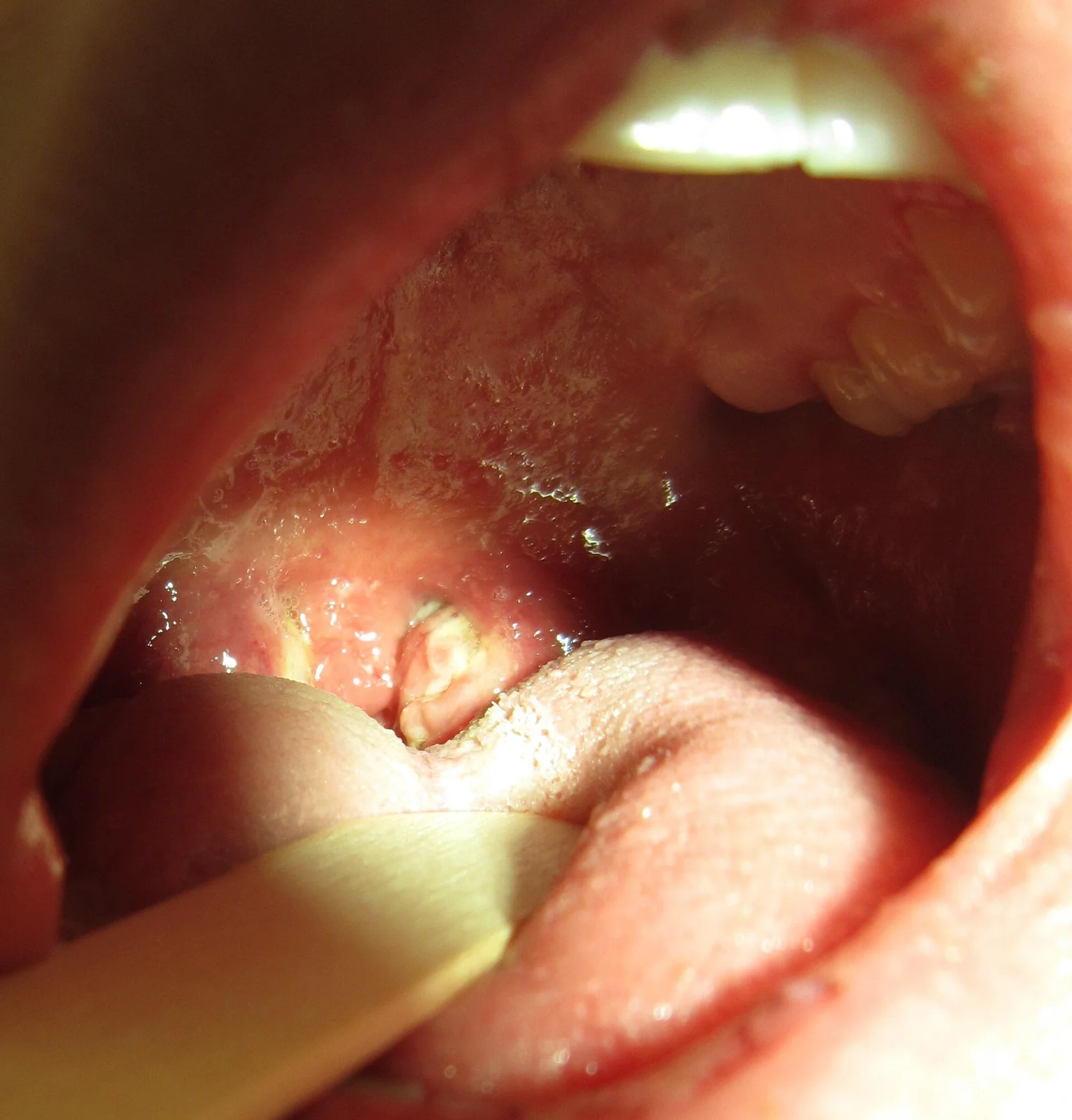
Long description
Exudative pharyngitis in someone with mononucleosis.

- Adenopathy: symmetric cervical chain enlargement; feel both anterior and posterior triangles. [4]
- Splenomegaly: occurs in about 50% of cases and is usually maximal in the second and third week; the spleen is most often only a barely palpable tip. Gentle palpation only. [4]
- Mild hepatomegaly and hepatic percussion tenderness are common. [4]
- Periorbital oedema and palatal petechiae are characteristic associated findings. [4]
- Rash: less frequent overall; a maculopapular eruption can occur in up to roughly 20% of individuals, more often than older descriptions suggested, and the evidence that ampicillin or amoxicillin further increases the rash rate is weaker than traditionally taught, though these drugs are still avoided in mononucleosis because the rash remains very common when they are given. [4] [1] [6]
- Very high atypical lymphocyte counts are typically seen only in primary EBV and cytomegalovirus infection. [4]
Palpate the spleen with particular care and stop if there is pain; remember that the tip is often only just palpable despite significant enlargement.
A pink or measles-like rash is much more likely in a person with infectious mononucleosis who receives ampicillin or amoxicillin for a presumed throat infection. [6] The teaching point for students is behavioural as much as biochemical: do not prescribe amoxicillin or ampicillin for sore throat until streptococcal infection has been confirmed, [6] and if the patient already has confirmed or probable mononucleosis, choose another agent if antibiotics are truly required. [1]
Two questions matter: which illnesses look like EBV mononucleosis, and, once a mass or test result is available, which one explains it. Posterior cervical or generalized adenopathy or hepatosplenomegaly suggests mononucleosis rather than streptococcal infection, but there is overlap, and finding streptococci in the throat does not exclude mononucleosis. [4]
| Condition | Features that separate it from EBV mononucleosis |
|---|---|
| Streptococcal pharyngitis | Abrupt sore throat, fever, tonsillar exudate and tender anterior cervical nodes; no posterior cervical chain enlargement, no splenomegaly, atypical lymphocytosis absent or mild. A positive rapid strep test does not exclude co-existing EBV infection. [4] |
| Cytomegalovirus mononucleosis | Atypical lymphocytosis and splenomegaly with hepatitis, but usually without severe pharyngitis or marked tonsillar exudate. [4] |
| Primary HIV infection | Clinically can resemble acute EBV infection, including fever, pharyngitis, lymphadenopathy and rash. Patients with risk factors must have quantitative HIV RNA and combined antibody and p24 antigen testing rather than antibody alone, which is usually negative in acute infection. [4] |
| Toxoplasmosis | Mononucleosis-like illness with fever and lymphadenopathy, but usually without severe pharyngitis. [4] |
| Hepatitis A, B or C | Elevated liver enzymes, often far higher than the modest elevations of EBV, and atypical lymphocytes may be present. [4] |
| Rubella, adenovirus and other viruses | Can produce mononucleosis syndromes; distinguished by epidemiology and specific testing. [1] |
Laboratory diagnosis usually involves a full blood count and EBV serology. Lymphocytes that are morphologically atypical account for up to 30% of the white blood cells, and very high atypical counts are typically seen only in primary EBV and cytomegalovirus infection. [4] Atypical lymphocytes may also be present in HIV, CMV, hepatitis B, influenza B, rubella and other viral illnesses, so a clinical suspicion must be confirmed serologically. [4] Other possible findings include mild thrombocytopenia or neutropenia, and abnormal liver function. [1]
Heterophile antibodies are measured by agglutination card (Monospot) tests. They are present in only about half of patients aged 2 to 5 years and in 85 to 90% of adolescents and adults with infectious mononucleosis. [4] [5] The titre and prevalence rise during the second and third week of illness, so if the clinical picture fits and an early test in the first week is negative, repeating it about 7 days later is worthwhile. [4] False positives occur in a small minority, and the test can be false positive in some patients with acute HIV infection. [4]
The CDC does not recommend heterophile antibodies to diagnose primary EBV infection because of the risk of false results, [4] while noting that a positive heterophile test in the right clinical context generally suffices to confirm primary EBV. [4] In practice this means: use it as a supportive test, and if the patient is very ill, immunocompromised, a young child, or has a confusing result, move straight to specific EBV serology.
A useful performance summary from the serology literature is that heterophile antibodies are positive in roughly half of patients in the first week and 60% to 90% in the second and third weeks, with peak positivity at 2 to 5 weeks. [5]
Three parameters do most of the work: IgM and IgG to the viral capsid antigen (VCA), and IgG to the EBV nuclear antigen 1 (EBNA). [5] In an immunocompetent patient, VCA IgM plus VCA IgG without EBNA IgG indicates acute infection, while VCA IgG plus EBNA IgG without VCA IgM indicates past infection. [5]
| Marker | What it means |
|---|---|
| Anti-VCA IgM | Appears early in infection; usually disappears within 4 to 6 weeks (CDC) or up to 3 months depending on assay (Merck). A negative early result does not exclude infection. [2] [4] |
| Anti-VCA IgG | Appears in the acute phase, peaks at 2 to 4 weeks, then persists for life. [2] |
| Anti-EBNA IgG | Not seen in the acute phase; appears 2 to 4 months after onset and persists for life. A positive EBNA with negative VCA IgM means past infection. [2] [4] |
| Anti-EA IgG | Appears in acute illness and falls to undetectable over 3 to 6 months; found in about 85% of patients with acute infection for up to 3 months. About 20% to 30% of healthy previously infected people retain it. [2] [5] |
Important edges that students should know:
- Children and adults with primary infection are not always VCA IgM positive. [5]
- About 5% of people do not produce EBNA-1 IgG after EBV infection, so isolated VCA IgG can represent either acute infection (VCA IgM early, short-lived and missed) or past infection with loss of EBNA. [5]
- VCA IgM may persist for several months or reappear during reactivation, creating a transient phase in which VCA IgM, VCA IgG and EBNA IgG are all positive. [5]
- In rare cases active EBV infection occurs without detectable EBV-specific antibodies. [2]
- EBV DNA by PCR is sensitive and appears in blood within 14 days of symptom onset, falling to undetectable in serum or plasma after 3 to 4 weeks. It is not usually needed in healthy immunocompetent patients because serology suffices, but it is useful with doubtful serology or strong clinical suspicion. [5]
- HIV testing should be performed in patients with risk factors, using quantitative HIV RNA and combined antibody and p24 antigen testing, because antibody-only tests can be negative in acute HIV infection. [4]
- Liver function tests are abnormal in about 90% of patients. [4]
- If symptoms persist beyond 4 weeks, reconsider the diagnosis; if beyond 6 months without a confirmed diagnosis, evaluate other causes of chronic illness or chronic fatigue. [2]
Treatment of infectious mononucleosis is supportive. [4] [6]
- Rest during the acute phase, with gradual return to activity as fever, pharyngitis and malaise resolve. [4]
- Hydration and analgesia or antipyretics such as acetaminophen or ibuprofen. [6]
- Salt-water gargles and soft diet for severe tonsillitis. [6]
- Airway assessment in patients with massive tonsillar enlargement; if obstruction threatens, the airway must be secured and definitive treatment arranged.
Because the spleen may be enlarged, patients should avoid heavy lifting and contact sports for 1 month after presentation, and for as long as splenomegaly persists, which can be monitored by ultrasound. [4] Contact sports include football, rugby, hockey, martial arts, and any activity with risk of abdominal trauma. [1] [6]
- Most patients return to work or school within 2 weeks, although fatigue may persist for several more weeks and in up to 10% of cases for months. [4]
- A gradual return to light aerobic activity is reasonable as symptoms settle, with contact and heavy lifting delayed until the restriction period and resolution of splenomegaly. [4]
- The spleen can rupture even without trauma: splenic enlargement and capsular swelling peak 10 to 21 days after presentation and about half of ruptures occur without a history of trauma. [4]
- Patients should know the red flag of sudden severe left upper quadrant or abdominal pain, referred shoulder pain, dizziness or collapse, and seek emergency care.
Glucocorticoids hasten resolution of fever and relieve pharyngitis but generally should not be used in uncomplicated disease; their use in routine mononucleosis is discouraged. [4] [6] They are helpful for complications:
- Impending or actual upper airway obstruction from tonsillar or lymphoid enlargement. [4] [5]
- Severe thrombocytopenia. [4]
- Haemolytic anaemia. [4]
Treatment of airway obstruction with steroids is often rapid in effect, but the threshold for involving senior clinicians and anaesthetic support is low, because swelling can progress and because bacterial superinfection or peritonsillar abscess can coexist. [3]
Antiviral therapy: oral or intravenous acyclovir decreases oropharyngeal shedding of EBV but has no convincing clinical benefit, so it is not used in routine mononucleosis. [4] [6]
Antibiotics: they are not indicated for viral mononucleosis, and ampicillin or amoxicillin should be avoided because of the high risk of a generalised rash. [1] [6] If group A streptococcus is confirmed by testing and treatment is required, an alternative agent such as phenoxymethylpenicillin or a macrolide in penicillin-allergic patients is chosen, rather than amoxicillin. [4] The detection of streptococci in the oropharynx does not exclude co-existing EBV infection. [4]
- Suspected splenic rupture, haemodynamic instability, or severe abdominal pain.
- Threatened or actual airway obstruction.
- Severe cytopenias: significant thrombocytopenia or haemolytic anaemia.
- Neurological complications, including encephalitis, meningitis, Guillain-Barre syndrome or cranial nerve palsies.
- Jaundice or marked liver dysfunction, which may be another hepatitis or a more severe EBV hepatitis and needs assessment.
- Diagnostic uncertainty in immunocompromised patients or in patients with HIV risk factors.
Although recovery is usually complete, complications can be dramatic.
- Transient mild neutropenia or thrombocytopenia occurs in about 50% of patients. [4]
- Severe cases with bleeding or secondary bacterial infection are much less common. [4]
- Autoimmune haemolytic anaemia, often mediated by cold agglutinin (anti-i) antibodies, is an important complication and is one of the established indications for glucocorticoids. [4] [6]
Splenic rupture is uncommon but potentially fatal. It results from splenic enlargement and capsular swelling that are maximal 10 to 21 days after presentation. [4] A history of trauma is present only about half of the time, and rupture is usually painful but occasionally presents as painless hypotension. [4] Immediate surgical or interventional radiology involvement is required.
- Upper airway obstruction from pharyngeal or paratracheal lymphadenopathy is rare but serious and may respond rapidly to glucocorticoids. [4]
- Peritonsillar abscess, acute bacterial sinusitis, suppurative lymph nodes, sialadenitis and mastoiditis are bacterial complications. [3]
Elevated serum aminotransferases, about 2 to 3 times the upper limit of normal, occur in about 90% of patients and return to baseline over 3 to 4 weeks. [4] Jaundice and more marked enzyme rises are uncommon and should prompt a search for other causes of hepatitis. [4] [6]
Neurological complications are rare but include encephalitis, seizures, Guillain-Barre syndrome, peripheral neuropathy, viral meningitis, myelitis, cranial nerve palsies and psychosis. Encephalitis may manifest with cerebellar dysfunction or can be global and rapidly progressive, but is usually self-limited. [4]
Overwhelming EBV infection is rare and may cluster in families, particularly in X-linked lymphoproliferative syndrome; survivors may later develop agammaglobulinaemia or lymphoma. [4] Hemophagocytic lymphohistiocytosis occurs occasionally and is life-threatening. [3]
Infectious mononucleosis is usually self-limited. The acute phase lasts about 2 weeks; many patients return to school or work within that time, while fatigue may linger for several more weeks and in up to 10% of cases for months. [4] [6] Median data from MedlinePlus note that fever usually settles within 10 days and swollen lymph glands and spleen within 4 weeks, with nearly everyone recovering completely. [6] Mortality is very rare and mostly results from complications such as encephalitis, splenic rupture or airway obstruction. [4]
Mononucleosis is not a gateway to chronic fatigue syndrome: EBV is not the cause of chronic fatigue syndrome, although a rare chronic active EBV syndrome with prolonged fever, interstitial pneumonitis, pancytopenia, hepatitis or uveitis can occur. [4]
There is no vaccine against infectious mononucleosis. [1] Because EBV spreads mainly through saliva, practical advice includes avoiding kissing and sharing drinks, food or personal items such as toothbrushes with a person who has the illness, and a person with mononucleosis may be contagious for some weeks and sometimes months. [1] [6] Routine screening is not used.
EBV is statistically associated with, and considered likely to have a causal role in, several malignancies and in multiple sclerosis. [4] [3] These associations matter for understanding the biology rather than for the management of an acute illness:
- Burkitt lymphoma (endemic, sporadic and immunodeficiency-related variants). [3] [4]
- Hodgkin lymphoma, particularly the mixed cellularity subtype. [3] [4]
- Nasopharyngeal carcinoma, especially in endemic regions. [3] [4]
- Post-transplant lymphoproliferative disorder and HIV-related B-cell lymphomas. [3] [4]
- Some large B-cell lymphomas, T-cell lymphomas, leiomyosarcomas and certain gastric cancers. [3] [4]
- Multiple sclerosis, with evidence of EBV involvement in its aetiology. [3] [4]
- Hemophagocytic lymphohistiocytosis, which can follow EBV infection, especially in the immunocompromised. [3] [4]
These are epidemiological associations and the mechanisms differ; only a tiny minority of people infected with EBV develop any of them. There is no screening programme indicated after an attack of mononucleosis.
- Children: primary infection is often asymptomatic. Young children with infectious mononucleosis have lower rates of heterophile positivity, so EBV-specific serology is needed more often. [5]
- Adolescents and students: the age group most likely to present with the full syndrome; plan for the fatigue that can outlast the fever by weeks. [4]
- Immunocompromised and transplant recipients: can have severe EBV disease, reactivation, post-transplant lymphoproliferative disorder and atypical serology; EBV DNA monitoring may be needed. [5]
- Pregnancy: management is supportive. Severe cases requiring airway or haematological intervention need specialist input; EBV DNA and serology can guide decisions in high-risk pregnancies.
- People with HIV: risk factors for HIV infection must be actively enquired about, and quantitative HIV RNA with p24 antigen testing performed when present, because primary HIV can mimic EBV infection. [4]
NoteLow-resource practice
Where EBV serology, imaging or tertiary care are unavailable, the diagnosis can usually be made clinically and managed safely with a few rules.
- Diagnose clinically: fever, exudative pharyngitis, posterior cervical lymphadenopathy, splenomegaly and atypical lymphocytosis on a blood film are enough in most cases; a monospot test, where available, supports but does not confirm the diagnosis. [1] [4]
- Avoid unnecessary antibiotics: reserve them for confirmed streptococcal infection, and when given, avoid amoxicillin and ampicillin because of the rash risk. [1] [6]
- Protect the spleen without ultrasound: advise no contact sports and no heavy lifting for at least 4 to 6 weeks and until the spleen is no longer palpable and the patient is fully recovered. [4]
- Use cheap airway vigilance: listen for snoring, stridor or drooling in the first two weeks; if obstruction threatens and steroids are available, glucocorticoids can produce a rapid improvement, but also arrange urgent transfer because airway compromise can evolve. [4]
- Take blood for haemolysis: where specific EBV serology is unavailable, a full blood count, peripheral smear and liver function tests, plus HIV testing in those with risk factors, can confirm the syndrome and exclude the dangerous mimics. [4]
- Counsel about contagion: advise no kissing or sharing of utensils while symptomatic. [1]
- Infectious mononucleosis is usually caused by primary EBV infection in adolescents and young adults; most EBV infections are silent, especially in young children. [4]
- It presents with a classic triad of fever, exudative pharyngitis and lymphadenopathy, with splenomegaly in about half of cases and fatigue that can outlast the acute illness. [4]
- EBV spreads mainly via saliva; only about 5% of patients catch it from someone with acute infection. [4]
- The heterophile (Monospot) test is insensitive in young children and can be falsely negative in the first week of illness; specific EBV serology should be used when doubt exists. [4] [5]
- Primary VCA IgM, VCA IgG without EBNA means acute infection; VCA IgG with EBNA, without VCA IgM means past infection, subject to the edges described above. [5]
- Complications include splenic rupture (peaking 10 to 21 days, often without trauma), airway obstruction, autoimmune haemolytic anaemia, severe thrombocytopenia, hepatitis and neurological disease. [4]
- Management is supportive; steroids are reserved for airway compromise, severe thrombocytopenia and haemolytic anaemia. [4]
- Avoid amoxicillin and ampicillin in suspected or confirmed mononucleosis because of a high rate of rash. [1] [6]
- Contact sports and heavy lifting should be avoided for 1 month and until splenomegaly resolves. [4]
- EBV is epidemiologically associated with Burkitt lymphoma, Hodgkin lymphoma, nasopharyngeal carcinoma, post-transplant lymphoproliferative disorder and multiple sclerosis, among others. [3] [4]
- There is no vaccine; prevention rests on avoiding saliva contact while symptomatic. [1]
Answer each one in your head first, then open the answer.
A 19-year-old university student has 5 days of fever, severe exudative pharyngitis and posterior cervical lymphadenopathy. The spleen is just palpable. A rapid strep test is positive. Why might infectious mononucleosis still be plausible, and what should you do next?
Show answerHide answer
EBV mononucleosis can mimic streptococcal pharyngitis perfectly and the two can coexist; finding streptococci in the throat does not exclude mononucleosis. Request EBV-specific serology if the clinical picture or blood film suggests it, and avoid amoxicillin or ampicillin until the streptococcal result is confirmed and the diagnosis clarified. [4]
The same patient's heterophile Monospot is negative on day 4 of illness. Does this rule out EBV mononucleosis, and when would you repeat it?
How do you distinguish acute EBV infection from past infection on the basic three-marker panel?
Show answerHide answer
Acute infection: VCA IgM positive and VCA IgG positive with EBNA negative. Past infection: VCA IgG positive and EBNA positive with VCA IgM negative. Remember the edges: VCA IgM can be negative in primary infection, EBNA may never be produced in about 5% of people, and reactivation can make all three positive. [5]
A 17-year-old with confirmed EBV mononucleosis asks when they can return to football. What is your advice?
Show answerHide answer
No contact sports and no heavy lifting for 1 month after presentation and until splenomegaly has resolved, which can be checked with ultrasound. The risk is splenic rupture, which peaks 10 to 21 days after presentation and happens without trauma about half of the time. [4]
A patient has severe tonsillar swelling with threatened airway obstruction but otherwise uncomplicated EBV mononucleosis. What pharmacological step is appropriate, and what drug class is not recommended?
A worried parent asks if their child with a past EBV infection is at high risk of cancer. What do you say?
Show answerHide answer
EBV is associated with several cancers, including Burkitt lymphoma and nasopharyngeal carcinoma, but only a tiny minority of infected people develop them, and there is no routine screening or special surveillance after mononucleosis. [4]
Cytomegalovirus; HIV infection; acute pharyngitis and streptococcal pharyngitis; hepatitis A, B and C; haemolytic anaemia; thrombocytopenia; Hodgkin lymphoma; Burkitt lymphoma; nasopharyngeal carcinoma; multiple sclerosis.
- Centers for Disease Control and Prevention. About Infectious Mononucleosis (Mono). CDC. Epstein-Barr Virus and Infectious Mononucleosis. Updated 9 May 2024.. Accessed .
- Centers for Disease Control and Prevention. Laboratory Testing for Epstein-Barr Virus (EBV). CDC. Epstein-Barr Virus and Infectious Mononucleosis. Updated 10 April 2024.. Accessed .
- Centers for Disease Control and Prevention. Clinical Overview of Epstein-Barr Virus (EBV). CDC. Epstein-Barr Virus and Infectious Mononucleosis. Updated 10 April 2024.. Accessed .
- Kenneth M. Kaye and Christina A. Muzny. Infectious Mononucleosis. Merck Manual Professional Edition. Reviewed January 2026; last updated August 2026.. Accessed .
- Massimo De Paschale and Pierangelo Clerici. Serological diagnosis of Epstein-Barr virus infection: Problems and solutions. World Journal of Virology 2012;1(1):31-43. doi:10.5501/wjv.v1.i1.31. Accessed .
- Jatin M. Vyas and A.D.A.M. Editorial Team. Mononucleosis. MedlinePlus Medical Encyclopedia. Updated 1 March 2026.. Accessed .
Figure credits
Educational content for medical students and doctors. It is not a substitute for clinical judgement or local guidelines.