Osteomyelitis
Osteomyelitis: how bone infection starts, how to confirm it, and how to treat acute, vertebral and diabetic foot disease.
- Osteomyelitis is an infection of bone. It most often reaches bone through the bloodstream, from an infected wound next door, or by direct contamination after trauma or an operation.
- Staphylococcus aureus is the organism isolated in most series. Setting changes the likely pathogen: Salmonella in sickle cell disease, Pseudomonas aeruginosa in puncture wounds and injecting drug use, Pasteurella multocida after bites, and mixed organisms in diabetic foot ulcers. [2]
- Plain radiographs can look normal for 10 to 14 days in adults [3] because a large fraction of bone mineral must be destroyed before the change is visible. Magnetic resonance imaging (MRI) is the most sensitive test and shows bone marrow change within days.
- Acute disease is managed with source control (debridement, abscess drainage, removal of dead or infected bone) plus antibiotics. Parenteral therapy classically lasts about 6 weeks [1][5], then an oral step-down with highly bioavailable agents is acceptable.
- Vertebral infection threatens the spinal cord: urgent MRI and surgical review are needed for new weakness, numbness, loss of bladder or bowel control, rapidly worsening pain, or paralysis.
By the end of this page you should be able to:
- Explain the three routes by which organisms reach bone and match a patient to the most likely route.
- Predict the likely organism from the clinical setting and choose sensible empiric coverage.
- Distinguish acute from chronic osteomyelitis and state why chronic infection persists.
- Select and interpret the first investigations for bone and vertebral infection, including plain radiograph timing, MRI, bone scan, probe-to-bone testing, blood cultures, inflammatory markers, and bone biopsy.
- Outline the management of osteomyelitis in adults, including duration of antibiotics, when to debride, and oral step-down.
- State the IDSA-related guidance for vertebral and diabetic foot osteomyelitis, including the follow-up that defines remission in the foot.
- Recognise the red flags of spinal epidural abscess and know when surgery must not be delayed.
- Describe how limited resources change the diagnosis and treatment plan.
Osteomyelitis is an infection of the bone that produces acute or chronic inflammation and destruction of bone and, in chronic disease, areas of necrosis. The infected unit may include bone marrow, cortex, periosteum, and surrounding soft tissues.[1]
A single term covers a spectrum of routes and anatomy, so clinicians also use descriptive labels:
- Classification by route (Waldvogel): haematogenous (through the blood), contiguous (from an adjacent infected focus), and osteomyelitis associated with vascular insufficiency.
- Classification by anatomy and host (Cierny-Mader): the extent of bone involvement (medullary, superficial, localised, or diffuse) and whether the host is normal, locally or systemically compromised, or so compromised that treatment would be worse than the disease.
- Classification by duration: acute (symptoms over days to weeks), subacute (about 2 weeks to 3 months), and chronic (months to years, with established bone necrosis and sequestration).[2]
Because osteomyelitis is defined by invasion of living bone, the aim of management is to remove non-viable and infected tissue and to deliver active antibiotic to viable bone. Infection does not always need an open wound: bone is usually resistant, but bacteremia, trauma, surgery, impaired perfusion, foreign material, or a nearby soft tissue infection can tip the balance in the microbe's favour.[3]
Early infection alters capillary permeability and tissue pH, leading to edema, cytokine release, trabecular destruction, and small-vessel thrombosis. As pressure rises in the medullary cavity, infection spreads through Haversian and Volkmann canals to the cortex, subperiosteal space, periosteum, and adjacent soft tissues.[3] Terms you will meet at the bedside include sequestrum (a piece of dead bone), involucrum (new bone formed around dead bone), cloaca (an opening in that shell), and Brodie abscess (a localised intraosseous abscess). These are not academic refinements: a sequestrum means antibiotic alone has little chance of sterilising the site.[3]
Osteomyelitis is uncommon but rising. In a population-based series from Olmsted County, Minnesota, incidence increased from 11.4 to 24.4 cases per 100,000 person-years between 1969-1979 and 2000-2009; the rise was concentrated in people over 60 years of age and was driven by diabetes-related infection. Incidence is bimodal, peaking in people under 20 and over 50 years, and is higher in developing settings. Bone infection also generates substantial disability and cost.[1]
The spine bears a disproportionate share in adults. Vertebral (native vertebral) osteomyelitis has an annual incidence of about 2.4 to 4.8 cases per 100,000 people, and it made up roughly 3% to 5% of all osteomyelitis cases in early series, with a later rise attributed to older and immunocompromised populations. Spinal epidural abscess remains rarer, with an estimated incidence of 1.8 per 100,000 persons per year.[1][4]
Three routes cover almost every case. The clinical history usually tells you which one is operating.
| Route | Typical setting | What usually happens |
|---|---|---|
| Haematogenous | Children, injecting drug use, infected long-term vascular devices, endocarditis, urinary tract infection, bacteraemia | Organisms seed bone from blood and settle in metaphyseal capillary loops in children, or the avascular disc in adults |
| Contiguous spread | Diabetes, vascular ulcers, pressure sores, adjacent soft tissue or joint infection | Bacteria spread from skin breakdown through fascia and compartments to underlying bone |
| Direct inoculation | Open fracture, penetration, animal or human bite, surgery, orthopaedic hardware | Organisms are implanted or introduced at the time of trauma or an operation |
- Haematogenous infection in children usually affects the metaphysis of long tubular bones, most often the femur, tibia, or humerus. The metaphyseal capillary loops are a natural settling point for blood-borne bacteria. In neonates and infants the metaphyseal and epiphyseal circulations communicate through transphyseal vessels, so spread to the growth plate and joint is possible.[2]
- Haematogenous infection in adults shows a strong preference for the spine, where the avascular disc and end-artery supply favour seeding. Adults who inject drugs are a classic risk group for vertebral osteomyelitis, and for Pseudomonas aeruginosa in particular.[1][4]
- Contiguous spread is the route behind a diabetic foot ulcer, a vascular ulcer, and pressure-related decubitus infection. The compartments of the foot encourage proximal spread along tendons, and infection with soft tissue compromise accelerates the process. Diabetes, extremes of age, vascular insufficiency, and abscess or infected ulcer are the risk factors.[1]
- Direct inoculation follows trauma (open fracture), a penetrating wound, a bite, or an operation. Open fractures, especially of the tibia, are at risk of osteomyelitis from contamination, severity and quality of debridement and antibiotic prophylaxis matter.
Other triggers are post-traumatic, iatrogenic, or vascular: prior orthopaedic surgery and indwelling hardware, human or animal bites, and chronic hypoxia or small-vessel disease. Always note the patient's host factors as well as the route, because the same organism can behave differently in a well person and in an immunocompromised host.[1]
S. aureus is isolated most often overall. The mechanism and risk profile usually determine the rest; a sickle cell patient with fever and bone pain, an injecting drug user with back pain, or a diabetic foot ulcer with exposed bone should each trigger a different coverage plan.
| Setting or risk factor | Likely or typical organisms |
|---|---|
| Haematogenous disease in general | Staphylococcus aureus most common; streptococci and gram-negative bacilli also occur, particularly in older or chronically ill adults |
| Sickle cell disease | Staphylococcus aureus, Salmonella species, Streptococcus pneumoniae |
| Trauma or direct inoculation | Polymicrobial mixes of gram-positive, gram-negative, and anaerobic bacteria |
| Injecting drug use | Staphylococcus aureus, Pseudomonas aeruginosa, Candida species |
| Human or animal bites | Pasteurella multocida, Eikenella corrodens, and oral anaerobes |
| Puncture wound (often plantar through a shoe) | Staphylococcus aureus, Pseudomonas aeruginosa |
| Diabetes (contiguous foot infection) | Polymicrobial: Staphylococcus aureus common, coagulase-negative staphylococci, streptococci, enterobacteriaceae, anaerobes |
| Vertebral infection (native) | Staphylococcus aureus predominates; streptococci in older people and those with diabetes; Pseudomonas aeruginosa with injecting drug use; Mycobacterium tuberculosis and Brucella in relevant exposures |
| Immunocompromise | Bartonella henselae, Mycobacterium avium complex, Aspergillus, Candida, and other unusual organisms |
| Prosthetic or hardware infection | Coagulase-negative staphylococci and other device-associated organisms |
A few habits make these predictions safer. In spinal infection, a multilevel process with subligamentous extension and paraspinal involvement can suggest tuberculosis. Culture-negative infection is common, especially when antibiotics have already been started, so the final regimen rests on tissue sampling and repeat assessment rather than the first guess.
Acute osteomyelitis evolves over days to weeks and inflames viable bone. Chronic disease has usually been present for weeks, months, or years and is marked by osteonecrosis, sequestrum, and a higher risk of recurrent infection.[1][2]
Useful distinctions from bedside to imaging:
- Acute: pain with warmth, swelling, and sometimes systemic upset; blood cultures often positive when the route is haematogenous; inflammation visible on MRI within days; treatment is still straightforward if a source is found.
- Subacute: symptoms for 2 weeks to about 3 months [2], with poorly defined local signs. A Brodie abscess, a single well-circumscribed lucency with surrounding sclerosis, is the classic example.
- Chronic: recurrent or unremitting infection with dead or avascular bone, sinus tracts, and biofilm. Antibiotics penetrate poorly into sequestrum, so debridement matters as much as drug selection. Recurrences can appear months or years later.[1][3][5]
Laboratory evolution mirrors the biology. In children, C-reactive protein (CRP) falls over a mean of 7 to 10 days, while the erythrocyte sedimentation rate (ESR) takes 2 to 3 weeks or more to return to normal, which is why CRP is preferred for tracking acute response and ESR for chronic courses [2].
Presentation is a mixture of nonspecific local symptoms and risk-factor clues, and many patients have no fever. A non-healing, repeatedly infected, or malodorous wound should raise the question of deeper bone involvement.
Typical local features are pain at the site, warmth, swelling, erythema, and limited movement; in chronic wounds one may see non-purulent drainage, friable or discoloured granulation tissue, undermining edges, and a sinus tract. Children with haematogenous disease often have fever, focal pain, reluctance to use the limb or to bear weight, and limited range of movement. Adults with hematogenous bone infection, particularly when spinal, may have only fever and back pain. Patients with diabetes often have muted signs because of neuropathy and vascular disease.[1][2]
Hematogenous osteomyelitis in adults has an anatomical pattern: long bones in children, spine in adults. Contiguous spread points to the foot, where diabetic and vascular ulcers are likely. Direct inoculation follows trauma or surgery, often adjacent to a large joint.[1]
- Back or neck pain in more than 90% of patients [1], often severe, worse at night, and not relieved by rest or usual analgesia.
- Fever in 35% to 60% [1]. Many elderly or immunocompromised patients have little or no fever.
- A third or more have neurologic deficits: radiculopathy, weakness, sensory loss, paralysis, urinary retention, or sphincter disturbance.
- Symptoms often precede diagnosis by 2 to 4 months; more than a third of cases are initially misdiagnosed, and delay is common over the age of 65.[1][4]
Vertebral infection can extend into the epidural or paravertebral space, the psoas, the retropharyngeal space, and the thorax.
A spinal epidural abscess (SEA) is pus in the spinal canal. It may accompany spondylodiscitis and can progress rapidly to irreversible neurologic injury. The classic triad of fever, back pain, and neurologic deficit occurs in only a minority of patients, so the diagnosis is easily missed. Staged progression runs from focal pain and tenderness with fever, through radicular pain and reflex change, to weakness, sensory loss, and bladder or bowel dysfunction, and finally to paralysis. Suspect SEA in any patient with new or worsening back pain plus systemic signs, immunosuppression, recent bacteraemia, injecting drug use, diabetes, renal impairment, or a recent procedure. New weakness, sensory loss, sphincter disturbance, or any progression along these stages is an emergency.[1][4]
The differential depends on the route. Bone pain is not always infection, and infection is not always obvious.
| Presentation | Main alternatives | Feature that helps separate them |
|---|---|---|
| Focal bone pain with fever | Septic arthritis, cellulitis, fracture, tumour, crystal arthritis | Joint aspirate, targeted imaging, cultures, lack of bone marrow signal change |
| Vertebral pain | Degenerative disc disease, Modic type I change, metastatic disease, fracture, inflammatory spondyloarthritis | Predominantly one end plate suggests degeneration; multilevel or disc destruction suggests infection; MRI claw sign favours degeneration |
| Diabetic foot ulcer with bone exposure | Soft-tissue infection alone, Charcot neuroarthropathy, gout, fracture | Probe-to-bone test, plain radiograph, ESR/CRP; MRI when doubt remains [6] |
| Chronic draining sinus | Squamous carcinoma in a sinus, tuberculosis, atypical infection | Histology of biopsy, culture, tuberculosis screening |
| Paediatric limb pain | Septic arthritis, leukaemia, bone tumour, sickle cell bone infarction | Age, sickle genotype, blood counts, MRI pattern |
A chronic osteomyelitis mimic can look plausible on imaging; degenerative Modic type I change, in particular, is a common trap in the spine. Where the diagnosis is not secure, repeat imaging, take cultures, and obtain histology.
A diagnosis of osteomyelitis rests on the clinical picture plus imaging and, whenever possible, tissue. Blood cultures are often positive in haematogenous disease; in contiguous and post-operative infection, deep samples matter more than surface swabs.
- Full blood count, CRP, and ESR are the minimum laboratory screen.[1] Leukocytosis may be absent, even in acute or vertebral infection, so a normal white cell count does not exclude disease.[1][4]
- CRP rises and falls quickly, with a half-life of about 19 hours. In children, the mean time to normalisation is 7 to 10 days; ESR takes 2 to 3 weeks or more. Use CRP to follow acute disease and ESR for chronic courses.[2]
- ESR is especially useful in vertebral infection: an ESR or CRP above the normal range, together with compatible back pain, has sensitivity of 94% to 100% for spondylodiscitis. In bacterial vertebral infection, more than 91% of patients have an initial ESR above 50 mm/h.[4] [1]
- In diabetic foot osteomyelitis, a highly elevated ESR of 70 mm/h or more has sensitivity of 81%, specificity of 80%, and area under the curve of 0.84 for the diagnosis.[6]
- Superficial wound cultures correlate poorly with bone isolates, matching bone culture in only 19% to 36% of diabetic foot cases. Do not rely on them.[1]
- Blood cultures: obtain at least two sets when haematogenous or vertebral infection is suspected. They are positive in about 58% of native vertebral osteomyelitis cases, in 25% to 59% they identify the causative organism, and in a minority they remove the need for biopsy. Previous antibiotics reduce yield. Vertebral osteomyelitis of a typical organism, such as Staphylococcus aureus (including MRSA), Staphylococcus lugdunensis, or Brucella, may be established by blood culture or serology without biopsy.[1][4] [2]
- Other tests when indicated: brucella serology and cultures, tuberculosis screening (purified protein derivative or interferon-gamma release assay with high negative predictive value) in endemic or exposed patients, fungal serology and antigen tests in immunocompromised patients, and tests for sexually transmitted infection where septic arthritis with Neisseria is plausible in a young sexually active patient.[2][4]
- Gram-stained smear is cheap and can guide empiric therapy in a low-resource setting without culture availability.[6]
Figure 1Spondylodiscitis LWK 34 82M - MR und CT - 001
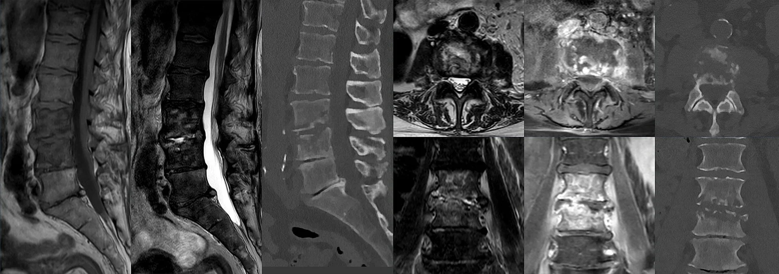
Long description
Composite image of an 82-year-old man's L3-L4 spondylodiscitis shown by MR (sagittal T1 and T2, axial and coronal T2/STIR, T1 fat-sat after contrast) and by CT in three planes.

Image in every suspected case. The strength of each modality depends on what you need: early diagnosis, extent, complications, or guidance of a biopsy.
Plain radiography remains the first study. It is cheap, widely available, and rules out fracture, gross destruction, and some tumours. Bony changes are late. In adults, changes may not be obvious for 10 to 14 days after the start of infection; in children, 5 to 7 days; in vertebral disease, subtle end-plate change may take 2 to 8 weeks. Osteomyelitis must involve about 1 cm of bone and reduce mineral content by 30% to 50% before it is visible. Sensitivity is only about 43% to 75% in meta-analytic and review data. Nuclear medicine can detect osteomyelitis 10 to 14 days before plain radiographic change.[2][3][4] [5]
MRI is the most sensitive and most specific investigation for osteomyelitis. It shows bone marrow oedema within 2 to 5 days, defines soft-tissue and intramedullary extension, and guides surgery. Reported sensitivity and specificity vary by site and source: about 90% and 83% for limb osteomyelitis in one emergency medicine review, 97% to 100% and 92% in a paediatric review, 82% to 100% and 75% to 96% in a radiology review, and 97% and 92% for vertebral infection. MRI can be repeated in 2 to 4 weeks if early findings are equivocal, and routine follow-up MRI in a clinically improving patient is unnecessary because imaging resolution lags behind clinical improvement.[1][2][3][4]
Bone (radionuclide) scintigraphy is sensitive and detects osteomyelitis days before plain radiographs, but its specificity is poor because many lesions take up tracer. Three-phase technetium scanning has sensitivity around 85% but specificity around 25%; combined bone and gallium imaging raises specificity to roughly 80% at the cost of sensitivity and convenience. A scan that is clearly negative is reassuring when MRI is unavailable, but a positive scan usually requires further imaging.[3]
CT excels at cortical and trabecular detail: sequestra, cloacas, involucra, intraosseous gas, soft-tissue abscess, and spinal enumeration. It is useful when MRI is contraindicated or when Modic type I degenerative change is a genuine alternative. CT-guided biopsy has sensitivity of about 50% for vertebral sampling. CT is less desirable than MRI for soft-tissue contrast and carries radiation exposure.[1][3][4]
Ultrasound can show periosteal elevation, fluid collections, soft-tissue abscess, and foreign bodies several days before plain radiographs, particularly in children, and can guide aspiration or biopsy without radiation. It cannot cross cortical bone, so its depth is limited.[3]
Probe-to-bone test is a bedside manoeuvre. A sterile blunt probe is gently passed into a wound or ulcer; if it reaches rigid bone, the test is positive. In a well-selected diabetic foot ulcer this has a positive likelihood ratio of about 6.4, and visible bone in the ulcer bed has a positive likelihood ratio of 9.2. The test is not perfect, but in combination with plain radiographs and ESR or CRP it is a sensible first step. The IWGDF and IDSA guidance recommends combining probe-to-bone testing, plain radiographs, and ESR, CRP, or procalcitonin as the initial assessment of suspected diabetic foot osteomyelitis.[1][6]
Bone biopsy with histopathology and microbiologic culture is the definitive diagnostic method. The practical question is when to obtain it and how.
- In a stable patient without systemic illness, it is reasonable to withhold empiric antibiotics until blood cultures and a bone biopsy are obtained, so the organism and susceptibilities can be identified.
- In unstable or septic patients, obtain cultures first, start empiric therapy, and biopsy as soon as it is safe.
- Blood cultures should be paired with bone sampling because they often yield the organism in haematogenous disease.
- Percutaneous, image-guided biopsy (CT or fluoroscopy) targets bone, disc, or adjacent spinal tissue. Cumulative yield in meta-analyses is about 48% to 52% [4], range 30.4% to 91%; yield improves with elevated CRP, a larger needle, more specimens, and aspiration of fluid collection. The effect of recent antibiotics is uncertain.
- Open (surgical) biopsy yields around 76% [4] and is preferred when percutaneous sampling is inconclusive or when debridement is already indicated.
- A negative first biopsy can be repeated, ideally at least 3 days later [4] [4], or followed by an open biopsy.
- Specimens should be sent for aerobic and anaerobic culture; histology shows acute inflammatory cells in 69% to 95% of cases [4]. Add fungal, mycobacterial, and zoonotic studies for immunocompromised patients, culture-negative cases, or relevant exposure.
- In diabetic foot disease, IDSA guidance specifies bone samples, not soft tissue samples, obtained intraoperatively or percutaneously. Bone biopsy sampling also helps distinguish infection from mimickers in the spine. [6]
Osteomyelitis has no single universally agreed diagnostic score. In practice the diagnosis is compatible clinical findings plus suggestive imaging and, ideally, positive culture or histology and radiologic evidence of infection. For native vertebral infection, the overall picture plus imaging and laboratory findings are used; in difficult or culture-negative cases repeated imaging and biopsy are required. In diabetic foot disease, the IWGDF and IDSA classify the infection using the IWGDF/IDSA scheme and indicate bone involvement with (O) after the severity grade, because the diagnosis, treatment, and prognosis of osteomyelitis differ from soft-tissue infection alone.
The aim is to control infection in the bone: remove dead and infected tissue, drain abscesses, restore blood supply, and give effective antibiotics. Long-term success usually needs the combined medical-surgical approach; medical therapy alone can suffice in selected stable cases without sequestrum or instability.
Approach to suspected osteomyelitis
- Suspected osteomyelitisRisk factors, route, focal pain, warmth, swelling, non-healing or infected wound, back pain
- Assess by routeHaematogenous, contiguous spread, direct inoculation, vascular insufficiency
- Question: Systemically unwell, sepsis, or new neurologic deficit?
Yes
- Take blood cultures first
- Start empiric antibioticsGram-positive (including MRSA where likely), Gram-negative and anaerobic cover; escalate for risk factors
- Urgent imaging and organ supportMRI for suspected cord compression; admit
No
- Delay empiric antibiotics if clinically safeMaximise culture yield; obtain blood cultures
- Plain radiograph and MRIMRI most sensitive; detects change within days
- Biopsy for culture and histologyPercutaneous or open; bone samples for diabetic foot
- Tailored long-term antibiotic therapyThe usual long-term duration; often IV first; oral step-down when suitable
- Escalate for source controlAbscess, necrotic or dead bone (sequestrum), instability, retained infected hardware, epidural or paravertebral abscess, evolving neurologic compromise
If the patient is stable, delaying empiric antibiotics until biopsy and cultures are obtained improves yield and tailoring. If unstable or septic, empiric therapy is justified, ideally after cultures. Empiric cover should include Gram-positive organisms (including MRSA where risk or local prevalence warrant it), Gram-negative bacilli, and anaerobes when a mixed chronic wound or diabetic foot infection is likely. Piperacillin-tazobactam plus vancomycin increases nephrotoxicity risk, so avoid that combination when an alternative exists.[1]
The standard recommendation for chronic osteomyelitis is 6 weeks of parenteral therapy, but this evolved from custom as much as evidence, and there is no convincing evidence that more than 4 to 6 weeks improves outcomes compared with shorter regimens. Nonrandomised adult studies of chronic osteomyelitis report cure rates of 60% to 90% with 4 to 6 weeks of parenteral beta-lactam therapy.[5]
- Adult acute/long-bone disease: long-term intravenous therapy usually lasts about 6 weeks initially.[1]
- Oral step-down: oral therapy with highly bioavailable agents (fluoroquinolones, trimethoprim-sulfamethoxazole, linezolid, clindamycin, metronidazole, rifampin as adjunct) achieves cure rates similar to parenteral therapy and avoids catheter complications and cost; it is most suitable after clinical improvement, debridement, and ideally organism identification. Adjunctive rifampin improves cure rates, especially for Staphylococcus aureus and device infections. Clinicians should individualise duration according to clinical and radiographic response, monitoring after treatment ends.[5]
- Vertebral infection: long-term antibiotic therapy for at least 6 weeks.
- Diabetic foot osteomyelitis (IDSA/IWGDF 2023): 6 weeks when there is no bone resection or amputation; up to 3 weeks after minor amputation with a positive bone margin culture. Define remission only after at least 6 months of follow-up after completing antibiotics. Mild and moderate foot soft-tissue infection antibiotics are given for 1 to 2 weeks, longer if extensive or slow to resolve.[6]
- Complicated disease (abscess, MRSA, pelvic or spinal involvement, bacteremia, poor host status) may require longer courses and operative source control.
- Source control is the key surgical goal: debridement of infected and non-viable tissue, drainage of abscess, management of dead space, tissue coverage, and stabilisation of unstable bone.
- Chronic osteomyelitis requires surgical debridement because antibiotics penetrate poorly into dead or avascular bone; debridement combined with medical therapy appears to improve cure rates, although not every case requires an operation.
- Vascular insufficiency demands revascularisation, otherwise systemic antibiotics cannot reach the site.
- Vertebral infection: medical management with at least 6 weeks of antibiotics is the mainstay. Surgery is needed for epidural or paravertebral abscess, evolving neurologic deficit, or spinal instability.
- Diabetic foot: draining abscess, necrosis, compartment involvement, gangrene, limb or life-threatening infection, or failure of appropriate therapy within 4 weeks requires surgical review. Early surgery (within 24 to 48 hours) is considered for moderate and severe infection. Surgical resection of infected bone is a reasonable approach, and forefoot osteomyelitis without immediate incision and drainage need, without poor perfusion, and without exposed bone can sometimes be treated with antibiotics alone.
- Open fractures: remove gross contaminants, irrigate, cover the wound, and give systemic antibiotic prophylaxis within 6 hours, tailoring cover to Gustilo-Anderson grade as advised by trauma teams.
Monitor clinically and with CRP, which is the preferred marker for acute response; ESR and MRI have slower resolution and can lag behind improvement. Repeat blood tests and imaging according to local practice. Educate patients about recurrent pain, fever, or drainage months or years later.
- Acute complications include bacteraemia and sepsis; adjacent septic arthritis (especially where metaphyseal infection crosses the physis in infants); soft-tissue necrosis; sinus tracts; and pathologic fracture.
- Chronic complications include persistent sequestrum, recurrent infection, bone deformity, joint stiffness, chronic pain, fistula, and limb loss.
- Vertebral complications include paravertebral, psoas, or retropharyngeal abscess, disc destruction, spinal instability, epidural abscess, and cord compression with paralysis.
- Delayed diagnosis worsens outcomes; neurologic compromise in spinal infection can be irreversible.
Relapse is the hallmark risk of chronic osteomyelitis and can occur long after apparently successful treatment because bacteria persist in biofilm and devitalised bone. Prognosis improves with culture-directed therapy, adequate debridement, restored perfusion, and removal of infected foreign material. There are no robust biomarkers of cure, so patients need clinical follow-up and a low threshold for re-evaluation.
- Good diabetic foot care, offloading, perfusion assessment, and early treatment of ulcers and soft-tissue infection reduce contiguous bone infection.
- Prompt treatment of bacteraemia, skin infection, and endocarditis reduces haematogenous seeding.
- In open fractures, irrigation and timely antibiotic prophylaxis reduce post-traumatic osteomyelitis. In total joint replacement, infection-prevention strategies reduce periprosthetic disease, although this is a related rather than identical problem.
- There is no population screening for osteomyelitis. Diagnosis is clinical, triggered by symptoms and risk factors.
- Children: acute haematogenous infection of the metaphysis predominates; Staphylococcus aureus is the cultured organism in about 80% of cases, followed by group A streptococcus. Short courses can suffice for uncomplicated methicillin-sensitive S. aureus infection, but MRSA or complicated disease may need longer treatment and surgery. A third-generation cephalosporin should be added in sickle cell disease to cover Salmonella.
- Sickle cell disease: bone infarction and infection look similar; fever and pain out of proportion to usual crisis should prompt imaging and culture-directed therapy.
- Immunocompromised patients: broader or unusual organisms, including mycobacteria, fungi, and Bartonella. Consider tuberculosis and endemic fungi when exposure supports it.
- Older adults and people with diabetes: gram-negative organisms, muted signs, and vascular compromise are more common; take foot and back pain seriously in these groups.
- People who inject drugs: vertebral infection and Pseudomonas are classic considerations, but S. aureus and MRSA remain common.
A full algorithm can be adapted without losing the essentials:
- Clinical suspicion comes first: pain, warmth, swelling, a non-healing or malodorous wound, and risk factors such as diabetes, immunosuppression, or injection drug use.
- Plain radiographs, bedside probe-to-bone testing, and inflammatory markers (CRP and ESR) are available in most settings and are genuinely useful first tests.
- Where MRI is unavailable, conventional radiograph plus bone scan or CT, and a high index of suspicion, must carry the diagnosis; repeated films over 2 to 3 weeks [2][3] help.
- Blood cultures should be taken before antibiotics when feasible.
- Without ready access to laboratories, a Gram-stained smear from the ulcer or wound is inexpensive and can visualise the likely class of pathogens and guide empiric therapy.
- Oral, highly bioavailable antibiotics are a legitimate step-down once the patient is improving and organism coverage is appropriate, avoiding prolonged intravenous treatment and the catheter complications that accompany it.
- Surgery and revascularisation remain important but may require referral; early recognition and timely referral matter more than technology.
- Osteomyelitis is bone infection reached by haematogenous, contiguous, or direct routes; the route points to the likely organism and to the needed treatment.
- Staphylococcus aureus is the most commonly isolated organism; Salmonella in sickle cell disease, Pseudomonas in puncture wounds and injecting drug use, Pasteurella after bites, polymicrobial infection in diabetic and traumatic wounds, and tuberculosis in the spine are the classic associations.
- A patient can have osteomyelitis with a normal white cell count. ESR and CRP are useful screens; ESR of 70 mm/h or more in a diabetic foot [1][6] and compatible back pain with a high ESR are particularly informative. CRP tracks acute response better than ESR.
- Plain radiographs in adults may not show change for 10 to 14 days [3]; vertebral change may take 2 to 8 weeks [4]; MRI is the most sensitive test and shows marrow change within days. [2]
- Bone biopsy with culture and histology establishes the diagnosis and guides treatment. Blood cultures are often positive in haematogenous disease; superficial swabs are not. [2]
- Treatment is source control plus antibiotics: IV therapy usually starts the course and typically lasts about 6 weeks for chronic osteomyelitis, parenteral therapy for vertebral disease is continued for at least 6 weeks, and diabetic foot disease without resection needs about 6 weeks (up to 3 weeks after minor amputation with a positive bone margin). [6]
- Oral step-down with highly bioavailable agents is acceptable after clinical improvement and appropriate debridement.
- Spinal infection is a medical emergency when neurology changes: triad is absent in most, and paralysis can be permanent.
- In chronic disease, dead bone and biofilm explain recurrence; debridement and vascular status matter as much as antibiotics.
- Low-resource care still works well with careful clinical diagnosis, radiographs, probe-to-bone testing, cultures, early antibiotics, and timely referral.
Answer each one in your head first, then open the answer.
A 7-year-old boy has refusal to bear weight on one leg, fever, and focal tibial tenderness. What is the most likely organism, and which test localises the infection early?
A 55-year-old man with a plantar puncture wound through a trainer now has calcaneal pain. Which organisms are most likely, and what guides long-term antibiotics?
Show answerHide answer
Staphylococcus aureus and Pseudomonas aeruginosa. A bone biopsy or deep tissue sample for culture and histology is needed, because the superficial wound does not predict the bone isolate.
Why can a patient with chronic osteomyelitis relapse decades after seemingly successful treatment?
Show answerHide answer
Bacteria can persist intracellularly and as slow-growing persisters within biofilm, often protected by devitalised bone and poor local blood supply. Debridement, perfusion, and culture-directed antibiotics reduce but do not eliminate this risk.
In a patient with a diabetic foot ulcer and suspected osteomyelitis, what three bedside or simple studies make a sensible first diagnostic combination?
Show answerHide answer
Probe-to-bone testing, plain radiographs, and an inflammatory marker (ESR or CRP; procalcitonin is an alternative). MRI is reserved for doubt, and bone sampling is needed for definitive culture.
A woman with 3 weeks of back pain that is worse at night and not relieved by rest has an ESR of 62 mm/h and no fever. Does this exclude vertebral osteomyelitis?
Show answerHide answer
No. Back pain is present in over 90% of cases [1] and is often the only early symptom; many patients, especially older adults, have no fever. An elevated ESR or CRP with compatible back pain is sensitive for spondylodiscitis, so MRI is required.
How long is a usual antibiotic course for adult chronic osteomyelitis, and what oral step-down strategy is acceptable?
Show answerHide answer
About 6 weeks initially [1][5], with no clear benefit beyond 4 to 6 weeks beyond debridement [5]. Highly bioavailable oral agents (such as fluoroquinolones, trimethoprim-sulfamethoxazole, clindamycin, linezolid, or metronidazole, with rifampin as an adjunct) can be used once the patient is improving, ideally after culture and debridement.
A patient with a diabetic foot osteomyelitis completes surgery with minor amputation and has a positive bone margin culture. What duration and follow-up does current IDSA-related guidance recommend?
Show answerHide answer
Up to 3 weeks of antibiotic therapy after the amputation, and remission is assessed only after a minimum of 6 months of follow-up after completing antibiotics [6]. If no resection is performed, about 6 weeks are used.
What four clinical changes in a patient with back pain and fever should make you treat spinal epidural abscess as an emergency?
Show answerHide answer
Development of weakness, sensory loss, bladder or bowel dysfunction, or progression to paralysis. The classic triad is present in only a minority, so deterioration of neurology is the red flag that must not be missed.
Sickle cell disease, tuberculosis, infective endocarditis, sepsis, septic arthritis, diabetes mellitus, diabetic foot infection, musculoskeletal (plain text).
- Kolinsky DC, Liang SY. Musculoskeletal infections in the emergency department. Emerg Med Clin North Am 2018;36(4):751-766. doi:10.1016/j.emc.2018.06.006. Accessed .
- Thakolkaran N, Shetty AK. Acute hematogenous osteomyelitis in children. Ochsner J 2019;19(2):116-122. doi:10.31486/toj.18.0138. Accessed .
- Pineda C, Espinosa R, Pena A. Radiographic imaging in osteomyelitis: the role of plain radiography, computed tomography, ultrasonography, magnetic resonance imaging, and scintigraphy. Semin Plast Surg 2009;23(2):80-89. doi:10.1055/s-0029-1214160. Accessed .
- Maamari J, Tande AJ, Diehn F, Tai DBG, Berbari EF. Diagnosis of vertebral osteomyelitis. J Bone Jt Infect 2022;7(1):23-32. doi:10.5194/jbji-7-23-2022. Accessed .
- Spellberg B, Lipsky BA. Systemic antibiotic therapy for chronic osteomyelitis in adults. Clin Infect Dis 2012;54(3):393-407. doi:10.1093/cid/cir842. Accessed .
- Senneville E, Albalawi Z, van Asten SA, et al. IWGDF/IDSA Guidelines on the Diagnosis and Treatment of Diabetes-Related Foot Infections. Clin Infect Dis 2023; ciad527. doi:10.1093/cid/ciad527. Accessed .
Figure credits
Educational content for medical students and doctors. It is not a substitute for clinical judgement or local guidelines.