Toxoplasmosis
Toxoplasma gondii infection: usually silent, dangerous in pregnancy and the immunosuppressed; cerebral, ocular and congenital disease.
- Toxoplasmosis is infection with the single-celled parasite Toxoplasma gondii, which about one-third of people worldwide carry, usually without ever knowing it. [5]
- Most healthy adults have no symptoms or a brief illness with swollen lymph nodes; the parasite then forms dormant tissue cysts, mainly in brain, muscle and eye.
- The parasite's sexual cycle runs only in cats, the definitive host. Humans are infected mainly by eating tissue cysts in undercooked meat or by swallowing oocysts from cat faeces in soil, water or unwashed food.
- Two situations make toxoplasmosis dangerous: pregnancy, where a newly infected mother can infect the fetus, producing the classic triad of hydrocephalus, cerebral calcifications and chorioretinitis; and advanced immunosuppression, especially AIDS with CD4 counts under 100 cells per microL, where dormant cysts reactivate and cause cerebral toxoplasmosis with headache, focal signs and seizures. [4][5]
- In cerebral toxoplasmosis, brain MRI usually shows multiple ring-enhancing lesions in the basal ganglia and corticomedullary junction, and the serum Toxoplasma IgG is positive.
- Standard treatment for severe disease is pyrimethamine plus sulfadiazine plus folinic acid; for AIDS-related cerebral disease, imaging and clinical improvement after 10 to 14 days of treatment supports the diagnosis before a biopsy is considered. [4] [5]
- All Toxoplasma drugs act only against the active (tachyzoite) stage, so tissue cysts are never eradicated and long-term maintenance treatment is needed in the immunosuppressed.
By the end of this page you should be able to:
- State where T. gondii comes from and the three routes by which humans are infected.
- Explain why toxoplasmosis is usually harmless in healthy adults but dangerous in pregnancy and in the immunosuppressed.
- Recognise the presentation of cerebral toxoplasmosis in AIDS and interpret the MRI and serology.
- Tell cerebral toxoplasmosis apart from primary CNS lymphoma and from other ring-enhancing lesions.
- Plan treatment with pyrimethamine, sulfadiazine and folinic acid, including dose by body weight, the response to expect at two weeks, and when to biopsy. [4]
- Manage toxoplasmosis in pregnancy and the newborn.
- Give the indications for primary and secondary prophylaxis and apply prevention in a low-resource setting.
DefinitionToxoplasmosis
Toxoplasmosis is disease caused by the obligate intracellular protozoan Toxoplasma gondii. It occurs in four principal clinical forms: (1) acute or latent (usually asymptomatic) infection in immunocompetent people; (2) lymphadenopathic, mononucleosis-like infection; (3) cerebral (neurotoxoplasmosis) disease, mainly reactivation in the immunosuppressed; and (4) congenital infection of the fetus. A fifth form, ocular toxoplasmosis, produces chorioretinitis in both immunocompetent and immunosuppressed people. [3]
The active, rapidly dividing form is the tachyzoite; the dormant, slowly dividing form packaged in cysts is the bradyzoite. Current drugs affect only tachyzoites: no drug clears the tissue cysts, so latent infection persists for life unless the immune system keeps it in check.
Toxoplasma gondii infects roughly one-third of the world's population.[5] Seroprevalence varies greatly by region and reflects diet, hygiene and climate. It is common in western Europe, South America and parts of Africa; data from Germany in 2008 to 2011 found seroprevalence rising from about 20% in women aged 18 to 29 years to nearly 50% in those aged 40 to 49 years.[7] Over 40 million people in the United States are infected.[1] Infection rates in immunocompromised people run higher than in the general population (about 36% versus 25% in one large review).[6]
In people living with HIV, reported seroprevalence ranges from about 26% in high-income countries to about 55% in low-income countries, and cerebral toxoplasmosis is a leading cause of central nervous system disease where HIV is uncontrolled and antiretroviral therapy or co-trimoxazole prophylaxis is unavailable.[5][6]
| Group | Approximate seroprevalence | Note |
|---|---|---|
| Healthy adults worldwide | about one-third | latent, usually asymptomatic |
| Women of childbearing age (Germany, older ages) | up to about 50% | age-related rise |
| USA population | over 40 million infected | most asymptomatic |
| PLWH, high-income settings | about 26% | reactivation risk if CD4 under 100 |
| PLWH, low-income settings | about 55% | highest burden where prophylaxis is absent |
Acquisition occurs through four routes:
- Foodborne. Eating raw or undercooked meat (especially pork, lamb and game) containing bradyzoite tissue cysts, or unwashed fruit, vegetables or water contaminated with sporulated oocysts. Raw shellfish can also be a source.
- Cat-related. Accidentally swallowing sporulated oocysts from cat faeces in soil, sand or litter; cats are the definitive host.
- Congenital. A mother infected for the first time during (or just before) pregnancy can transmit tachyzoites across the placenta.
- Iatrogenic. Organ or stem-cell transplantation from an infected donor, or, rarely, blood transfusion.
Risk factors include feeding cats raw meat; handling soil or cat litter without gloves; eating undercooked meat or unwashed produce; living in warm, humid climates where oocysts survive; drinking untreated water; and low health literacy. Pregnant women and immunocompromised people must take the greatest care.[1][6]
There is no human vaccine and no way to eradicate the cyst stage.
Toxoplasma gondii has three life-cycle stages relevant to human disease.[6]
- Oocyst (containing sporozoites): the product of sexual reproduction, formed only in the intestine of cats and other felids. An infected cat may shed millions of these oocysts for a non-recurring period of up to about three weeks. Oocysts sporulate in fresh air within two to three days (depending on conditions) and then survive in the environment for a year or more. [6]
- Tachyzoite: the rapidly dividing stage. After a person ingests cysts or oocysts, bradyzoites or sporozoites are released in the gut, convert to tachyzoites and disseminate throughout the body. Tachyzoites cross the blood-brain barrier by at least three routes: paracellular migration through tight junctions, direct infection of cerebral endothelial cells, and carriage inside infected dendritic cells and monocytes ("Trojan horse").
- Bradyzoite in a tissue cyst: the slowly dividing, dormant form that evades immune clearance and persists for life, most often in brain, skeletal and cardiac muscle and retina. Bradyzoites become active again when immunity falls.
The outcome depends almost entirely on the host's cell-mediated immunity, driven by interferon-gamma, CD4+ and CD8+ T cells and microglia. In immunocompetent people, tachyzoites are controlled and the cysts stay dormant; in the immunosuppressed, bradyzoites reactivate and become tachyzoites again, producing focal necrotic brain lesions. In pregnancy, tachyzoites cross the placenta and infect fetal tissues directly.
Brain disease targets the basal ganglia and the corticomedullary junction. Cyst dormancy is why no drug can cure latent infection.
Most infections are silent. In a small minority (less than 10% in one large review), a mononucleosis-like illness develops after an incubation period of one to three weeks, with fever, headache, cervical or occipital lymphadenopathy, malaise and fatigue; symptoms can persist for several weeks.[6][7] Hepatosplenomegaly, rash, arthritis, myocarditis or myositis are uncommon but occur.[4] Lymphadenopathic toxoplasmosis is usually self-limiting and, in healthy adults, rarely needs treatment unless symptoms are severe or persistent.[3]
Ocular toxoplasmosis (chorioretinitis) can occur even in immunocompetent hosts, either as congenital disease that presents later or after postnatal infection. Recurrences are common and can threaten vision.
This is the most important form. It usually arises by reactivation of latent cysts in people whose CD4 count has fallen under 100 cells per microL.[4][5] Risk is highest without co-trimoxazole prophylaxis and without effective antiretroviral therapy.[6]
Presentation is subacute and focal: headache, hemiparesis, cranial nerve palsies, ataxia, altered mental status, seizures and visual changes; fever may or may not be present and should not be relied on to exclude the diagnosis.[4] Involvement of the basal ganglia can produce abnormal movements such as chorea or ballismus. Less commonly there is a diffuse, rapidly fatal encephalitic picture, or necrotising ventriculitis.
On brain MRI, the typical findings are multiple ring-enhancing lesions with surrounding oedema in the basal ganglia, frontal and parietal lobes; CT is less sensitive but more available and shows hypodense enhancing lesions with mass effect. The characteristic eccentric target sign and concentric target sign may be seen.[5]
Diagnosis is made by the combination of compatible clinical signs, appropriate imaging and, crucially, a serum Toxoplasma IgG that is positive: IgG rises within one to two months of infection and remains high for life, so it marks risk of reactivation. Polymerase chain reaction for T. gondii DNA in the cerebrospinal fluid is specific but insensitive; a positive test confirms the diagnosis, a negative test does not exclude it. A positive CSF PCR raises the probability of cerebral toxoplasmosis from 87% to 96% in the right clinical setting.[5][6]
The disease is presumptively diagnosed when a patient has CD4 under 100 cells per microL, multiple ring-enhancing lesions, positive Toxoplasma IgG and no prophylaxis, and then shows clinical and radiographic improvement after 10 to 14 days of empirical treatment.[4][5] About 85% of responders improve within the first week and 90% by two weeks, so failure to improve by then raises suspicion of another cause and a biopsy should be considered.[4]
Transmission from mother to fetus can occur if a previously seronegative woman is infected during or just before pregnancy (reactivation in a severely immunocompromised pregnant woman is a much rarer route). The classical symptomatic triad, described by Sabin in the 1950s, is retinochoroiditis, intracranial calcifications and hydrocephalus.[7] Serious neurological outcomes such as hydrocephalus appear mainly with infection in the first or second trimester, whereas chorioretinitis can follow infection at any gestational time.[7] Most congenitally infected children are asymptomatic at birth and may develop retinochoroiditis or other problems years later. Transmission rate rises with gestational age.[7]
Acute infection in a pregnant woman is usually asymptomatic or mild; less than 10% of immunocompetent adults develop mononucleosis-type symptoms with cervical or occipital lymphadenopathy.[7] In immunocompromised pregnant women, severe encephalitis, myocarditis, pneumonitis or hepatitis can occur.[7]
The differential is usually between cerebral toxoplasmosis, primary CNS lymphoma and a tuberculoma or bacterial brain abscess. Both toxoplasmosis and lymphoma cause contrast-enhancing mass lesions, so imaging alone cannot always separate them. Features that favour lymphoma include a single lesion, a subependymal location, hyperattenuation on unenhanced CT and uptake on thallium-201 SPECT (in toxoplasmosis, uptake is absent); SPECT has reported sensitivity of 92% and specificity of 85% for distinguishing CNS lymphoma from other focal lesions in people living with HIV.[6]
| Feature | Cerebral toxoplasmosis | Primary CNS lymphoma | Tuberculoma / tuberculous abscess |
|---|---|---|---|
| Number of lesions | Usually multiple | Often single, may be multiple | Single or few |
| Favoured sites | Basal ganglia, corticomedullary junction | Subependymal, periventricular | Variable |
| MRI enhancement | Multiple ring-enhancing with oedema | Ring or nodular enhancement | Ring-enhancing |
| Serum Toxoplasma IgG | Positive in almost all | May be positive by chance | Not specific |
| CD4 count | Usually under 100 | Usually under 100 | Variable |
| Other clues | Responds over 10 to 14 days | Hyperattenuation on unenhanced CT; SPECT uptake | TB history, systemic disease |
| CSF PCR | Specific but insensitive | Often clonal B-cell markers | TB PCR possible |
- Lymphadenopathy in healthy adults can mimic mononucleosis, cytomegalovirus, Epstein-Barr virus, HIV seroconversion, sarcoidosis or lymphoma. Toxoplasmosis is usually distinguished by positive Toxoplasma IgG or IgM, mild disease and, if treated, response.
- Congenital infection must be differentiated from other infections that cause hydrocephalus, calcifications or chorioretinitis (cytomegalovirus, rubella, herpes simplex, syphilis).
- Ocular toxoplasmosis must be distinguished from other causes of posterior uveitis such as sarcoidosis, tuberculosis, syphilis, presumed ocular histoplasmosis and toxocariasis.
Figure 1BrainToxoplasmosis MRI 2 07
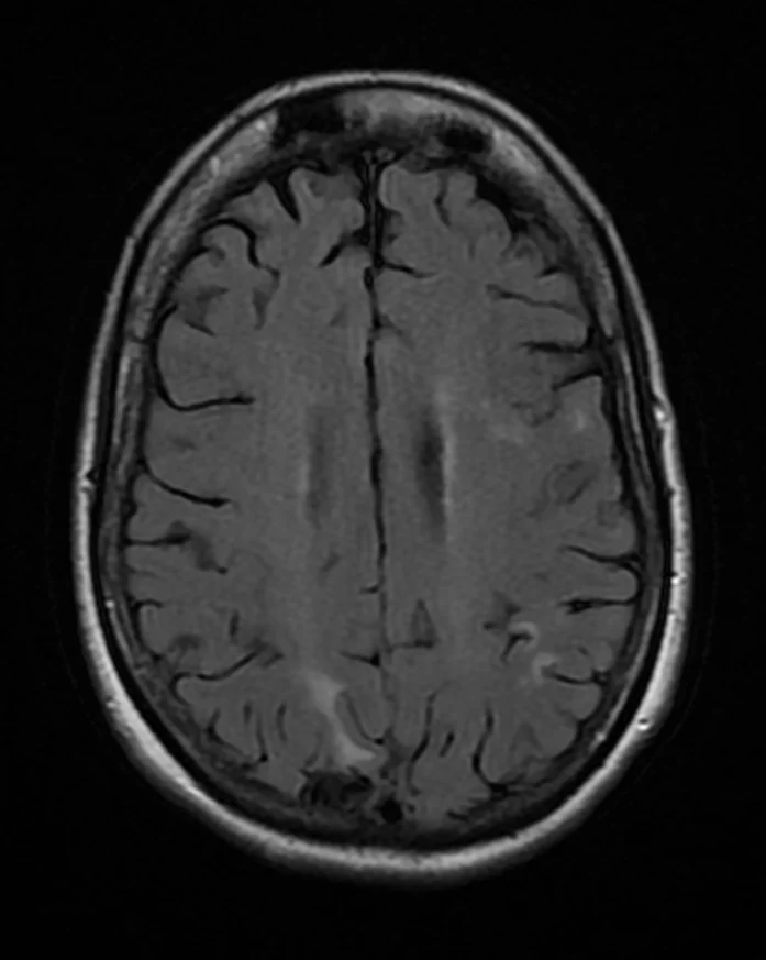
Long description
MRI of cerebral toxoplasmosis with primary involvement in the right occipital lobe in a 48-year-old woman with AIDS.

- Serology. Toxoplasma-specific IgG and IgM are the main tests. IgG appears within weeks, peaks at one to two months and stays high for life. IgM can persist for a year, so a positive IgM does not prove recent infection; absence of IgM rules out recent infection. In the immunosuppressed, serology is unreliable and must be interpreted with clinical findings.[2][3] [6]
- Avidity. Low IgG avidity suggests recent infection; rising avidity over time helps date seroconversion, which matters in pregnancy.
- PCR. T. gondii DNA by PCR is highly specific. It can be run on blood, CSF, amniotic fluid or tissue. Sensitivity in CSF is low, so a negative CSF PCR does not exclude cerebral toxoplasmosis. Amniotic fluid PCR from 18 weeks of gestation (and at least 4 weeks after maternal infection) is used to diagnose fetal infection.[2][7] [3]
- Direct detection. Stained tissue, CSF or biopsy can show tachyzoites but is rarely needed; parasites are hard to find.
- Ocular exam. Ocular toxoplasmosis is diagnosed mainly by slit-lamp and fundus examination (lesion appearance, course, symptoms), supported by serology.[2]
- Imaging. Brain MRI is far more sensitive than CT for cerebral toxoplasmosis and can reveal the target signs. CT remains useful in low-resource settings.
- Other tests. In pregnancy, fetal ultrasound looks for ventriculomegaly, intracranial calcifications and growth restriction; MRI is sometimes added.
Acute infection in an immunocompetent adult is usually a clinical and serological diagnosis: compatible mononucleosis-like illness plus positive IgG or IgM, with confirmation by a reference laboratory when needed.
Cerebral toxoplasmosis in AIDS is diagnosed presumptively by: (a) advanced HIV with CD4 under 100 cells per microL; (b) positive serum Toxoplasma IgG; (c) one or more (usually multiple) ring-enhancing brain lesions on imaging; (d) no co-trimoxazole prophylaxis; and (e) clinical and radiographic improvement after 10 to 14 days of empirical therapy. Definite diagnosis needs histology or a positive CSF PCR, but biopsy is reserved for seronegative patients or non-responders.[4][5]
Congenital infection is proven by detecting parasite DNA in amniotic fluid (PCR) and confirmed in the neonate by serology (persistence of IgG after maternal transfer wanes) and imaging plus ophthalmology.
Pregnancy dating. Whether maternal infection is acute or pre-existing is settled by IgG, IgM and IgG avidity, ideally before 18 weeks; IgM can persist for years and is not on its own proof of recent infection. [3]
Start empirical treatment promptly; do not delay for biopsy in seropositive patients with typical findings, because delay worsens outcome.[4]
Induction (at least 6 weeks):
- Body weight 60 kg or more: pyrimethamine 200 mg once, then 75 mg daily, plus sulfadiazine 1.5 g every 6 hours, plus folinic acid 10 to 50 mg daily. [4]
- Body weight under 60 kg: pyrimethamine 200 mg once, then 50 mg daily, plus sulfadiazine 1 g every 6 hours, plus folinic acid 10 to 50 mg daily.[4]
Alternative induction regimens when pyrimethamine or sulfadiazine cannot be used:[3][4]
- Pyrimethamine plus folinic acid (as above) plus clindamycin 600 mg every 6 hours; clindamycin carries a risk of Clostridioides difficile colitis. [4]
- Trimethoprim-sulfamethoxazole 10/50 mg per kg per day (TMP component) in divided doses (higher doses of 15/75 or 20/100 mg per kg per day are sometimes used). [4]
- Atovaquone 1,500 mg twice daily, ideally combined with pyrimethamine and folinic acid or with sulfadiazine, and taken with a high-fat meal. [4]
- Pyrimethamine plus folinic acid plus azithromycin 1,000 mg daily or clarithromycin 500 mg twice daily; experience is limited and azithromycin has a high relapse rate in maintenance. [4]
Maintenance (after induction, continued while immunosuppression persists):
- Pyrimethamine 50 mg daily (25 mg if under 60 kg) plus sulfadiazine 1 g every 6 hours (0.5 g every 6 hours if under 60 kg) plus folinic acid 10 to 25 mg daily. [4]
- Maintenance can be stopped once the CD4 count has been above 200 cells per microL for at least 6 months on effective antiretroviral therapy, provided the patient responded well.[4]
- Co-trimoxazole 5/25 mg per kg per day in divided doses is an acceptable maintenance option; pyrimethamine-based combinations other than pyrimethamine plus dapsone also cover Pneumocystis. [4]
Response to expect. About 85% of those who ultimately respond do so within the first week and 90% by two weeks. Failure to improve within the first one to two weeks should raise suspicion of an alternative diagnosis such as lymphoma and prompt brain biopsy. Radiological improvement lags behind clinical improvement.[4]
Other measures in AIDS. Start or optimise antiretroviral therapy without delay; immune reconstitution inflammatory syndrome is uncommon after treatment for cerebral toxoplasmosis. Corticosteroids are reserved for significant brain oedema and should be used judiciously. In AIDS, cerebral toxoplasmosis is often fatal if untreated, so treatment must not be withheld while awaiting confirmation.
Treatment depends on the ophthalmologist's assessment of lesion activity, size, location (especially near the macula or optic disc), visual acuity and inflammation. Quiescent healed lesions are not treated. A common regimen is pyrimethamine 100 mg as a single loading dose, then 25 to 50 mg daily, plus sulfadiazine 2 to 4 g daily for two days then 500 mg to 1 g four times daily, plus folinic acid 5 to 25 mg with each dose of pyrimethamine, for 4 to 6 weeks, with corticosteroids as needed for inflammation.[3] Alternatives include TMP-SMX, atovaquone, clindamycin and azithromycin, and intravitreal clindamycin with dexamethasone for selected cases. [4][7]
Lymphadenopathic disease is usually self-limiting and rarely needs drugs. Treatment for 2 to 4 weeks is reasonable when symptoms are severe or persistent, when visceral disease is evident, for laboratory-acquired infection, or for ocular disease. The regimen is pyrimethamine 100 mg daily for one or two days then 25 to 50 mg daily, plus sulfadiazine 1 g every 6 hours, plus folinic acid 10 to 20 mg daily, or TMP-SMX 5/25 to 10/50 mg per kg per day in divided doses, or atovaquone 1,500 mg twice daily. A 4- to 6-week course is sensible if symptoms have developed.[4] [3]
Approach varies by centre and guideline. The consensus principles:
- Before 18 weeks' gestation, or when fetal infection is not suspected or proven, spiramycin 3 g daily (for example 1 g three times daily) is used to reduce transmission to the fetus; it is most effective within 8 weeks of seroconversion. [3] [7]
- At or after 18 weeks' gestation, or when fetal infection is suspected or documented (amniotic fluid PCR positive), use pyrimethamine 50 mg daily (with a 50 mg dose on day 1 in many European schedules), sulfadiazine (commonly 2 times 1.5 g daily) and folinic acid; pyrimethamine and sulfadiazine are avoided in the first trimester because of teratogenicity. [4] [3]
- These drugs are usually continued to delivery.
- Amniocentesis for PCR is not performed before 18 weeks and at least 4 weeks after maternal infection; sensitivity is 69 to 92% and specificity 98 to 100%, so a negative PCR does not fully exclude fetal infection. [3] [7]
- Spiramycin does not cross the blood-brain barrier, which is one reason combination therapy is preferred once fetal infection is proven.
Regional schedules differ markedly (for example, German and Austrian regimens start pyrimethamine at 25 mg daily with 3 g sulfadiazine daily, while North American schedules use pyrimethamine 50 mg daily with sulfadiazine up to 4 g daily). Guidelines disagree, and the exact doses should be taken from the local protocol.[7][3]
Infants with congenital toxoplasmosis are generally treated for 12 months with pyrimethamine, a sulfonamide and leucovorin, using weight-based doses: pyrimethamine 2 mg per kg per day orally in two divided doses for the first two days, then 1 mg per kg daily to age 2 months (or 6 months if symptomatic), then 1 mg per kg three times weekly; sulfadiazine 100 mg per kg per day in two divided doses; and folinic acid (leucovorin) 10 mg three times weekly.[3]
- Primary prophylaxis. In HIV, it is indicated when the CD4 count is under 100 cells per microL and the serum Toxoplasma IgG is positive. Co-trimoxazole (TMP-SMX) at prophylactic doses (for example 1 single-strength or 1 double-strength tablet daily) is the standard choice; dapsone plus pyrimethamine plus leucovorin, or atovaquone, are alternatives. It can be stopped when the CD4 count rises above 200 cells per microL for more than three months on antiretroviral therapy.[5][6]
- Secondary (maintenance) prophylaxis. After treated cerebral toxoplasmosis, continue maintenance therapy until the CD4 count is stable above 200 cells per microL for at least 6 months on effective therapy.[4]
- Transplant settings. Haematopoietic stem-cell and solid-organ (especially heart) transplant recipients who are Toxoplasma-seropositive should receive prophylaxis during the period of highest risk; TMP-SMX started at engraftment reduces reactivation. Screening donors and recipients before transplantation identifies those at risk.
- Pregnancy. Prevention of maternal infection (below) is the main strategy; there is no routine screening in all countries.
Preventive advice applies to everyone, and is essential for pregnant women and immunocompromised patients (and for people who are Toxoplasma-seronegative and living with HIV).[1][6]
- Cook meat to a safe internal temperature (and freeze meat for several days at sub-zero (0F) temperatures before cooking to greatly reduce risk).
- Do not eat raw shellfish; rinse fruit and vegetables under running water.
- Do not drink unpasteurised goat's milk.
- Wash hands with soap and water after touching raw meat, soil, cats or anything that might be contaminated.
- Wear gloves when gardening or handling soil or sand; wash hands afterwards.
- Avoid emptying or cleaning cat litter if immunocompromised; if unavoidable, do it daily, wear disposable gloves and wash hands; keep cats indoors and do not feed them raw meat; do not adopt stray cats.
- Health education about congenital toxoplasmosis should be given to all pregnant women; some authorities add periodic serological screening.
- Pregnancy. As above.
- Children and congenital infection. 12 months of therapy; shunting for hydrocephalus if needed, early placement improves outcomes. Long-term ophthalmology follow-up because chorioretinitis can develop years later.
- Immunocompromised non-HIV patients (transplant, CD4-depleting therapy, high-dose corticosteroids, haematological malignancy) are treated with the same regimens as AIDS-related cerebral disease; tissue diagnosis is preferred more often because the range of possible pathogens is wider.
- Older adults. Immune senescence lowers resistance to reactivation; the same precautions apply.
- Cerebral toxoplasmosis: seizures, focal deficits, hydrocephalus from aqueduct obstruction, raised intracranial pressure, coma and death if untreated.
- Retinochoroiditis with permanent visual loss; late recurrences.
- Congenital: intracranial calcifications, mental retardation, epilepsy, motor impairment, hydrocephalus or microcephaly, and ocular disease.
- Bone-marrow suppression and rash from pyrimethamine and sulfonamides; crystalluria and renal injury from sulfadiazine if hydration is inadequate; Clostridioides difficile colitis with clindamycin.
- Reactivation of ocular disease in the immunocompetent.
Untreated cerebral toxoplasmosis is almost universally fatal. With induction therapy, about 70 to 80% of AIDS patients show partial or complete clinical response. Mortality and relapse have improved dramatically with effective antiretroviral therapy. Over half of survivors of cerebral toxoplasmosis have residual neurological deficits, and cognitive impairment may be more common than after other CNS opportunistic infections.[5][6]
Congenital disease outcomes range from asymptomatic to severe disability; early treatment and early shunting for hydrocephalus improve outcomes. Healed ocular lesions still recur in more than half of patients.
- Co-trimoxazole-based regimens are much less expensive than pyrimethamine-based ones and are used successfully in many developing countries; pyrimethamine prices in some markets are prohibitive.
- CT is often the only available brain imaging and is less sensitive than MRI, so multiple hypodense enhancing lesions with mass effect in the basal ganglia, combined with positive Toxoplasma IgG and CD4 under 100 cells per microL, still justify empirical treatment.
- Systemic screening for toxoplasmosis antibodies early in prenatal care is not universal; where screening is unavailable, rely on maternal seroconversion testing when feasible and treat acutely infected mothers.
- Prevention is low-cost and effective: cook meat thoroughly, wash produce, avoid untreated water, wear gloves for gardening and soil contact, and have a non-pregnant, immunocompetent person change cat litter daily.
- Folic acid supplementation is different from folinic acid: patients on pyrimethamine need leucovorin (folinic acid) to protect the bone marrow.
- Drug-specific monitoring (full blood count, creatinine, liver function) should be done where possible; sulfadiazine requires good hydration to avoid crystalluria.
Brain lesions with mass effect in a patient with advanced HIV
- PLWH with focal neurological symptomsHeadache, seizure, focal deficit
- CD4 count and Toxoplasma IgGRisk highest when CD4 is under 100 cells per microL
- Question: Serum Toxoplasma IgG positive?
Yes
- Brain MRI or CTMultiple ring-enhancing lesions with oedema
- Question: Typical, multiple lesions and CD4 under 100?
Yes
- Start empirical pyrimethamine plus sulfadiazine plus folinic acid
- Reassess clinically and radiologically at 10 to 14 daysIf improved, confirms cerebral toxoplasmosis
- If no improvement, consider biopsy or alternate diagnosis (CNS lymphoma)
No / atypical
- Consider brain biopsy, CSF PCR, SPECT or other diagnostics for CNS lymphoma, tuberculosis or others
No
- Cerebral toxoplasmosis unlikely; investigate other causes (lymphoma, tuberculosis, etc.)
- Toxoplasma gondii infects about one-third of people globally and persists for life as tissue cysts; disease comes from reactivation or acute infection in pregnancy.
- Most healthy adults are asymptomatic; lymphadenopathic disease is self-limiting and rarely needs treatment.
- Cats are the only definitive host; humans are infected by tissue cysts in undercooked meat, oocysts from cat faeces, transplacentally, or rarely by transplant or transfusion.
- Congenital disease classically presents with retinochoroiditis, intracranial calcifications and hydrocephalus, depending on the trimester of maternal infection.
- In AIDS with CD4 under 100 cells per microL, cerebral toxoplasmosis presents with headache, focal signs and seizures, usually as multiple ring-enhancing lesions on MRI, with positive Toxoplasma IgG.
- Cerebral toxoplasmosis is distinguished from primary CNS lymphoma by lesion number and distribution, hyperattenuation on unenhanced CT, and absent thallium-201 uptake.
- First-line therapy is pyrimethamine plus sulfadiazine plus folinic acid; a 200 mg loading dose of pyrimethamine is used for one day, followed by 75 mg (or 50 mg under 60 kg) daily, with at least 6 weeks of induction and weight-adjusted maintenance thereafter. [4]
- A characteristic 10 to 14 day clinical and radiological response to empirical therapy supports the diagnosis; failure to respond by then points to another cause.
- Ocular, immunocompetent and congenital forms use the same drugs with adjusted doses; pyrimethamine and sulfadiazine are avoided in the first trimester of pregnancy.
- Prophylaxis is given in HIV when CD4 is under 100 cells per microL and Toxoplasma IgG is positive, and after treated disease until CD4 stays above 200 cells per microL for 6 months on therapy.
- Prevention relies on thoroughly cooking meat, washing produce, avoiding untreated water and careful cat-litter hygiene.
- No drug eradicates tissue cysts; latent infection persists for life unless immunity controls it.
Answer each one in your head first, then open the answer.
A 35-year-old man with untreated HIV presents with two weeks of headache, right-sided weakness and a single generalised seizure. His CD4 count is 48 cells per microL, serum Toxoplasma IgG is positive and he takes no medications. Brain MRI shows three ring-enhancing lesions in the basal ganglia. What is the most likely diagnosis, and what should happen if he is not clearly better after 14 days of treatment?
Show answerHide answer
Cerebral toxoplasmosis (toxoplasmic encephalitis) is most likely, given advanced immunosuppression, positive Toxoplasma IgG, no prophylaxis and multiple basal ganglia lesions. Empirical pyrimethamine plus sulfadiazine plus folinic acid should be started immediately. If there is no clinical or radiological improvement after 10 to 14 days, cerebral toxoplasmosis is unlikely and brain biopsy (and reconsideration of primary CNS lymphoma, tuberculosis and other causes) is indicated. [4]
How does pyrimethamine plus sulfadiazine act, and why must a patient taking pyrimethamine also take folinic acid?
Show answerHide answer
Pyrimethamine inhibits dihydrofolate reductase and sulfadiazine inhibits dihydropteroate synthase, blocking T. gondii folic acid synthesis; together they act synergistically. Pyrimethamine also inhibits human folate metabolism and can suppress bone marrow, so folinic acid (leucovorin) is given to rescue human cells without rescuing the parasite.
A woman in her 14th week of pregnancy has newly diagnosed Toxoplasma infection. The amniotic fluid PCR is negative. Which drug would you choose and why?
Show answerHide answer
Spiramycin, because before 18 weeks or when fetal infection is not proven, spiramycin reduces transmission to the fetus without the teratogenic risk of pyrimethamine and sulfadiazine, which are avoided in the first trimester. If infection were documented in the fetus (positive amniotic PCR or suggestive ultrasound), or if infection were acquired at or after 18 weeks, pyrimethamine, sulfadiazine and folinic acid would be used instead. [4]
A 2-month-old infant has congenital toxoplasmosis. Which three drug components and for how long are standard, and what doses are used?
Show answerHide answer
Pyrimethamine, sulfadiazine and folinic acid (leucovorin) for 12 months: pyrimethamine 2 mg per kg per day in two divided doses for two days then 1 mg per kg daily; sulfadiazine 100 mg per kg per day in two divided doses; folinic acid 10 mg three times weekly, with later reductions as per local protocol. [4]
What clinical and imaging clues make primary CNS lymphoma more likely than cerebral toxoplasmosis in a patient with advanced HIV?
Show answerHide answer
A single rather than multiple lesion, a subependymal or periventricular location, hyperattenuation on unenhanced CT and uptake on thallium-201 SPECT (toxoplasmosis shows no uptake) favour lymphoma. A negative Toxoplasma IgG makes toxoplasmosis unlikely.
What CD4 threshold triggers primary prophylaxis for Toxoplasma gondii in HIV, and when can it be safely stopped?
Show answerHide answer
Immunocompromised host, HIV infection, opportunistic infections, pregnancy and infections, posterior uveitis, neonatal infections, TORCH infections, congenital infections.
- Centers for Disease Control and Prevention. About Toxoplasmosis. CDC; March 11, 2025. Accessed .
- Centers for Disease Control and Prevention. Clinical Overview of Toxoplasmosis. CDC; January 22, 2024. Accessed .
- Centers for Disease Control and Prevention. Clinical Care of Toxoplasmosis. CDC; May 15, 2026. Accessed .
- Dunay IR, Gajurel K, Dhakal R, Liesenfeld O, Montoya JG. Treatment of Toxoplasmosis: Historical Perspective, Animal Models, and Current Clinical Practice. Clinical Microbiology Reviews 2018;31(4):e00057-17. Accessed .
- Dian S, Ganiem AR, Ekawardhani S. Cerebral toxoplasmosis in HIV-infected patients: a review. Pathogens and Global Health 2023;117(1):14-23. Accessed .
- Elsheikha HM, Marra CM, Zhu XQ. Epidemiology, Pathophysiology, Diagnosis, and Management of Cerebral Toxoplasmosis. Clinical Microbiology Reviews 2021;34(1):e00115-19. Accessed .
- Schneider MO, Faschingbauer F, Kagan KO, Gross U, Enders M, Kehl S. Toxoplasma gondii Infection in Pregnancy - Recommendations of the Working Group on Obstetrics and Prenatal Medicine. Geburtshilfe und Frauenheilkunde 2023;83(12):1431-1445. Accessed .
Figure credits
Educational content for medical students and doctors. It is not a substitute for clinical judgement or local guidelines.