Pneumocystis jirovecii pneumonia
Fungal pneumonia of immunocompromised people: subacute dry cough, exertional hypoxia and ground-glass opacities; diagnosed by PCR on BAL; treated with.
- Pneumocystis jirovecii is a fungus that causes pneumonia only when the host's cellular immunity is badly impaired; it does not produce disease in a healthy adult, although colonisation can occur.
- The illness is a subacute, diffuse pneumonia: dry cough, steadily worsening breathlessness, fever and marked hypoxia on exertion, with bilateral interstitial or ground-glass shadowing on imaging.
- It is still one of the classic AIDS-defining illnesses, and it also affects transplant recipients, people on high-dose corticosteroids and other immunosuppressed patients.
- The diagnosis is made by finding the organism in a respiratory sample, usually induced sputum or bronchoalveolar lavage (BAL) fluid, by polymerase chain reaction (PCR) or by silver staining.
- First-line treatment is trimethoprim-sulfamethoxazole (TMP-SMX, also called co-trimoxazole) for 21 days, with adjunctive corticosteroids for moderate to severe disease, defined by a partial pressure of arterial oxygen (PaO2) under 70 mmHg on room air or an alveolar-arterial (A-a) oxygen gradient of 35 mmHg or more.
- Prophylaxis with TMP-SMX is highly effective and is given to all adults with HIV whose CD4 (helper T-cell) count is under 200 cells per mm3, and to many other at-risk groups.
- Where HIV is present, antiretroviral therapy (ART) should be started early, usually within two weeks of starting anti-Pneumocystis treatment, because this improves long-term outcomes without increasing harms.
- The disease remains common in settings with limited access to antiretroviral drugs and to prophylaxis, and it should be considered in any immunocompromised patient with unexplained hypoxic pneumonia.
By the end of this page you should be able to:
- Name the patients at risk of Pneumocystis jirovecii pneumonia (PJP) and the immunological threshold that defines high risk in HIV.
- Describe the typical clinical picture and explain why the physical signs are often few despite severe hypoxia.
- Interpret the chest radiograph and the computed tomography (CT) findings, and state what serum lactate dehydrogenase (LDH) and beta-D-glucan add.
- Choose the first-line diagnostic specimen and test, and recognise when bronchoscopy is needed.
- Write the first-line regimen for PJP with dose, route and duration, and give the steroid taper used in moderate to severe disease.
- List the alternative regimens and the monitoring each needs, including glucose-6-phosphate dehydrogenase (G6PD) deficiency.
- Give the indications and drugs for PJP prophylaxis.
- Adapt diagnosis, treatment and prevention to a low-resource setting.
DefinitionPneumocystis jirovecii pneumonia (PJP, often still called PCP for Pneumocystis pneumonia)
A diffuse fungal pneumonia caused by the organism Pneumocystis jirovecii, an atypical fungus that cannot be grown in routine culture. It occurs almost entirely in people with impaired cellular immunity. The organism was formerly named Pneumocystis carinii, and older literature and guidelines still use the terms PCP and P. carinii.
The fungus responds poorly, or not at all, to the drugs used for most systemic mycoses: it has no ergosterol in its cell membrane and little chitin in its wall, so azoles and amphotericin B are not useful. [1] This is an important exam point, because PJP is one of the few "fungal" infections treated with an antibacterial, TMP-SMX, and not with an antifungal.
Two clinical forms are useful to separate:
- HIV-associated PJP usually evolves over two to four weeks, with well-preserved lung architecture until late.
- Non-HIV PJP (after transplantation, during cancer treatment, or on immunosuppressive drugs) tends to run a much faster course, with rapid deterioration over five to six days rather than over twenty-five to twenty-eight days. [1]
Both are the same infection; the difference reflects the host immune response rather than the fungus.
PJP was one of the defining illnesses of the early HIV epidemic and, before effective antiretroviral therapy, affected perhaps three-quarters of people with advanced HIV disease. [3] Routine prophylaxis and ART have reduced its incidence dramatically in people with HIV in well-resourced settings, while the rate has been rising among non-HIV immunocompromised patients receiving chemotherapy, biologics and high-dose steroids. [1]
In the United States, PJP still caused an estimated 10,590 hospitalisations in 2017, and in countries with limited access to ART and prophylaxis it remains one of the commonest AIDS-defining infections. [3]
P. jirovecii is acquired by the respiratory route. Transmission is from person to person, and healthy adults can carry the organism without symptoms. [3] Disease represents reactivation of latent infection or recent acquisition in a host unable to mount a CD4 T-cell response.
Risk is governed by cellular immunity:
- HIV. A CD4 count under 200 cells per mm3 is the classic threshold at which PJP prophylaxis is started and at which disease risk rises sharply; a high HIV viral load, weight loss, recurrent bacterial pneumonia and a prior episode of PJP add risk. [1]
- Transplantation. Solid organ and haematopoietic stem cell transplant recipients are at high risk, particularly in the first six months, or where T-cell-mediated rejection has been treated and immunosuppression intensified. [1]
- Malignancy and chemotherapy. Haematological malignancies (especially lymphoma and chronic lymphocytic leukaemia), solid tumours receiving radiotherapy or concurrent chemoradiotherapy, and the lymphopenia they produce are settings in which PJP is increasingly recognised. [1][4]
- Immunosuppressive drugs. High-dose corticosteroids (studies in rheumatic disease have implicated doses above 40 mg per day), calcineurin inhibitors, cyclophosphamide, rituximab, alemtuzumab and tumour necrosis factor inhibitors all predispose; systemic lupus erythematosus and dermatomyositis are over-represented. [1]
- Other immune defects. Severe combined immunodeficiency, hyper-IgM syndromes, Good's syndrome (thymoma with hypogammaglobulinaemia) and primary antibody deficiency can present with PJP. [1]
- Chronic lung disease and chronic corticosteroid use for non-malignant disease are additional risk factors noted in population studies. [3]
After inhalation the organism attaches to type I pneumocytes in the alveolus and proliferates, filling alveolar spaces with a foamy, protein-rich exudate. [1] Alveolar macrophages normally clear it. When CD4 T-cell help fails, the organism is not eliminated, yet the inflammatory response, dominated by CD8 T cells, damages tissue and impairs gas exchange. [1]
The result is a pneumonia that is more physiological than anatomical: the lungs are diffusely involved but not consolidated, so gas transfer across the alveolar-capillary membrane fails and hypoxia precedes most physical signs. A surface antigen, the major surface glycoprotein (MSG), also alters surfactant handling and worsens oxygenation. [1]
Histology shows eosinophilic amorphous material in alveoli with a honeycomb appearance plus interstitial lymphocytic infiltration, which explains the reticulonodular pattern seen on imaging. [1]
PJP has a fairly stereotyped presentation, although the pace varies with the host. [1]
- Dry cough in around ninety-five per cent of HIV-infected patients.
- Progressive dyspnoea in around ninety-five per cent.
- Low-grade fever in around eighty per cent.
- Exertional (or resting) hypoxia that is out of proportion to the findings on auscultation.
- Chest pain and pneumothorax in about two to four per cent. [1]
In HIV infection the illness usually builds over two to four weeks: a gradually worsening dry cough, then breathlessness on stairs, then on the flat, then at rest. In non-HIV immunocompromised patients the same syndrome can reach respiratory failure in five to six days. [1]
Constitutional upset (fatigue, weight loss) often accompanies the pulmonary symptoms, and co-infection with cytomegalovirus (CMV) is common in advanced HIV disease and adds to the severity.
The striking finding is how normal the chest sounds despite significant disease. Breath sounds are usually clear or show only fine basal crepitations; the severity is revealed by pulse oximetry and arterial blood gas rather than by auscultation. Look for:
- tachypnoea at rest,
- intercostal recession and accessory muscle use,
- cyanosis,
- fever,
- diffuse crepitations in the more advanced cases.
Because the disease is diffuse and alveolar, lobar signs are absent, which is one of the features that separates PJP from routine bacterial pneumonia.
PJP is a diagnosis of probability and then of proof. The common mimics are listed because missing them in a hypoxic immunocompromised patient is common. [1]
| Condition | Feature that separates it from PJP |
|---|---|
| Bacterial or viral pneumonia | Acute onset, productive cough, focal consolidation, raised procalcitonin |
| Pulmonary tuberculosis | Upper-lobe disease, cavitation, night sweats, weight loss, granulomas |
| Cytomegalovirus pneumonitis | Very immunocompromised host (transplant), CMV viraemia, diffuse ground-glass with nodules |
| Drug-induced lung injury or radiation pneumonitis | Temporal link to a drug or radiotherapy; responds to drug withdrawal and steroids |
| Acute respiratory distress syndrome (ARDS) | Identifiable trigger, bilateral dependent consolidation, worse oxygenation |
| Pulmonary oedema or heart failure | Elevated natriuretic peptides, cardiomegaly, effusions, wheeze |
| Lymphocytic interstitial pneumonia | Chronic course, occurs in HIV and Sjogren disease, cysts on CT |
| COVID-19 or other viral pneumonia | Viral swabs, epidemic context, rapid onset |
A sudden pneumothorax in a patient with known PJP should raise suspicion of cystic change and cavitation rather than a labelled "spontaneous" pneumothorax.
Figure 1PCP CAP CXR
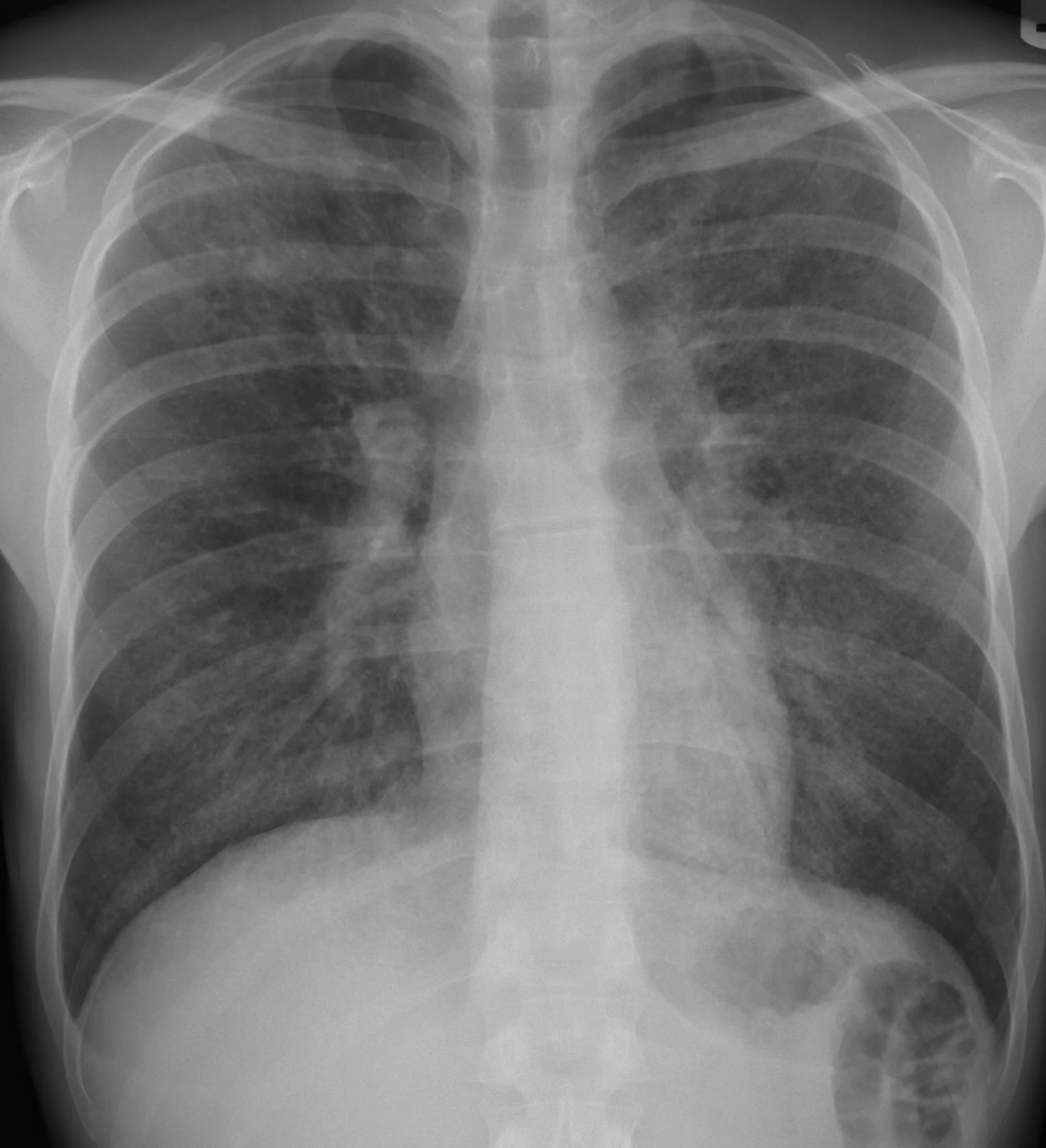
Long description
PA chest radiograph of a patient with Pneumocystis jiroveci pneumonia showing reticular interstitial markings in all lung fields.

The classic picture is bilateral, diffuse, symmetrical interstitial shadowing: a finely granular, reticular or ground-glass pattern, often perihilar and spreading outward. [1] The radiograph can be normal early in the illness, especially in non-HIV patients, and may show cysts, nodules or pneumothorax later.
When the radiograph is inconclusive but the clinical picture fits, high-resolution CT of the chest is the most sensitive imaging test. [1] The hallmark is ground-glass opacification, often with a mosaic or diffuse distribution and a perihilar predominance. Cystic change is more common in HIV infection, and nodular or solitary-lesion presentations are reported but rare. [1]
The absence of ground-glass opacities on CT makes PJP unlikely, although atypical infiltrates occur. [1]
- Lactate dehydrogenase (LDH). An elevated level is common and is a marker of disease severity rather than a diagnostic test in its own right; a high value supports the diagnosis of PJP and a very high value correlates with poor outcome, but a normal value does not exclude the disease. [1]
- Beta-D-glucan. A serum biomarker of fungal cell-wall components; it is elevated in PJP and can be used when an invasive procedure is not possible. [1] It is not specific, because it is also raised in invasive aspergillosis, candidaemia and several other conditions, and it is falsely elevated by some wound dressings, haemodialysis membranes and certain antibiotics. [1]
- HIV testing and CD4 count, where the risk group or HIV status is unknown.
Figure 2Pneumocystis carinii 01
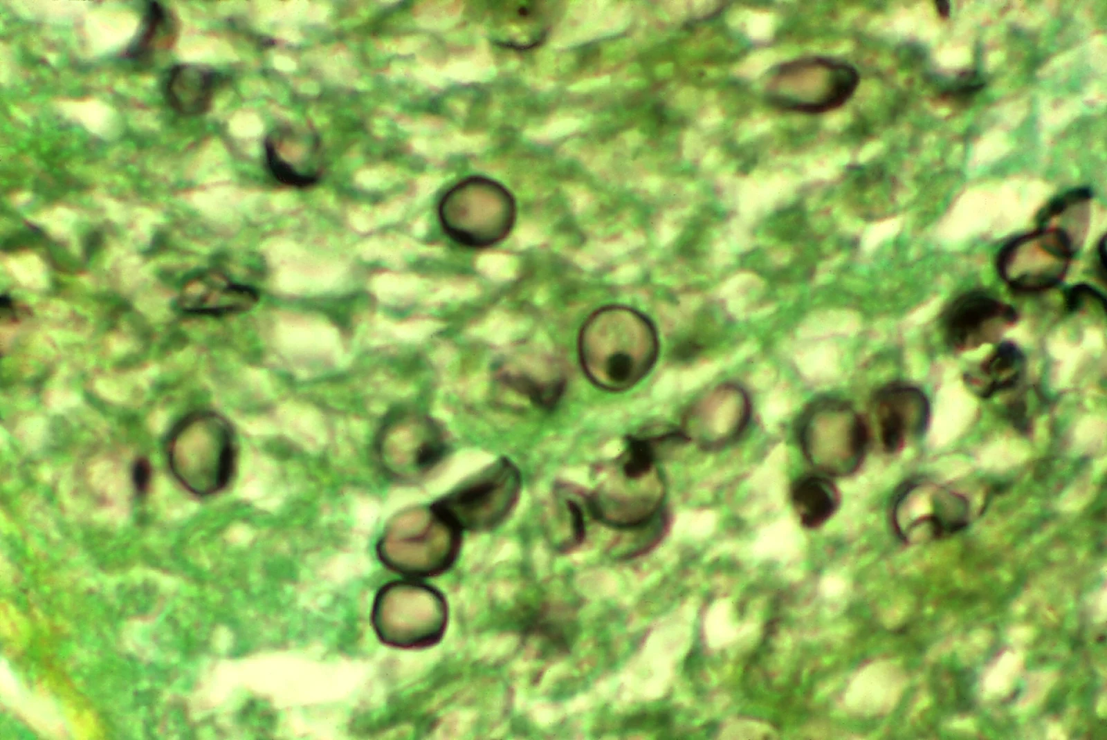
Long description
Methenamine silver stain of Pneumocystis jiroveci cysts in tissue from an AIDS patient, showing thick-walled Pneumocystis jirovecii organisms including characteristic cup-shaped forms and dot-like cysts.

Routine culture does not grow P. jirovecii; definitive diagnosis requires molecular or direct visualisation methods on a respiratory specimen. [1]
- Induced sputum is the non-invasive first step: the patient inhales hypertonic saline after nebulisation and produces a deep cough specimen.
- Bronchoalveolar lavage has a better diagnostic yield and is used when induced sputum is negative or cannot be obtained; bronchoscopic washings or biopsy are occasionally needed.
- PCR on induced sputum or BAL fluid is highly sensitive (97 to 100 per cent in many series in HIV-infected patients), although it may be less useful in non-HIV immunocompromised patients, where specificity falls and the test can detect colonisation as well as disease. [1]
- Silver staining (Grocott-Gomori methenamine silver) and immunofluorescence (direct or indirect fluorescent antibody) demonstrate the cysts on stained preparations of induced sputum or BAL fluid. [1]
Clinical practice therefore requires correlating respiratory symptoms, imaging and laboratory results; a positive PCR in an immunocompetent, asymptomatic person may represent colonisation rather than pneumonia. [1]
There are no universally accepted diagnostic criteria for PJP beyond a compatible clinical picture plus microbiological or histological evidence. [1] Severity is best judged clinically.
Moderate to severe disease is defined for treatment decisions (mainly for steroids) as either:
- PaO2 under 70 mmHg on room air, or
- an A-a oxygen gradient of 35 mmHg or more. [1]
Non-HIV PJP is graded more sharply than ordinary pneumonia; standard pneumonia scores such as CURB-65 underestimate the risk, so the Miller severity system, which incorporates oxygen saturation, respiratory rate, age and comorbidity, is preferred in this group. [1]
Approach to suspected Pneumocystis jirovecii pneumonia
- Suspect PJPImmunocompromised host, subacute dry cough, dyspnoea, fever, hypoxia
- ImagingChest radiograph; CT chest if radiograph inconclusive or presentation atypical
- Question: Ground-glass opacities or bilateral interstitial infiltrates present?
No
- Look for alternative diagnosesBacterial pneumonia, tuberculosis, pulmonary oedema, drug-induced lung injury
Yes
- Send baseline testsHIV test and CD4 count where indicated; LDH; serum beta-D-glucan; arterial blood gas
- Question: Can the patient produce sputum or undergo bronchoscopy?
Induced sputum first
- PCR and silver or immunofluorescence stain on induced sputum
Too unwell for bronchoscopy
- Treat empirically while optimising supportReassess if no improvement after about eight days; repeat imaging and bronchoscopy when stable
- Severity assessmentPaO2 under 70 mmHg on room air or A-a gradient at least 35 mmHg means moderate to severe disease
- First-line treatmentTMP-SMX 15 to 20 mg/kg per day of the TMP component, divided, intravenously or orally, for 21 days; add adjunctive corticosteroids if moderate to severe
- Adjunctive corticosteroids (if criteria met)Prednisone 40 mg twice daily days 1 to 5, 40 mg daily days 6 to 10, 20 mg daily days 11 to 21; start within 72 hours
- Reassess at day 8Persistent fever or worsening imaging warrants repeat bronchoscopy to look for co-infection; note that persistent PCR positivity alone does not prove treatment failure
Management is best delivered by a multidisciplinary team: infectious diseases, respiratory medicine, intensive care, pharmacy, nursing and respiratory therapy. [1]
- Oxygen therapy to keep the saturation adequate; early non-invasive or invasive ventilation when the disease is severe.
- Fluids and nutrition, and treatment of precipitating illnesses.
- Search for co-infection (CMV, tuberculosis, bacterial pneumonia), especially in HIV with a very low CD4 count.
TMP-SMX is first-line for PJP whatever the underlying condition. [1]
- Moderate to severe disease: TMP 15 to 20 mg per kg per day plus SMX 75 to 100 mg per kg per day, intravenously in three or four divided doses, converting to oral once the patient improves.
- Mild to moderate disease: the same total dose by mouth, or double-strength tablets (160 mg TMP with 800 mg SMX) two to three times daily. [1]
Most units treat for 21 days, although at least fourteen days are used for mild to moderate disease and longer courses are given when improvement is slow. [1]
In moderate to severe HIV-associated PJP (PaO2 under 70 mmHg on room air or A-a gradient of 35 mmHg or more), adjunctive corticosteroids halves the risk of death and is started within 72 hours of anti-Pneumocystis therapy. [1]
Standard taper: [1]
- Days 1 to 5: prednisone 40 mg by mouth twice daily.
- Days 6 to 10: prednisone 40 mg once daily.
- Days 11 to 21: prednisone 20 mg once daily.
Intravenous methylprednisolone is an acceptable alternative where oral therapy is not possible. [1]
In non-HIV PJP the role of steroids is contested. Some retrospective studies report shorter intensive care unit stays and ventilator times, but a meta-analysis found no clear survival benefit, and several cohort studies found no effect on mortality or ventilation. [1][4] In solid organ transplant recipients, some guidelines recommend prednisone 40 to 60 mg twice daily for five to seven days started within 72 hours for moderate to severe disease, tapered over the following one to two weeks. [1] Steroids are never a substitute for effective anti-Pneumocystis treatment.
These are used when TMP-SMX is contraindicated, not tolerated or fails. [1]
| Alternative | Dose (adult) | Notes |
|---|---|---|
| IV pentamidine | 4 mg per kg once daily (3 mg per kg if toxicity develops) | Avoid in pancreatic and islet cell transplant recipients because of islet cell necrosis [1] |
| Oral atovaquone | 750 mg twice daily (three times daily in some regimens) | Less effective than TMP-SMX; useful for mild to moderate disease or prophylaxis failures [1] |
| Primaquine plus clindamycin | Primaquine 30 mg by mouth once daily plus clindamycin 600 mg intravenously every 6 hours or 900 mg every 8 hours, or 450 mg by mouth every 6 hours | Good second-line results in HIV-associated disease; screen for G6PD deficiency before primaquine [1] |
| Dapsone plus trimethoprim | Dapsone 100 mg once daily plus TMP 15 mg per kg per day | Oral option; avoid or monitor carefully in G6PD deficiency [1] |
| Clindamycin-primaquine salvage | As above, combined | Used after TMP-SMX failure |
| TMP-SMX plus caspofungin | TMP-SMX 15 to 20 mg per kg per day plus caspofungin 70 mg on day one then 50 mg daily | Salvage option in haematology patients [1] |
TMP-SMX remains the most effective agent; second-line drugs are clearly inferior in most comparisons and should not be chosen lightly. [1]
- Common TMP-SMX effects: rash, fever, nausea and marrow suppression. [3][1] Hypersensitivity reactions and bone-marrow suppression are particularly common in children on chronic therapy. [1]
- Sulphonamide-specific problems: hyperkalaemia (TMP is potassium-sparing), renal impairment and crystal deposition in concentrated urine; maintain good hydration, take renal impairment into account and reduce the dose in renal failure.
- G6PD deficiency: test for it before primaquine or dapsone, and choose another alternative if the patient is deficient.
- Pentamidine: pancreatic, renal and cardiac toxicity; monitor glucose, renal function and blood pressure.
- Atovaquone: variable absorption with food; take with fatty meals.
Response is assessed clinically (oxygen requirements, fever) and radiologically. If there is no improvement after about eight days, repeat investigations including CT and bronchoscopy to look for co-infection. [1] Persistent PCR positivity in a repeat BAL sample does not indicate treatment failure. [1]
PJP in a patient not on ART is an emergency in both directions: treat the pneumonia and start ART. Early initiation of ART, within two weeks of anti-Pneumocystis treatment, produced no increase in mortality, AIDS-defining events, immune reconstitution inflammatory syndrome (IRIS) or adverse events compared with deferring, and many early deaths occurred in patients who never reached ART at all. [2] This supports starting ART within two weeks of treatment, tailored to the patient's ability to absorb oral medication and to the clinical course. [2]
A transient worsening can occur when ART is started, sometimes with a new systemic inflammatory response (IRIS). This is rare in PJP, is treated with continued therapy and targeted supportive care, and does not usually mean treatment failure.
- Respiratory failure and need for mechanical ventilation, with higher failure rates of non-invasive ventilation in non-HIV PJP. [1]
- Pneumothorax from cystic change, which can occur spontaneously or with ventilation.
- Disseminated infection is not typical; extrapulmonary PJP is rare.
- Drug toxicity (see above) may force a change of regimen.
- Co-infection with CMV, bacterial pneumonia and tuberculosis, particularly in late HIV.
- Treatment failure with resistant (DHPS mutant) organisms is reported but has not changed first-line empirical practice. [1]
Outcomes differ markedly by host. Mortality is around ten to twenty per cent in HIV-associated PJP and thirty to sixty per cent in non-HIV PJP, largely because the latter group presents later and with more respiratory failure. [1] Need for intensive care, mechanical ventilation, pneumothorax, a high LDH and a low CD4 count predict poor outcomes; the degree of hypoxaemia (the A-a gradient) tracks response to treatment in HIV-associated disease. [1] In HIV, early anti-Pneumocystis treatment and early ART substantially improve survival.
- HIV: adults and adolescents with a CD4 count under 200 cells per mm3; HIV-infected children according to age-based CD4 thresholds (under 500 cells per mm3 at one to five years, under 200 cells per mm3 at six to twelve years) also require prophylaxis. [1]
- Non-HIV patients at high risk: prolonged high-dose corticosteroids (more than 20 mg prednisone equivalent for three weeks or more in lung cancer, for example), combined immunosuppression, absolute lymphopenia, transplant recipients (especially the first six months and after rejection treatment), autoimmune disease on cyclophosphamide or rituximab, and haematological malignancy. [1]
- Secondary prophylaxis is given after an episode of PJP until the underlying immunosuppression resolves.
TMP-SMX is the agent of choice for both primary and secondary prophylaxis. [1]
| Drug | Adult dose | Notes |
|---|---|---|
| TMP-SMX | 80 mg TMP with 400 mg SMX once daily, or 160 mg TMP with 800 mg SMX once daily, or 160 mg TMP with 800 mg SMX three times weekly | First line; co-trimoxazole also protects against toxoplasmosis and some bacterial infections [1] |
| Dapsone | 100 mg once daily | Alternative where TMP-SMX is not tolerated; G6PD screening recommended; only primary prophylaxis is established [1] |
| Aerosolised pentamidine | 300 mg inhaled once every 28 days | Good safety profile but less effective than TMP-SMX; dose to the lower respiratory tract only [1] |
| Atovaquone | 1,500 mg once daily | Expensive alternative; breakthrough infections reported; take with food |
TMP-SMX is markedly more effective than the alternatives and should be used wherever it is tolerated. [1][3] Where a patient has a history of a mild sulphonamide rash, desensitisation or re-challenge is often attempted under specialist guidance.
Pregnant patients need specialist obstetric and infectious disease input, because drug choices for treatment and prophylaxis, especially in the first trimester, differ from those for other adults. Follow local guidance.
The clinical picture, complications and radiology are close to those in adults. The treatment of choice is TMP-SMX, 15 to 20 mg of the TMP component per kg per day with 75 to 100 mg per kg per day of SMX, intravenously or by mouth in three to four divided doses for 21 days. [1] Pentamidine and atovaquone are the main alternatives; caspofungin added to TMP-SMX has shown good results in immunocompromised children. [1] Hypersensitivity reactions and bone-marrow suppression are common with prolonged therapy. [1]
There is no change in recommended regimen, but drug toxicity and renal impairment need particular attention; dose adjustment in renal failure is standard.
- The disease is common where ART and routine prophylaxis are not universally available, and it should be considered in every immunocompromised patient with an unexplained hypoxic pneumonia. [3]
- Induced sputum and PCR, bronchoscopy, CT and serum beta-D-glucan are frequently unavailable. In that situation the diagnosis is made clinically from risk factors, presentation and the plain chest radiograph, and treatment is started empirically.
- Cotrimoxazole (TMP-SMX) is cheap, widely available and effective by mouth; intravenous therapy is reserved for severe disease or inability to take oral medication. The same oral regimen used for treatment can be completed as an outpatient once the patient is stable.
- Adjunctive corticosteroids require clinical oxygen assessment (room-air saturation or arterial blood gas), not imaging alone, to identify moderate to severe disease.
- HIV testing must be offered, because PJP is often the presenting illness of undiagnosed HIV; linking the patient to HIV services and starting ART promptly is central.
- Local tuberculosis prevalence means TB should be actively excluded in every patient, since the two can coexist and TB can mimic PJP.
- Prophylaxis with TMP-SMX is one of the cheapest, most effective interventions available; health systems should ensure it reaches all adults with HIV whose CD4 is under 200 and anyone starting potent immunosuppression.
- Pneumocystis jirovecii causes pneumonia only in the setting of impaired cellular immunity; it does not respond to azoles or amphotericin B.
- The hallmark is a subacute, diffuse, hypoxic pneumonia with a dry cough and remarkably few chest signs.
- In HIV the illness takes two to four weeks to reach peak; in non-HIV immunocompromised patients it can progress in five to six days.
- Chest radiograph shows bilateral interstitial infiltrates; high-resolution CT shows ground-glass opacities, which may be the only abnormality.
- Elevated LDH supports the diagnosis and predicts severity; serum beta-D-glucan is a useful but non-specific marker.
- Diagnosis is by induced sputum or BAL fluid with PCR or silver staining; PCR is highly sensitive but cannot always separate infection from colonisation.
- Moderate to severe disease is defined by a PaO2 under 70 mmHg on room air or an A-a gradient of at least 35 mmHg.
- First-line treatment is TMP-SMX 15 to 20 mg per kg per day of the TMP component for 21 days, with adjunctive corticosteroids (a 21-day taper) when the PaO2 threshold is met.
- Alternative drugs include intravenous pentamidine, atovaquone, and primaquine plus clindamycin; test for G6PD deficiency before primaquine or dapsone.
- Prophylaxis is TMP-SMX for all adults with HIV with CD4 under 200, and for high-risk non-HIV patients; the alternatives are dapsone, inhaled pentamidine and atovaquone.
- Start ART within two weeks of anti-Pneumocystis treatment in HIV, as early treatment does not add harm and reduces late mortality.
- Mortality is about 10 to 20 per cent with HIV and 30 to 60 per cent without HIV.
Answer each one in your head first, then open the answer.
A 34-year-old man with newly diagnosed HIV (CD4 90 cells per mm3) has had a dry cough and low-grade fever for three weeks and is now breathless on climbing a flight of stairs. His chest radiograph shows diffuse bilateral interstitial infiltrates and his PaO2 on room air is 64 mmHg. What is the most likely diagnosis and what treatment should be started?
Show answerHide answer
Pneumocystis jirovecii pneumonia. Given moderate to severe disease (PaO2 under 70 mmHg), start high-dose TMP-SMX intravenously (TMP 15 to 20 mg per kg per day with SMX 75 to 100 mg per kg per day in divided doses) for 21 days plus adjunctive corticosteroids within 72 hours (prednisone 40 mg twice daily for five days, then 40 mg daily for five days, then 20 mg daily for eleven days), together with oxygen and an assessment for HIV-related co-infection. [1]
Why can a patient with severe PJP have clear breath sounds on auscultation?
Show answerHide answer
The disease is a diffuse, alveolar interstitial pneumonia rather than a lobar consolidation. Hypoxia arises from impaired gas exchange across the alveolar-capillary membrane and from altered surfactant, so very little wheeze or crepitation is produced until the disease is advanced.
A patient with PJP has a PaO2 of 72 mmHg on room air and an A-a gradient of 38 mmHg. Does this patient meet the criteria for adjunctive corticosteroids?
Show answerHide answer
Yes. Moderate to severe disease is defined by a PaO2 under 70 mmHg or an A-a gradient of 35 mmHg or more, so the raised A-a gradient alone qualifies. Start prednisone 40 mg twice daily for five days, then 40 mg daily for five days, then 20 mg daily for eleven days, within 72 hours of anti-Pneumocystis therapy.
Before starting primaquine as an alternative regimen for PJP, what must be checked and why?
Show answerHide answer
Glucose-6-phosphate dehydrogenase (G6PD) deficiency must be checked, because primaquine (and dapsone) can cause haemolysis in G6PD-deficient patients. If the patient is deficient, choose a different alternative such as intravenous pentamidine or atovaquone.
What is each of the following used to test for PJP, and how reliable is it: serum beta-D-glucan, PCR on induced sputum, and silver staining of BAL fluid?
Show answerHide answer
Serum beta-D-glucan is a supportive marker of fungal cell-wall material: raised in PJP but not specific, as it is also raised in other invasive fungal infections. PCR on induced sputum is highly sensitive (97 to 100 per cent in many HIV-infected series) but can detect colonisation and is less reliable in non-HIV immunocompromised patients. Silver staining (Grocott methenamine silver) on BAL fluid demonstrates the cysts directly and provides a morphological diagnosis with lower sensitivity than PCR.
A 28-year-old woman with newly diagnosed HIV and a CD4 count of 120 cells per mm3 is admitted with moderate PJP that is being treated. When should antiretroviral therapy be started?
Show answerHide answer
Within two weeks of starting anti-Pneumocystis treatment. Early ART does not increase mortality, AIDS-defining events, immune reconstitution inflammatory syndrome or adverse events in this setting, and it reduces late mortality by avoiding the situation in which the patient dies before ART can be started.
What are the three standard prophylactic regimens for PJP in adults, and which is preferred?
Show answerHide answer
TMP-SMX is preferred, given as 80 mg/400 mg daily, 160 mg/800 mg daily, or 160 mg/800 mg three times weekly. The alternatives are dapsone 100 mg daily, aerosolised pentamidine 300 mg every four weeks, and atovaquone 1,500 mg daily. TMP-SMX is markedly more effective than these alternatives.
Pneumocystis jirovecii pneumonia intersects with HIV infection (risk threshold, ART and prophylaxis), transplant immunosuppression, haematological malignancy, interstitial lung disease and corticosteroid use. Readers should also revise acute respiratory failure, pneumothorax, hypoxaemia and opportunistic infections in immunocompromised hosts.
- Ibrahim A, Chattaraj A, Iqbal Q, Anjum A, Rehman MEU, Aijaz Z, Nasir F, Ansar S, Zangeneh TT, Iftikhar A. Pneumocystis jiroveci Pneumonia: A Review of Management in Human Immunodeficiency Virus (HIV) and Non-HIV Immunocompromised Patients. Avicenna Journal of Medicine 2023;13(1):23-34. Accessed .
- Zeng YM, Li Y, Lu YQ, et al. Initiating antiretroviral therapy within 2 weeks of anti-Pneumocystis treatment does not increase mortality or AIDS-defining events in patients with HIV-associated moderate to severe Pneumocystis pneumonia: results of a prospective observational multicenter study. BMC Pulmonary Medicine 2022;22:323. Accessed .
- Centers for Disease Control and Prevention. Pneumocystis Pneumonia Basics. CDC; reviewed 2026-10-21. Accessed .
- Rhoads S, Maloney J, Mantha A, Van Hook R, Henao-Martinez AF. Pneumocystis jirovecii Pneumonia in HIV-Negative, Non-transplant Patients: Epidemiology, Clinical Manifestations, Diagnosis, Treatment, and Prevention. Current Fungal Infection Reports 2024;18(2):125-135. Accessed .
Figure credits
Educational content for medical students and doctors. It is not a substitute for clinical judgement or local guidelines.