Herniated disc, spinal stenosis and low back pain
Low back pain, disc herniation and spinal stenosis: red flags, radiculopathy, claudication, MRI and step-by-step management.
- Most low back pain is non-specific and improves over weeks; it is rarely caused by a single, surgically correctable structural problem.
- The dangerous presentations are red flags for malignancy, fracture, infection, cauda equina syndrome and progressive neurological deficit, and a clear root syndrome with persistent or worsening weakness.
- A lumbar disc herniation causes radiculopathy: pain, numbness and weakness in a dermatomal pattern, often with a positive straight leg raise.
- Lumbar spinal stenosis causes neurogenic claudication: pain or heaviness in the buttocks or legs brought on by standing or walking and eased by sitting or bending forward.
- Imaging is not routine; use MRI when it will change management, usually after 6 weeks of symptoms, or earlier with a neurological deficit.
- Conservative care comes first for most people: advice, exercise, non-steroidal anti-inflammatory drugs, and time. Epidural steroid injection and surgery are reserved for selected, persistent or severe cases.
By the end of this page you should be able to:
- Define non-specific low back pain, lumbar disc herniation with radiculopathy and symptomatic lumbar spinal stenosis.
- List the red flags for serious pathology in back pain and state which change urgent management.
- Explain why a herniated L4-L5 or L5-S1 disc produces its particular sensory, motor and reflex findings, and interpret the straight leg raise.
- Separate neurogenic from vascular claudication using history and simple bedside clues.
- Choose when to image, what to image with, and what the result means.
- Outline conservative, injective and surgical management according to current guidelines, including when guidelines disagree.
- Recognise cauda equina syndrome as an emergency and know the appropriate cross-check in neurology.
This page covers three presentations that are usually assessed together: non-specific low back pain, radiculopathy from a herniated lumbar disc, and symptomatic lumbar spinal stenosis. Together they account for most back pain seen in practice. The book groups them on the same pages (B2:260-261), so they are taught here as one topic, with cross-references to the neurology discussion of cauda equina syndrome.
The teaching order follows how a clinician meets the patient: what the problem is, who gets it, why it hurts, what the examination shows, when a serious cause must be excluded, what investigations help, and what treatment the current guidelines support.
A herniated lumbar disc is the localised displacement of intervertebral disc material beyond the normal margins of the disc space, with pain, weakness or numbness in a dermatomal or myotomal distribution. [4] The disc itself is not always symptomatic: small bulges and protrusions are common on MRI of people without symptoms, especially over the age of 50. [4] The diagnosis is therefore clinical first, supported by imaging.
Common descriptors are:
- Bulge/protrusion: broad-based displacement, often contained.
- Extrusion: focal displacement with a neck narrower than the displaced material.
- Sequestration: a free fragment separated from the parent disc.
The level is usually named by the disc and the root affected. L4-L5 disc herniation most often affects the L5 root; L5-S1 herniation most often affects the S1 root.
Lumbar spinal stenosis is a narrowing of the spinal canal, lateral recess or intervertebral foramen, so that the neural and vascular elements have less space than they need. [3] When symptomatic it produces a slowly progressive syndrome, most often in older adults, of back and buttock or leg pain and fatigue that is brought on by standing or walking and relieved by sitting or bending forward. [3] [6]
Non-specific low back pain is back pain not attributed to a recognisable specific pathology. It is the common, self-limiting mechanical pain that constitutes the majority of back pain presentations worldwide. [7]
For day-to-day decisions it is more useful to classify a patient by syndrome than by imaging:
| Presentation | Dominant symptom | Typical age | Key clue |
|---|---|---|---|
| Non-specific low back pain | Aching back pain, variable with movement | Adults | No neurological signs; no red flags |
| Radiculopathy (disc) | Sharp pain down the leg in a dermatome | Third to fifth decade [4] | Positive straight leg raise, dermatomal sensory or motor change |
| Spinal stenosis | Heaviness or aching in the legs on standing or walking | Older adults [3] | Relief on sitting, bending forward or pushing a trolley (shopping cart sign) |
| Cauda equina syndrome | Back and leg pain with saddle numbness and sphincter disturbance | Any age | Emergency: urgent MRI and decompression |
Low back pain is the leading cause of disability worldwide. [1] It is usually brief: episodes most often improve over weeks to months. [1]
A lumbar herniated disc has a lifetime risk of about 30%, but the risk of a symptomatic herniation is only 1 to 3%, and 60 to 90% of symptomatic herniations resolve with non-operative care. [4] In a Norwegian national study the incidence of lumbar disc herniation was about 5 to 20 cases per 1000 adults per year, and among surgical patients the male-to-female ratio was 2 to 1, with cases concentrated in the third to fifth decades of life. [4] Among adults aged 25 to 55, more than 95% of herniations occur at L4-L5 or L5-S1. [4]
Symptomatic disc degeneration of some degree is estimated to affect about 403 million people, or 5.5% of the world's population. [4]
Risk factors that are reasonably well supported include: [4]
- Age (peak 30 to 50 years).
- Male sex.
- Strenuous physical work or lifting (the strongest predictor in a long Copenhagen cohort study).
- Smoking: relative risk about 1.27 (95% confidence interval 1.15 to 1.40) in a review of 12 studies. [4]
- A substantial genetic contribution (twin studies).
- Certain occupations (healthcare workers including surgeons, drivers, astronauts, athletes) and visceral adiposity.
- Certain recreational loads.
Body weight alone has not been consistently linked to herniation in all cohorts, so advise on overall lifestyle rather than weight-based blame.
Spinal stenosis is a disease of ageing. It is uncommon in younger adults and becomes more frequent with each decade. [3] Mild to moderately symptomatic stenosis has a generally favourable course: about one third to one half of patients improve or stabilise, and rapid or catastrophic neurological decline is rare. [3]
The intervertebral disc has a soft, water-rich nucleus pulposus surrounded by a tough annulus fibrosus. With ageing the nucleus loses water and the annulus becomes less elastic. Repeated flexion and rotation, especially with load, can then produce a tear in the annulus, and part of the nucleus can travel through it. [4]
Two mechanisms make the patient symptomatic:
- Mechanical compression. The displaced material physically compresses or deforms the adjacent nerve root or cauda equina. Higher lumbar discs more often require central protrusion to affect multiple roots; far-lateral fragments at L5-S1 affect the exiting L5 root before it enters the canal.
- Chemical irritation. Nucleus pulposus material is antigenic. It releases phospholipase A2, nitric oxide, and cytokines that inflame and sensitise the nerve root even when imaging compression looks modest. This is why pain severity does not always match the size of the herniation.
A third mechanism, discogenic back pain, occurs when annular tearing itself is painful but without root compression. The patient then has axial low back pain, not leg pain.
Stenosis is different. Its mechanism is position-dependent compression. In extension the spinal canal narrows as the ligamentum flavum buckles inward and the facets override; in flexion the canal widens. That is why walking uphill (slight flexion) is easier for a stenosis patient than walking downhill (extension), and why leaning on a shopping trolley (flexion) relieves the pain.
Most episodes of back pain start abruptly after a bending or lifting effort, or for no clear reason. The pain is diffuse, aching and worsened by movement, and the patient has difficulty continuing daily activities for some days. This is the typical picture of non-specific low back pain.
Painless night pain, fever, weight loss, or pain that is unaffected by movement or position are not typical, and should raise the possibility of another cause.
Serious pathology behind back pain is uncommon (fracture occurs in 1 to 4% of patients presenting with low back pain in primary care; malignancy in fewer than 1%). [7] Red flags exist to make clinicians stop and look harder, not because every red flag proves serious disease. Evidence for the accuracy of individual red flags is limited, and the lists in different guidelines do not fully agree. [7]
The widely used red flags, with their associated serious pathologies, are: [1] [7]
| Red flag | Most serious conditions it raises |
|---|---|
| New onset saddle or perineal numbness; recent bladder (retention, altered sensation, incontinence) or bowel disturbance; loss of anal sphincter tone | Cauda equina or conus medullaris syndrome (emergency) |
| Progressive or severe motor weakness, or widespread neurological deficit | Cord compression, cauda equina |
| History of cancer, particularly breast, lung, gastrointestinal, prostate, kidney or thyroid | Metastatic disease to bone |
| Unexplained weight loss | Malignancy |
| Fever, recent infection, immunosuppression, intravenous drug use, indwelling catheter, HIV | Spinal infection (discitis, epidural abscess) |
| Significant trauma, or minor trauma in older, osteoporotic or steroid-treated patients | Vertebral fracture |
| Chronic corticosteroid use or osteoporosis | Fracture, infection |
| Age under 20 or over 55 with new, progressive pain | Malignancy, inflammatory axial disease [7] |
| Pain that is worse at night or at rest, or that is unremitting regardless of position | Malignancy, infection, inflammatory disease |
| Thoracic pain, severe and unremitting pain | Malignancy, infection, cord compression |
| Morning stiffness that improves with exercise | Spondyloarthritis |
| Known abdominal aortic aneurysm, or vascular risk with abdominal symptoms | Vascular causes |
The red flags with the best evidence are a history of cancer, which raised the post-test probability of spinal malignancy to about one third in one emergency department study, and prolonged corticosteroid use, severe trauma and a contusion or abrasion, which are the stronger pointers to spinal fracture. [7] Older age, unexplained weight loss and failure to improve after a month are weaker cancer pointers, with post-test probabilities under 3%. [7] A combination of red flags is more informative than a single one. [7]
NICE NG59 still tells clinicians to think about and exclude specific causes such as cancer, infection, trauma or inflammatory disease, and notes that the guideline does not cover progressive neurological deficit or cauda equina syndrome, which require referral. [1]
Inspection looks for a list, a flattened lordosis from muscle spasm, scoliosis and gait. Palpation of the spinous processes and paraspinal muscles is performed standing and lying. Range of movement in flexion, extension, lateral flexion and rotation is assessed. Forward flexion (touching the toes) normally obscures the lumbar lordosis; inability to do so may be due to pain or hamstring tightness.
Straight leg raising is described below. The femoral nerve stretch test (patient prone, knee flexed; pain in the anterior thigh is positive) is more sensitive for upper lumbar (L2-L4) roots.
Neurological examination of the legs documents motor power in every myotome, sensation in every dermatome and reflexes at the knee and ankle, so that a level can be assigned.
The nerve roots most often compressed are L5-S1, L4-L5 and L3-L4. [5] A disc prolapse at one of these levels produces a predictable (if not perfect) pattern:
| Root | Typical disc level | Pain distribution | Key sensory area | Key motor loss | Reflex |
|---|---|---|---|---|---|
| --- | --- | --- | --- | --- | --- |
| L3 | L2-L3 or L3-L4 | Anterior thigh, groin | Anterior thigh | Hip flexion and knee extension | Knee jerk may be reduced |
| L4 | L3-L4 | Anterior thigh to medial calf | Medial calf, medial foot | Ankle dorsiflexion, knee extension | Knee jerk |
| L5 | L4-L5 | Lateral thigh and leg, dorsum of foot | Dorsum of foot, big toe | Great toe extension, ankle dorsiflexion/eversion | No reliable deep tendon reflex (internal hamstring sometimes) |
| S1 | L5-S1 | Posterior thigh and calf to lateral foot and sole | Lateral foot and sole | Ankle plantarflexion | Ankle jerk |
| L2 | L1-L2 or L2-L3 | Upper anterior thigh | Anterior thigh | Hip flexion | None reliable |
Three or more concordant findings from history (dermatomal pain), sensory change, reflex or motor weakness and a positive straight leg raise make a lumbar disc herniation more likely than any single test. [4] An isolated abnormality is a weak pointer because imaging often shows disc changes in people without symptoms. [4]
The straight leg raise (Lasegue test) is performed with the patient lying supine while the examiner raises the straight leg. It is positive when radicular pain is reproduced below the knee at an elevation of under 60 degrees. [5] It works by pulling on the sciatic nerve and its roots; the mechanism is dural tension rather than simple hip pain. [5] In the literature its reported sensitivity ranges from 0.35 to 0.97 and its specificity from 0.10 to 1.00, with better performance in younger patients. [8] A crossed (well) straight leg raise is more specific but less sensitive. [5] [8] The slump test (sitting slumped with neck flexed, then extending the knee) is more sensitive but less specific than the supine straight leg raise. [5]
Neurogenic claudication from lumbar stenosis is a postural and positional phenomenon. [3] [6] The patient reports pain, aching, heaviness or cramping in the buttocks, thighs or legs that is brought on by standing upright or walking and is relieved by sitting down, bending forward at the waist or leaning on a support (the shopping cart sign). [3] [6] Key bedside distinctions from systemic causes of exercise limitation are: [6]
| Feature | Neurogenic claudication (stenosis) | Vascular (arterial) claudication |
|---|---|---|
| Trigger | Standing or walking, often unpredictable | Walking a set distance |
| Relief | Sitting or bending forward | Standing still, regardless of posture |
| Walking uphill | Often easier | Worse |
| Walking downhill | Often worse | Easier |
| Cycling | Usually painless | Usually provokes pain |
| Distribution of worst symptoms | Often buttocks and thighs (above the knee) | Often calves (below the knee) |
| Neurological features | Numbness, tingling, heaviness common | Uncommon |
| Peripheral pulses | Present | Often reduced or absent |
| Ankle-brachial index | Usually above 0.90 | 0.90 or less [6] |
| Skin changes | Absent | Trophic changes (hair loss, atrophy, ulcers) possible |
Single features in isolation are weak discriminators; a combination (symptoms triggered by standing, relieved by sitting, located above the knees, with a positive shopping cart sign) makes neurogenic claudication much more likely, with a positive likelihood ratio of about 13. [6] Symptoms relieved by standing alone and located below the knees make vascular claudication much more likely, with a positive likelihood ratio of about 20. [6] The two conditions also commonly coexist in older adults, so neither excludes the other. [6]
When a patient presents with back and/or leg pain, the main alternatives to non-specific low back pain and disc-related radiculopathy are:
- Hip osteoarthritis: groin and anterior thigh pain, worse with movement of the hip, limited internal rotation and flexion. The pain does not follow a dermatome and straight leg raise is negative.
- Sacroiliac joint pain: buttock pain below L5, often unilateral, no radiation below the knee; no neurological signs.
- Facet (zygapophyseal) joint pain: localised low back pain worsened by extension and rotation, relieved by flexion; no leg radiation.
- Piriformis syndrome / deep gluteal pain: buttock pain with possible distal radiation; hip external rotation provokes symptoms.
- Peripheral neuropathy: bilateral, distal, glove-stocking sensory loss with diabetes or alcohol, no back pain link.
- Spinal infection: fever, night pain, high inflammatory markers, recent infection or immunosuppression.
- Malignancy (primary or metastatic): night pain, weight loss, history of cancer, constant pain.
- Osteoporotic vertebral fracture: older age, steroid use, minor trauma, sudden back pain, local tenderness.
- Ankylosing spondylitis: young men, chronic morning stiffness, improvement with exercise, sacroiliac and spinal tenderness.
- Cauda equina or conus medullaris syndrome: saddle anaesthesia, bladder or bowel disturbance, bilateral leg symptoms or severe progressive deficit (emergency).
- Vascular claudication or abdominal aortic disease (see table above).
- Thoracic or abdominal cause referred to the back (renal colic, peptic ulcer, pancreatitis, aortic aneurysm).
Figure 1Hernie discale L4 L5
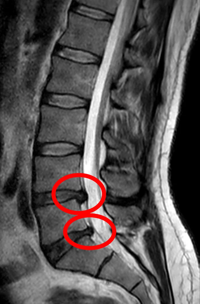
Long description
A lumbar spine MRI, including T1 and T2 Dixon sagittal slices, reported as revealing a herniated left paramedian disc at L4-L5 with apparent emergence of the dural sheath of the left L5 root.

Routine imaging is not offered in non-specialist settings for people with low back pain with or without sciatica. [1] Patients should understand that being referred for a specialist opinion does not mean imaging is needed. [1] In specialist care, imaging is considered only when the result is likely to change management. [1]
- MRI of the lumbar spine is the most appropriate non-invasive test to confirm a herniated disc or spinal canal narrowing. [4] [3] Reported overall diagnostic accuracy for disc herniation is up to 97%, although a pooled analysis gave sensitivity about 75% and specificity about 77%. [4] MRI can also be misleading: disc bulges and protrusions are found in about 60% and 36% respectively of asymptomatic people over 50, so imaging must be matched to the clinical picture. [4]
- Timing. Without neurological deficit and with persistent symptoms, radiological assessment is suggested between 6 and 12 weeks; with a motor deficit it should be requested earlier. [4] [5] Imaging (and electrodiagnostic studies) is also appropriate once symptoms have persisted more than 6 weeks. [5]
- CT or CT myelography. Used when MRI is contraindicated, inconclusive or unavailable. [3] [4] NASS places CT after MRI and CT myelography in the diagnostic chain. [3]
- Axial loading MRI or CT can be a useful adjunct when central or lateral stenosis is suspected but not verified on routine unloaded imaging, particularly with a dural sac area under 110 mm2. [3]
- Plain radiographs are not useful for routine low back pain. They may be used when specific pathology (fracture, suspected structural lesion) is a concern, or as an adjunct to differentiate causes. [4]
- Electrodiagnostic studies can confirm root compression, grade it and exclude mimics such as peroneal palsy or polyneuropathy; abnormalities may not appear until 2 or more weeks after symptom onset. [5]
Where red flags for infection or malignancy are present, MRI (for infection with ESR and CRP) or appropriate oncology work-up is arranged outside this pathway.
There is no single positive test for non-specific low back pain or for disc herniation; diagnosis is clinical. Imaging severity does not equal symptom severity. NASS uses symptom severity to guide management:
- Mild: manageable symptoms; consensus supports education and a limited course of active physical therapy. [3]
- Moderate: interventional medical treatment may be considered. [3]
- Moderate to severe: decompressive surgery is suggested to improve outcomes. [3]
For disc herniation, a motor deficit, dermatomal pain and a concordant level on imaging together make the source more certain. Sphincter dysfunction, saddle anaesthesia or progressive weakness are not severity markers: they are emergencies. [5]
Validity is limited: no single clinical test discriminates well in isolation, but three or more concordant findings make disc herniation more likely. [4]
Both NICE and ACP agree that most acute and subacute low back pain improves over time regardless of treatment, and that most first-line treatment should be non-invasive. [1] [2]
- Provide tailored advice and information to help the person self-manage at every step, including the nature of the pain and encouragement to continue normal activities. [1]
- Consider risk stratification (for example the STarT Back tool) to inform shared decisions about support intensity. [1]
- Promote and facilitate return to work and normal daily activities. [1]
- Consider a group exercise programme (biomechanical, aerobic, mind-body or combined) tailored to the person's needs and preferences. [1]
- NICE does not offer belts or corsets, foot orthotics, rocker-sole shoes, traction, acupuncture, ultrasound, PENS, TENS or interferential therapy for low back pain or sciatica. [1]
- ACP, in contrast, lists as option for acute or subacute low back pain (when non-pharmacological treatment is chosen) superficial heat, massage, acupuncture and spinal manipulation, with NSAIDs or skeletal muscle relaxants if pharmacological treatment is desired. [2] This is a genuine guideline difference: acupuncture, massage and spinal manipulation are endorsed by the ACP 2017 but excluded by NICE NG59. [1] [2]
- ACP lists for chronic low back pain, non-pharmacological options first (exercise, multidisciplinary rehabilitation, acupuncture, mindfulness-based stress reduction, tai chi, yoga, motor control exercise, progressive relaxation, EMG biofeedback, low-level laser therapy, operant therapy, cognitive behavioural therapy, spinal manipulation); then, if inadequate response, NSAIDs first-line pharmacologically, tramadol or duloxetine second-line, and opioids only after failure of other treatments, with a benefit-harm discussion. [2]
- Manual therapy is considered by NICE only as part of a package that includes exercise. [1]
- NSAIDs are the main pharmacological option considered for low back pain (NICE) and are first-line pharmacological therapy for chronic low back pain after non-pharmacological treatment (ACP). [1] [2] Use the lowest effective dose for the shortest possible period, with gastroprotection and monitoring of gastrointestinal, hepatic and cardio-renal risk. [1]
- Weak opioids may be considered for acute low back pain only if an NSAID is contraindicated, not tolerated or ineffective; they are not offered routinely, and opioids are not offered for chronic low back pain. [1]
- Paracetamol alone is not offered. [1]
- Antidepressants (SSRIs, SNRIs, tricyclics), gabapentinoids and other antiepileptics are not offered for low back pain. [1]
- Do not allow BMI, smoking status or psychological distress to influence whether a person is referred for a surgical opinion about sciatica. [1]
Sciatica is managed for up to 6 weeks conservatively in most patients. [5]
- Activity as tolerated. Acute pain can be eased by brief bed rest (24 to 48 hours, semi-Fowler position), followed by gentle, increasing activity. [5]
- Analgesia. NSAIDs, paracetamol and, where appropriate, medications acting on neuropathic pain (such as gabapentin, which is started at 100 to 300 mg at bedtime and titrated slowly, other anticonvulsants or low-dose tricyclic antidepressants) may be used. [5]
- Physical therapy (heat, cold, massage, guided exercise, and possibly a lumbosacral corset for temporary relief of distance) is recommended or optional in guidelines and consensus. [3] [5]
- NICE does not offer gabapentinoids, other antiepileptics, oral corticosteroids or benzodiazepines for sciatica because the overall evidence of benefit is lacking and there is evidence of harm, and opioids are not offered for chronic sciatica. [1]
- By contrast, an oral methylprednisolone taper (for example 24 mg once daily, reduced by 4 mg each day over six days) is sometimes used for radicular pain in some specialist practice, and oral glucocorticoids may produce slight short-term benefit for some patients. [5] This is a genuinely contested area: NICE 2020 has advised against oral corticosteroids for sciatica, while other sources recognise a small short-term effect. [1] [5]
- Epidural steroid injection. NICE considers epidural injection of local anaesthetic and steroid for acute and severe sciatica. [1] NASS recommends contrast-enhanced (fluoroscopic) guidance as standard, considers interlaminar epidural steroid injection useful for short-term relief (2 weeks to 6 months) of neurogenic claudication or radiculopathy, and notes conflicting evidence for long-term (21.5 to 24 months) relief. [3] It also suggests that a multiple-injection regimen of transforaminal or caudal epidural steroid can give medium-term (3 to 36 months) relief of radiculopathy or neurogenic intermittent claudication from stenosis. [3] Epidural injections should not be used for neurogenic claudication in people with central spinal canal stenosis. [1] [5]
- Surgery for disc herniation. NICE considers spinal decompression when non-surgical treatment has not improved pain or function and imaging is consistent with sciatic symptoms. [1] Surgery is indicated for cauda equina syndrome, or for unequivocal disc herniation with worsening or non-resolving weakness, other progressive neurological deficits, or intractable pain that has not improved after 6 weeks of conservative care. [5] The standard operation is discectomy through a limited laminotomy; microdiscectomy is used for localised herniation. [5]
- Poor predictors of surgical outcome include prominent mental health factors, symptom duration over 6 months, heavy manual labour, prominence of back rather than leg pain, and medicolegal secondary gain. [5]
- Post-operative recurrence of herniation occurs in about 9% of operated patients, with more than one third of recurrences within the first year. [4]
- Conservative approach: limited course of active physical therapy, lumbosacral corset (temporary evidence of increased walking distance and reduced pain while worn), education and analgesia; pharmacotherapy has insufficient evidence in NASS. [3]
- ACP-style general back pain measures (exercise, analgesia) are also reasonable where symptoms are mixed.
- Interventional: epidural steroid injection may give short-term relief (see above); contrast guidance is recommended. [3]
- Surgery: decompression is suggested for moderate to severe symptoms, with long-term functional benefit for 4 years or more, and can be considered in patients aged 75 years or older. [3] Decompression alone is suggested when symptoms are leg-predominant without instability. [3]
- Natural history is favourable for mild to moderate symptoms; rapid or catastrophic neurological decline is rare. [3]
United States
The ACP 2017 recommendations and the NASS 2013 guideline are North American, and reflect practice there: early imaging and earlier epidural or surgical options may be used than NICE's conservative pathway would support. Follow local guidance.
United Kingdom (NICE)
NICE NG59 (2016, last updated July 2026) does not offer spinal injections, fusion, disc replacement, traction, acupuncture, TENS, opioids for chronic sciatica or back pain, or NSAIDs beyond the lowest effective dose for the shortest time. It does offer manual therapy only within an exercise package, exercise programmes, and epidural steroid for acute severe sciatica. [1] [5]
A common pathway for low back pain with or without sciatica
- Adult with low back pain (with or without leg pain)Take history, examine (including saddle sensation, anal tone, lower limb power and reflexes); check red flags
- Question: Red flags present, especially saddle anaesthesia, bladder or bowel disturbance, progressive or severe weakness?
Yes
- Cauda equina or another emergency suspectedUrgent MRI of the lumbar spine; alert neurosurgical or spinal team; manage as emergency
No
- Clear radiculopathy or stenosis?Dermatomal pain, sensory or motor signs, or characteristic claudication
- Question: Motor deficit or neurological emergency?
Yes
- Image early (MRI or CT if MRI unavailable)
- Refer for surgical opinion; consider decompression
No
- Conservative care for 4 to 6 weeksAdvice, activity, exercise, NSAIDs (lowest effective dose, shortest time); consider risk stratification
- If symptoms persist or red flags develop, reconsider diagnosis and imagingMRI at 6 to 12 weeks if leg pain persists and surgery is contemplated
- For persistent disc herniation with concordant imaging and function-limiting pain, consider decompression
- For stenosis: epidural steroid for short-term relief, decompression for moderate to severe symptoms
- Follow-up and reviewPromote return to work; stop or avoid opioids, gabapentinoids and oral steroids for non-specific back pain per local guideline; reassess if new red flags
- Progression to cauda equina or conus compression: a missed disc herniation can cause permanent bladder, bowel, sexual and leg dysfunction.
- Persistent or recurrent radiculopathy: repeat herniation occurs in about 9% after surgery, more than one third within the first year. [4]
- Disability from chronic pain: up to one third of people report persistent, at least moderate-intensity pain one year after an acute episode needing care. [1]
- Spinal stenosis functional decline: falling, loss of walking distance, and deconditioning. [3]
- Complications of interventions: epidural steroid and surgery have recognised risks (infection, bleeding, nerve injury, dural tear, recurrent herniation); weigh them against expected benefit.
- Drug-related harm: long-term or high-dose NSAIDs (gastrointestinal, renal, cardiovascular), opioids (dependence) and gabapentinoids (sedation, falls, dependence).
- Missed cancer, infection or fracture when red flags are ignored.
Most acute low back pain resolves or returns to a tolerable level within weeks; many people have recurrent episodes. [1] Outcomes for disc herniation are good: 60 to 90% of symptomatic patients respond to non-operative treatment. [4] After surgery, most selected patients report substantial relief; predictors of a poor outcome are long symptom duration (more than 6 months), heavy manual work, prominent back rather than leg pain, mental health morbidity and medicolegal issues. [5] For stenosis, mild to moderate symptoms follow a generally favourable course in about one third to one half of patients; rapid or catastrophic neurological decline is rare. [3] Surgery tends to give better relief of leg symptoms than of back pain, with benefit lasting 4 years or more in many studies. [3]
There is no established primary prevention or screening test for these conditions.
- Avoid tobacco smoking, the one modifiable risk factor with consistent association (relative risk about 1.27). [4]
- Modify occupational and recreational loading (lifting technique, breaks from prolonged sitting or driving, regular physical activity). [4]
- No imaging screening of asymptomatic people: disc bulges are common in people without symptoms and do not predict future symptoms well. [4]
- Educate clinicians and patients to recognise cauda equina symptoms early; that is the closest thing to screening in practice. [1]
- Pregnancy. Acute low back pain is common in pregnancy and usually non-specific. Symptomatic disc herniation and stenosis in pregnancy are uncommon; management is conservative wherever possible, with analgesia and obstetric input chosen for fetal safety. Surgical decisions require multidisciplinary discussion.
- Children and adolescents. Lumbar disc herniation is uncommon; it should raise the possibility of underlying developmental or traumatic causes. Stenosis is rare. Surgery is reserved for progressive neurological deficit or intolerable, persistent pain.
- Older adults. Stenosis is the dominant cause of back and leg pain in older adults. Co-morbidity, osteoporosis, drug interactions (NSAIDs, gabapentinoids) and falls risk matter more. Surgical decompression may be considered in patients aged 75 and over when symptoms are moderate to severe. [3]
- Immunocompromised and systemically unwell patients. Infection must be excluded before labelling back pain non-specific; fever, night pain, weight loss and raised inflammatory markers are clues. [1] [7]
- History and examination, including saddle sensation and anal tone, are available anywhere and are the trigger for urgent referral in suspected cauda equina syndrome.
- Routine imaging should still be avoided unless it changes management. [1] Where MRI is not available, CT or CT myelography is the alternative. [3] [4]
- Conservative measures (education, exercise, NSAIDs) are inexpensive and remain first-line.
- Gabapentin and a short course of amitriptyline may be cheaper than advanced imaging for refractory radicular pain, but choose analgesia according to evidence (NICE advises against gabapentinoids for sciatica, while other sources use them for neuropathic pain). [1] [5]
- Surgery requires a spinal team and, for decompression or discectomy, a centre with appropriate anaesthesia and instrumentation. Refer early; avoid long delays once there is a neurological deficit or intractable pain.
- Injected or oral corticosteroid use should follow local availability and safety policy; contrast guidance improves the accuracy of epidural delivery but may not be available. [3]
Low back pain; sciatica; lumbar disc prolapse; spinal stenosis; neurogenic claudication; arterial (vascular) claudication; cauda equina syndrome (see neurology for emergency cross-check); peripheral arterial disease; spinal tumours and metastases; osteoporosis and vertebral fracture; ankylosing spondylitis; clinical neuroanatomy and spinal cord syndromes.
- Low back pain is the leading cause of disability worldwide and is usually self-limiting. [1]
- Always look for red flags first: cauda equina, malignancy, infection, fracture, inflammatory disease. [1] [7]
- Serious causes are uncommon (fracture 1 to 4%, malignancy under 1% in primary care). [7]
- A lumbar herniated disc has a lifetime risk of about 30%, but symptomatic herniation occurs in 1 to 3%, and 60 to 90% respond to non-operative care. [4]
- More than 95% of symptomatic herniations in adults aged 25 to 55 occur at L4-L5 or L5-S1. [4]
- Smoking raises the risk (relative risk about 1.27). [4]
- Three or more concordant clinical findings make a herniated disc more likely than any single test. [4]
- Straight leg raise is positive when radicular pain is reproduced under 60 degrees; it is sensitive but not specific. Crossed straight leg raise and femoral nerve stretch have higher specificity. [5] [8]
- L4-L5 herniation most often affects the L5 root; L5-S1 herniation most often affects the S1 root. [5]
- Stenosis causes neurogenic claudication: standing or walking brings on symptoms; sitting or bending forward relieves them. [3]
- NASS defines mild-moderate stenosis as having a generally favourable natural history, with rapid neurological decline rare. [3]
- MRI is the imaging test of choice, but only when it will change management, usually from 6 to 12 weeks without a deficit, or earlier with a motor deficit. [1] [4] [5]
- Routine imaging, NSAIDs beyond the lowest effective short course, opioids for chronic sciatica or back pain, and fusion or disc replacement for back pain are not recommended. [1]
- Conservative care (advice, activity, exercise, NSAIDs) is first-line for almost everyone. [1] [2]
- Epidural steroid is for severe acute sciatica; it is not for neurogenic claudication from central stenosis. Contrast guidance improves delivery. [1] [3]
- Surgery is indicated for cauda equina syndrome, progressive or non-resolving weakness, or intractable pain after 6 weeks of conservative care with concordant imaging. [1] [5]
- For stenosis, decompression improves outcomes for moderate to severe symptoms and can be considered in patients aged 75 and over. [3]
- Cauda equina syndrome is a surgical emergency: saddle numbness or bladder or bowel disturbance needs urgent MRI and decompression without waiting. [5]
Answer each one in your head first, then open the answer.
A man has six weeks of right-sided leg pain radiating from the buttock to the lateral foot, with tingling of the fourth and fifth toes and weakness of great-toe extension. Straight leg raise reproduces the pain at 35 degrees. What is the most likely level and first-line management?
Show answerHide answer
[5]L5 radiculopathy from an L4-L5 herniation (great-toe extension and dorsum-of-foot sensation). First-line management is conservative for up to 6 weeks: advice to stay active, appropriate analgesia and physical therapy. Urgent imaging is not needed unless there are red flags or progressive weakness. [4] [5]
An older man develops cramping leg pain and heaviness after walking a short distance. The pain forces him to sit down or bend forward, which relieves it rapidly. Walking uphill is easier than walking downhill. Pulses are present. What is the most likely diagnosis and what simple test supports the alternative?
A woman with a history of breast cancer has new, unremitting thoracic back pain that wakes her at night, with unexplained weight loss over two months. What must you do and why?
Show answerHide answer
Treat these as red flags for spinal malignancy (history of cancer, night pain, weight loss) and arrange early appropriate imaging and specialist referral. A history of cancer is the red flag with the highest post-test probability for malignancy in this context. [7]
According to NICE NG59 and ACP 2017, which treatments are recommended for uncomplicated acute low back pain, and where do the two guidelines disagree?
Show answerHide answer
Both support non-invasive care, activity, education and NSAIDs as the main drug. ACP 2017 also endorses superficial heat, massage, acupuncture and spinal manipulation for acute or subacute pain, and opioids only after failure of other treatment with cautious patient discussion. NICE NG59 does not offer acupuncture, TENS, opioids routinely, or NSAIDs beyond the lowest effective short course, and considers manual therapy only within an exercise package. [1] [2]
Why might a patient with a large, painless disc bulge on MRI have no symptoms, and why might a patient with severe pain have only a small herniation?
Show answerHide answer
Disc bulges and protrusions are common in asymptomatic people over 50 (about 60% and 36% respectively). Symptomatic pain comes from a combination of mechanical compression and chemical inflammation of the root, so the degree of pain does not reliably match the size of the herniation, and imaging must always be interpreted with the clinical picture. [4]
A patient with suspected cauda equina syndrome has back pain and newly reduced urinary stream, but no urinary retention yet. What is the correct next step?
Show answerHide answer
Do not wait for painless retention to develop. Document the red flags, perform and record saddle sensation and anal tone, alert the spinal or neurosurgical team, and arrange urgent lumbar MRI; proceed to emergency decompression if compression is confirmed. Early decompression improves the chance of recovery. [5]
- National Institute for Health and Care Excellence (NICE). Low back pain and sciatica in over 16s: assessment and management (NICE guideline NG59). NICE guideline NG59. Published 30 November 2016, last updated 29 July 2026. Accessed .
- Qaseem A, Wilt TJ, McLean RM, Forciea MA, et al. Noninvasive Treatments for Acute, Subacute, and Chronic Low Back Pain: A Clinical Practice Guideline From the American College of Physicians. Annals of Internal Medicine 2017;166(7):514-530. doi:10.7326/M16-2367 (PMID 28192789). Accessed .
- Kreiner DS, Shaffer WO, Baisden JL, et al. (North American Spine Society). An evidence-based clinical guideline for the diagnosis and treatment of degenerative lumbar spinal stenosis (update). The Spine Journal 2013;13(7):734-743. doi:10.1016/j.spinee.2013.04.022. Accessed .
- Pojskic M, Bisson E, Oertel J, et al. Lumbar disc herniation: epidemiology, clinical and radiologic diagnosis. WFNS spine committee recommendations. World Neurosurgery: X 2024;22:100279. doi:10.1016/j.wnsx.2024.100279 (PMC10911853). Accessed .
- Moley PJ; peer reviewed by Mandell BF (Merck Manual Professional Version). Lumbosacral radiculopathy (Sciatica). Merck Manual Professional Version, full review October 2026. Accessed .
- Nadeau M, Rosas-Arellano MP, Gurr KR, et al. The reliability of differentiating neurogenic claudication from vascular claudication based on symptomatic presentation. Canadian Journal of Surgery 2013;56(6):372-377. doi:10.1503/cjs.016512. Accessed .
- Downie A, Williams CM, Henschke N, et al. Red flags to screen for malignancy and fracture in patients with low back pain: systematic review. BMJ 2013;347:f7095. doi:10.1136/bmj.f7095. Accessed .
- Kaito T, Yamato Y. The Essence of Clinical Practice Guidelines for Lumbar Disc Herniation, 2021: 3. Diagnosis. Spine Surgery and Related Research 2022;6(4):325-328. doi:10.22603/ssrr.2022-0044 (PMC9381081). Accessed .
Figure credits
Educational content for medical students and doctors. It is not a substitute for clinical judgement or local guidelines.