Primary bone tumours in adults
Primary bone tumours in adults: benign and malignant bone tumours, how they present on imaging, how they are staged, and how specialist centres treat them.
- Primary bone tumours are rare. They make up about 0.2% of all cancers, with an annual incidence of around 8.5 cases per million in Europe, and only about 580 people a year are diagnosed with a primary malignant bone tumour or giant cell tumour of bone in England.[1] [4]
- In adults the real problem is that secondary tumours of bone (metastases from lung, breast, thyroid, kidney or prostate, or myeloma and lymphoma) outnumber primary bone tumours by a wide margin. A new bone lesion in an adult over 40 should be treated as metastatic disease until a primary bone tumour has been proved beyond reasonable doubt.[1]
- The benign tumours you will meet are osteochondroma, enchondroma, giant cell tumour of bone and osteoid osteoma. The malignant ones that dominate adult practice are chondrosarcoma (the commonest primary malignant bone tumour in adults), osteosarcoma (with a second peak in the seventh and eighth decades) and Ewing sarcoma (mainly of children and young adults, but it does occur in adults).[1]
- Night pain in a bone, progressive local pain, swelling or a pathological fracture is the red-flag cluster. A plain radiograph is the first investigation. Bone destruction, new bone formation, periosteal swelling or soft tissue swelling on that film always warrants further imaging and referral.[1]
- Once a primary bone tumour is suspected, the patient must be discussed with, and usually referred to, a specialist bone sarcoma multidisciplinary team before any biopsy is taken, because a poorly placed biopsy can compromise both limb salvage and cure.[1][2]
By the end of this page you should be able to:
- List the benign and malignant primary bone tumours that occur in adults, with their typical age, site and radiographic appearance.
- Explain why a bone lesion in an adult is treated as metastatic disease until proven otherwise, and which first investigations that assumption demands.
- Recognise the red-flag symptoms and radiographic signs of a primary malignant bone tumour and know the urgent referral criteria.
- Describe the standard investigation pathway from plain radiograph through MRI, staging CT and specialist biopsy.
- Outline how chondrosarcoma, osteosarcoma, Ewing sarcoma and giant cell tumour of bone are treated, and why management differs between them.
- Name the guideline bodies and the tumour classification whose editions underpin this topic.
DefinitionPrimary bone tumours
Primary bone tumours are tumours that arise from the tissues that make up bone itself: cartilage, bone-forming (osteoid-producing) cells, fibrous tissue, the bone surface and mesenchymal stroma. They are different from cancers that have spread to bone from elsewhere, and different again from tumours of the blood-forming marrow such as myeloma and lymphoma, which belong to haematological malignancy rather than to bone sarcoma.[1]
These tumours are uncommon at any age, but their behaviour varies far more with type than with size. Osteochondroma can sit unchanged for a lifetime; giant cell tumour of bone is histologically benign yet locally aggressive and occasionally metastasises; high-grade chondrosarcoma can be lethal. Classification by tissue of origin, not merely by benign or malignant, is what lets you predict what a lesion will do.
The World Health Organization classifies bone tumours by the tissue they resemble. The current standard is the 5th edition of the WHO Classification of Tumours of Soft Tissue and Bone, published in 2020 by the International Agency for Research on Cancer, and the UK bone sarcoma guidelines that this page follows adapt that scheme.[1] In it, malignant primary bone tumours fall into chondrogenic (chondrosarcoma and variants), osteogenic (osteosarcoma and variants), fibrogenic, vascular, notochordal (chordoma), other mesenchymal (Ewing sarcoma, CIC-rearranged and BCOR-altered sarcomas, undifferentiated pleomorphic sarcoma) and a few more. Giant cell tumour of bone sits in the osteoclastic giant-cell-rich group and is usually locally aggressive rather than frankly malignant.
| Tumour | Typical age and site | Typical radiographic appearance | Usual treatment |
|---|---|---|---|
| Osteochondroma | Children and young adults; metaphysis of long bones (distal femur, proximal tibia, proximal humerus) | Bony outgrowth continuous with the medullary cavity, with a cartilage cap | Observation; excision if symptomatic or if the cartilage cap thickens or enlarges in an adult |
| Enchondroma | Young to middle-aged adults; small bones of the hands and feet; medullary cavity of long bones | Central, well-defined lytic lesion with chondroid (rings and arcs) calcification | Usually none; curettage and grafting for symptoms, fracture or diagnostic doubt |
| Giant cell tumour of bone | 20 to 50 years (peak in the third decade); epiphysis of long bones around the knee; more common in women | Purely lytic, eccentric lesion with a non-sclerotic margin, extending to the articular surface; "soap bubble" trabeculation | Curettage (with risk of recurrence) or en bloc resection; denosumab if unresectable or metastatic |
| Osteoid osteoma | Teenagers and young adults; proximal femur is the commonest site | Small radiolucent nidus with surrounding dense osteosclerotic reaction; CT best shows the nidus | Non-steroidal anti-inflammatory drugs for pain; percutaneous thermal ablation or surgical excision of the nidus |
| Osteosarcoma (adult peak) | Second peak in the seventh and eighth decades; axial skeleton, pelvis, craniofacial bones and areas of Paget disease | Destructive metaphyseal lesion with permeative bone destruction, periosteal reaction (sunburst or Codman triangle) and soft tissue mineralisation | Neoadjuvant multi-agent chemotherapy, wide surgical excision (limb-sparing where possible), adjuvant chemotherapy; radiotherapy only for margins or unresectable disease |
| Chondrosarcoma | 30 to 60 years; long bones (femur, tibia, humerus) and flat bones (pelvis, ribs, scapula) | Lobulated medullary lesion with chondroid calcification, endosteal scalloping and cortical thickening or destruction | Wide surgical excision; chemotherapy only for dedifferentiated and mesenchymal variants; radiotherapy (often protons) for unresectable skull-base or sacral lesions |
| Ewing sarcoma | Adolescents and young adults (median age around 15 years, but adults are affected); long bones, pelvis, ribs, spine | Permeative, purely lytic lesion with layered "onion skin" periosteal reaction and soft tissue mass | Neoadjuvant chemotherapy, local control with surgery and/or radiotherapy, adjuvant chemotherapy |
CT detection rates for the nidus were 100% against 52.4% on plain radiography, and all 23 intra-articular lesions were accompanied by joint effusion and synovitis.[6] Giant cell tumour represents about 5% of all primary bone tumours and about 20% of benign skeletal tumours; it is most often diagnosed between 20 and 50 years, with roughly 80% of cases in the third decade, and is seen rather more often in women.[4]
Primary bone tumours are rare at every age. In Europe they account for 0.2% of all cancers, with an annual incidence of around 8.5 cases per million, and only about 580 people a year are diagnosed with a primary malignant bone tumour or giant cell tumour of bone in England. As a result, a general practitioner is unlikely to see one in a working lifetime and diagnostic delay is common.[1]
The mix of tumours changes sharply with age. In children the dominant malignant diagnoses are osteosarcoma and Ewing sarcoma. In adults, especially over 40 years, metastatic carcinomas (usually from lung, breast, thyroid, kidney or prostate) and haemopoietic malignancies such as myeloma and lymphoma in bone outnumber primary bone tumours by a large margin. Suspicion of a primary malignant bone tumour in an older adult is therefore a diagnosis that has to be earned: metastasis and myeloma must be actively excluded.[1] [2]
Within primary tumours, chondrosarcoma has become the commonest primary malignant bone tumour overall because of demographic ageing, with most patients presenting between 30 and 60 years.[1] Osteosarcoma is the second most frequent; as well as its adolescent peak there is a second peak in the seventh and eighth decades, chiefly secondary tumours arising in Paget disease or after irradiation. Ewing sarcoma is the second most common primary malignant bone tumour in children and adolescents but does occur in adults, with a median age at diagnosis around 15 years and lower incidence in people of Chinese or Black African origin.[1]
For most adults there is no modifiable risk factor; the causes are largely genetic. Recognised associations include:
- Osteosarcoma: previous ionising radiation, Paget disease of bone and germ-line abnormalities (Li-Fraumeni syndrome, Werner syndrome, Rothmund-Thomson syndrome, familial retinoblastoma).[1]
- Chondrosarcoma: malignant transformation of pre-existing cartilage lesions. Secondary central chondrosarcoma arises in an enchondroma, secondary peripheral chondrosarcoma in an osteochondroma. The risk is uncertain for solitary lesions but increased with multiple lesions or with an axial location, particularly in the pelvis. Differentiation between enchondroma and low-grade (atypical cartilaginous) chondrosarcoma can be difficult.[1]
- Ewing sarcoma: somatic FET::ETS gene fusions, most commonly EWSR1::FLI1. It is not usually inherited, although susceptibility loci are increasingly reported.[1]
- Giant cell tumour of bone: neoplastic mononuclear stromal cells recruit osteoclast-like giant cells through the RANK-RANKL pathway. It is locally aggressive despite benign histology, and occasionally metastasises to lung, especially after local recurrence.[4]
In osteosarcoma the defining feature is osteoid produced directly by malignant cells; conventional (high-grade) tumours are subtyped by predominant matrix into osteoblastic, chondroblastic and fibroblastic forms, with other variants such as telangiectatic, parosteal and periosteal osteosarcoma. In chondrosarcoma the matrix is cartilaginous and the grade (1 to 3, plus dedifferentiated, mesenchymal and clear-cell variants) drives prognosis. Ewing sarcoma is a small round blue cell tumour with CD99 positivity and EWSR1 rearrangement; molecular confirmation is required for diagnosis. Giant cell tumour comprises mononuclear stromal cells with numerous osteoclast-type giant cells. These biological differences are the reason the tumours are staged and treated so differently.
Presentation is dominated by symptoms and signs local to the lesion rather than by systemic illness. The most common symptoms of a primary malignant bone tumour are pain and/or a lump or swelling, and some patients present with a pathological fracture. Bone pain may vary in intensity, but night pain is a red flag requiring further investigation; a mass often develops later. Patients with spinal tumours may present with spinal cord compression or other neurological symptoms. Systemic symptoms are unusual and, when present, suggest metastatic disease or Ewing sarcoma. The average duration of symptoms is about 3 months, and a history of recent injury does not exclude a primary malignant bone tumour.[1]
Examination should establish the size, consistency, mobility and exact relation to bone of any mass, and the state of regional lymph nodes, adjacent joints and neurovascular structures. An urgent radiograph of the affected site is required.[1]
Differences between tumours:
- Osteochondroma is usually found incidentally or presents as a slow-growing, painless bony lump near a joint in a child or young adult. Pain, a bursa, fracture or a thickening cartilage cap are warning signs.
- Osteoid osteoma classically causes severe night pain relieved by non-steroidal anti-inflammatory drugs, with a normal peripheral blood count and inflammatory markers.
- Enchondroma is often asymptomatic and discovered after a minor fracture; the small tubular bones of the hand are the commonest sites.
- Giant cell tumour presents with pain, swelling, joint effusion, inability to bear weight on the limb and restricted movement; it is locally aggressive and may fracture.[4]
- Chondrosarcoma causes progressive ache or swelling, often in the proximal femur or pelvis, and can be mistaken for osteoarthritis.
- Osteosarcoma in an older adult can mimic osteomyelitis or metastasis; presentation with axial pain and fracture is not uncommon.
- Ewing sarcoma may present with fever, weight loss and a raised lactate dehydrogenase, simulating infection.
In adults the differential diagnosis is dominated by secondary bone disease, not by other primary tumours. A bone lesion should be assumed to be metastatic carcinoma, myeloma or lymphoma until it is proved to be a primary bone tumour, especially in a patient over 40. Infection and benign lesions remain possibilities at any age.[1]
| Lesion | Features that point away from (or toward) a primary bone tumour |
|---|---|
| Metastatic carcinoma (lung, breast, thyroid, kidney, prostate) | Multiple lesions, known primary cancer, patient over 40, no osteoid matrix; the most common bone malignancy in adults |
| Myeloma | Diffuse osteopenia, punched-out lytic lesions, monoclonal paraprotein; check blood tests including calcium and a marrow or electrophoresis study |
| Lymphoma of bone | May mimic Ewing sarcoma; systemic B symptoms, marrow infiltration |
| Osteomyelitis or Brodie abscess | Fever, raised inflammatory markers, central sequestrum, sinus; can look identical to a tumour on plain film |
| Benign bone lesions (fibrous dysplasia, aneurysmal bone cyst, simple bone cyst, non-ossifying fibroma) | Characteristic site and appearance, corticated margins, absence of aggressive periosteal reaction |
| Brown tumour of hyperparathyroidism | High calcium, raised parathyroid hormone, other skeletal features |
Figure 1Riesenzelltumor RÖ ap
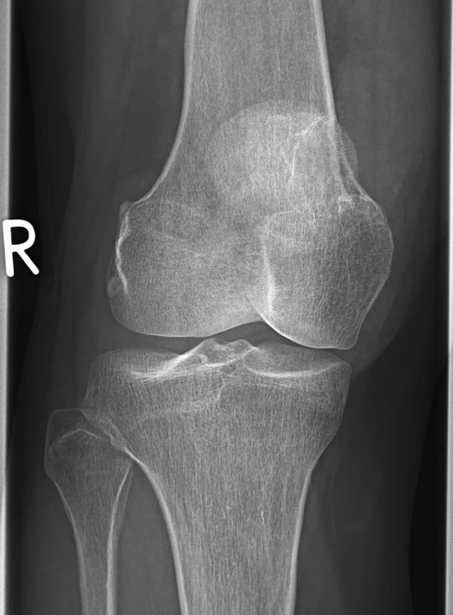
Long description
Anteroposterior radiograph described on Commons only as 'Riesenzelltumor' (giant cell tumor), with no stated affected bone or soap-bubble appearance in the description.

Figure 2Lytic tumour proximal femur - 001
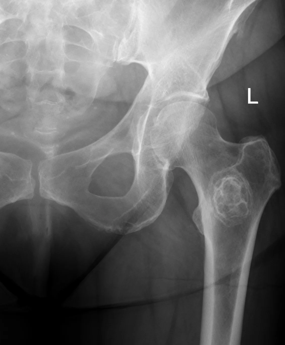
Long description
A radiograph of a lytic tumour in the proximal left femur with a calcified rim, suggestive of a liposclerosing myxofibrous tumour.

Figure 3Osteosarcoma of the tibia
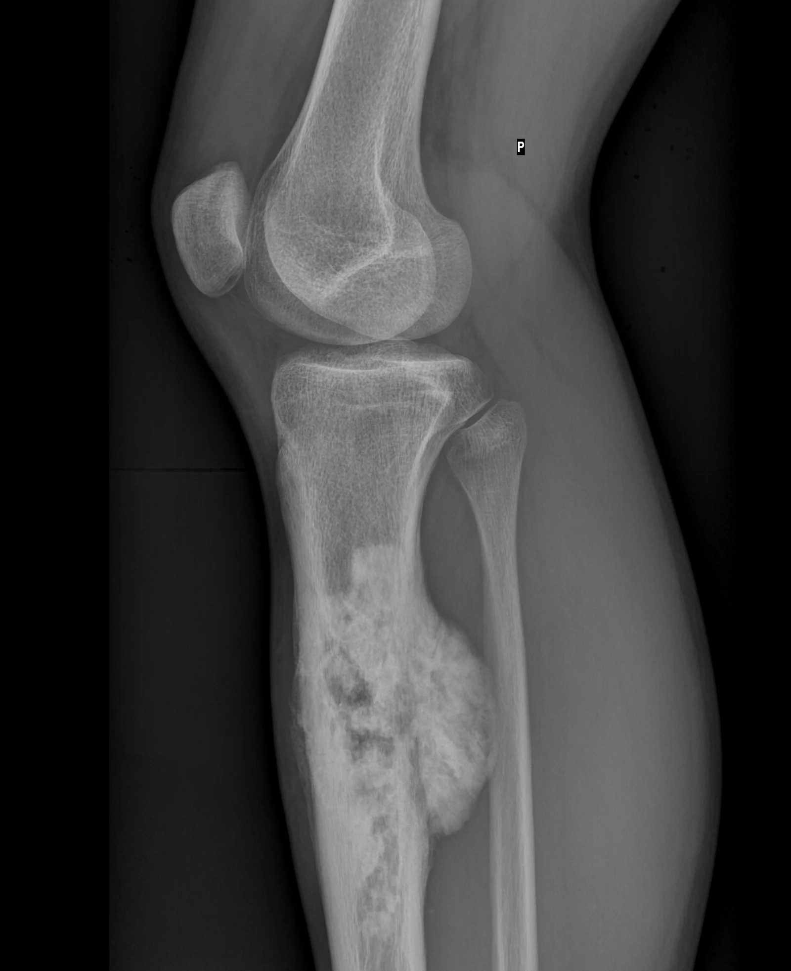
Long description
An X-ray of an osteosarcoma listed under the Commons category 'X-rays of osteosarcoma'; the file's own description only states 'Osteosarkom pistaly' (Slovak) without siting the lesion or mentioning a sunburst periosteal reaction.

The investigation pathway is deliberately staged, and each step has a reason.
- Plain radiograph (two planes) is the first test and the most informative single investigation. Lesions that need further investigation include bone destruction, new bone formation, periosteal swelling and soft tissue swelling. A normal radiograph does not exclude a primary malignant bone tumour, so persistent pain or a mass requires an urgent MRI.[1]
- MRI of the local site, including the whole anatomical compartment and the whole of the involved bone with the adjacent joints, is required before biopsy. It defines intramedullary and soft tissue extent, joint involvement, skip lesions and the relationship to neurovascular structures. For pelvic tumours CT and MRI are both routine.[1]
- CT is helpful when MRI is contraindicated or when there is diagnostic uncertainty; it shows microcalcification, periosteal bone formation and cortical destruction well, and it is essential for mapping a biopsy.[1]
- Staging investigations in a confirmed primary malignant bone tumour include CT of the chest. Whole skeleton (or whole-body) imaging is required in Ewing sarcoma and osteosarcoma, and whole-body MRI and PET-CT are replacing bone scintigraphy for staging and response assessment. Bone marrow biopsies are no longer mandated outside clinical trials, because modern whole-body imaging gives equivalent information. Skeletal metastases are rare in chondrosarcoma unless it is dedifferentiated.[1]
Laboratory tests are not diagnostic of a primary bone tumour, but erythrocyte sedimentation rate, alkaline phosphatase and lactate dehydrogenase may have prognostic value. In anyone over 60 with persistent bone pain, particularly back pain, or an unexplained fracture, myeloma must be excluded with a full blood count and blood tests for calcium, erythrocyte sedimentation rate or plasma viscosity, paraprotein and serum free light chains.[1][2]
Approach to a suspected primary bone tumour in an adult
- Symptoms and signsPersistent bone pain (especially night pain), swelling, pathological fracture or systemic upset
- Plain X-ray (two views)Look for bone destruction, new bone formation, periosteal swelling or soft tissue swelling
- Question: Does the film look suspicious or are symptoms persistent?
No, and symptoms resolve
- Safety-netting and reviewReassure; return if symptoms recur or change
Yes
- Urgent MRI of the whole boneLocal extent, skip lesions, neurovascular relations
- Question: Patient age?
Under 40
- Urgent referral to a bone sarcoma centreBefore biopsy; include X-ray and bloods (full blood count, ESR, alkaline phosphatase)
40 or over
- Investigate for metastatic carcinoma and myeloma firstCT chest/abdomen/pelvis, whole-body imaging, myeloma screen
- Refer proven solitary bone lesions
- Specialist core needle biopsyAfter cross-sectional imaging; track planned for later excision; tissue for histology and molecular testing
- Multidisciplinary team review and AJCC stagingImaging, histology and molecular results discussed together
There is no single blood test or imaging that diagnoses a primary bone tumour; diagnosis requires the correlation of imaging, histology and, for relevant tumours, molecular testing.
Enneking staging uses histological grade (I low, II high) and anatomical compartment (A intracompartmental, B extracompartmental); stage III is metastatic disease regardless of grade. AJCC/TNM staging uses grade, tumour size, skip lesions and metastases, and separates metastatic disease confined to the lung (stage 4A) from metastases at other sites (stage 4B), which carries a worse prognosis. The two systems are used side by side in musculoskeletal oncology.[1]
Campanacci grading (stages 1, 2 and 3) is used specifically for giant cell tumour of bone on plain radiographs and influences whether curettage or resection is appropriate.[4]
Every primary malignant bone tumour is managed by a bone sarcoma multidisciplinary team, with treatment delivered in specialist commissioned centres and patients allocated a key worker. Children, teenagers and young adults are also discussed at the relevant paediatric or young-adult tumour board. Management differs by tumour type rather than by a single bone-sarcoma protocol.[1]
Chondrosarcoma is usually treated surgically, because it is relatively radioresistant and insensitive to conventional chemotherapy. Low-grade lesions (atypical cartilaginous tumour in the long bones, grade 1 chondrosarcoma axially) may be treated by careful curettage or marginal excision; higher grades demand wide excision with clear margins, sometimes amputation or reconstruction depending on the site. Chemotherapy is considered for dedifferentiated and mesenchymal chondrosarcoma. Radiotherapy is used adjuvantly for high-risk resections and as primary treatment for unresectable lesions, particularly skull-base and sacral tumours treated with proton beam or carbon ion therapy. Avoiding both extremes (curetting a high-grade tumour, or widely excising a benign lesion) is a recurring trap, because distinguishing between the two is difficult on a small biopsy.[1]
Osteosarcoma follows the same broad sequence as in younger patients, adjusted to the adult's presentation. Treatment is neoadjuvant combination chemotherapy, definitive local surgery (wide excision, ideally limb-sparing with reconstruction when clear margins are achievable, or amputation), then adjuvant chemotherapy. Radiotherapy is reserved for positive or close margins, axial or unresectable disease and palliation, because osteosarcoma is relatively radioresistant. A pathological fracture does not automatically preclude limb-sparing surgery, because fractures often heal during neoadjuvant chemotherapy. Pulmonary metastases that remain or appear after treatment are resected when possible, with selective radiotherapy. Secondary osteosarcoma arising in Paget disease or after radiation carries a worse outlook and chemotherapy may need modification for cardiac and renal safety.[1]
Ewing sarcoma is a systemic disease treated intensively. The usual plan is neoadjuvant (induction) chemotherapy with multi-agent regimens, local control by surgery and/or radiotherapy (definitive radiotherapy at high dose when surgery is not possible or function-preserving), then adjuvant chemotherapy, with treatment of metastatic sites where possible (whole-lung irradiation after pulmonary metastases). Because the same tumour occurs in children and young adults, protocols are generally shared across age groups within specialist centres, with careful attention to cardiotoxicity, fertility preservation and late effects in adults.[1]
Giant cell tumour is usually treated surgically. Curettage, often with local adjuvants, is used for contained lesions with acceptable recurrence, and en bloc resection for more aggressive tumours or when the joint cannot be preserved. Recurrence is observed in about a quarter of patients overall, and in up to half after curettage alone.[4] Denosumab, a fully humanised monoclonal antibody against RANK ligand, is the standard treatment for unresectable or metastatic giant cell tumour requiring morbid surgery; neoadjuvant administration can enable resection in selected cases, although it should be used cautiously before joint-saving surgery because of high local recurrence rates. Where the tumour is unresectable or metastatic, denosumab is given for years or even lifelong, with injection-site and calcium monitoring.[5]
Benign lesions are managed for what they are, not by a sarcoma protocol. Enchondromas are usually left alone; curettage and grafting are reserved for symptomatic lesions, pathological fracture, or any doubt about malignancy, and the distinction from an atypical cartilaginous tumour is made in a multidisciplinary meeting. Osteoid osteoma is traditionally relieved by non-steroidal anti-inflammatory drugs; definitive treatment is percutaneous ablation (radiofrequency or other thermal ablation) or, where necessary, surgical excision of the nidus. Osteochondroma is excised only if it is painful, limits function, causes complications (bursa, fracture, neurovascular compression), or shows suspicious features in the cartilage cap. Malignant change in a solitary lesion is rare, but a thickened or growing cap in an adult crosses the threshold for excision and histology.
- Splint or immobilise and control pain in any suspected or pathological fracture; internal fixation is contraindicated until the diagnosis is established.[1]
- Refer before biopsy. A poorly placed biopsy can compromise both limb salvage and cure, and biopsy tracks must be planned so they can be excised.[1][2]
- Refer any patient under 40 with suspected primary malignant bone tumour urgently to a bone sarcoma centre; in patients over 40 investigate first for metastatic carcinoma and myeloma, but refer proven solitary bone lesions.[1][2]
- NICE NG12 recommends a suspected cancer pathway referral for adults when an X-ray suggests the possibility of bone sarcoma, a very urgent assessment within 48 hours for children and young people in the same situation, and a very urgent direct-access X-ray for unexplained bone swelling or pain in children and young people. The age, the referral urgency and the diagnostic route therefore differ markedly between adults and children.[2]
Follow-up is long. Reviews agree that every patient needs long-term imaging, because recurrence and new tumours can appear years later, but no single schedule commands universal agreement. Follow-up schedules are suggested by the UK guidelines, by the ESMO-EURACAN-GENTURIS-ERN PaedCan guideline and by local protocols, with imaging of the primary site and the chest at intervals tailored to tumour type, grade and response.[1][3]
- Local recurrence after resection, particularly in chondrosarcoma and after marginal excision.
- Metastasis, most often to the lung in osteosarcoma, Ewing sarcoma and metastasising giant cell tumour.
- Pathological fracture, which may alter surgery.
- Infection around a prosthesis or allograft and periprosthetic complications.
- Late effects of chemotherapy and radiotherapy: anthracycline cardiotoxicity, nephrotoxicity, secondary malignancy, infertility.
- Loss of function after amputation or major reconstruction, needing rehabilitation.
- Giant cell tumour left untreated becomes locally destructive; denosumab, used without timely surgery, can lead to uncontrolled disease if stopped.
Prognosis is governed more by tumour type, grade and stage than by patient age, although younger patients generally fare better.
- Chondrosarcoma. Most are low grade and have an excellent prognosis after complete excision; dedifferentiated chondrosarcoma behaves like a high-grade sarcoma and carries a poor prognosis.
- Osteosarcoma. High-grade conventional osteosarcoma in an adult carries a substantially worse outlook than in a child. Axial or pelvic primaries, older age, male sex, metastases at diagnosis and a poor histological response to preoperative chemotherapy all worsen outcome.
- Ewing sarcoma. Adults do less well than children, and pelvic and axial primaries carry a higher risk of metastasis and relapse.
- Giant cell tumour. Histologically benign but locally aggressive. About a quarter recur after treatment; lung metastases develop in roughly 5% of patients, usually after recurrence, and many indolent metastases can be observed or resected.[1]
- Survival differences across Europe have been documented, with patients in Ireland and the UK having lower five-year survival than those in Northern and Central Europe, which underlines why referral to a high-volume specialist centre matters.[1]
Children and adolescents. Osteosarcoma and Ewing sarcoma affect children and adolescents as well as adults, and paediatric oncology protocols apply. Children, teenagers and young adults are discussed at the relevant paediatric or young-adult tumour board in addition to the bone sarcoma team. Hereditary syndromes (retinoblastoma, Li-Fraumeni syndrome) are more likely in very young patients. The dedicated childhood bone tumours topic covers primary malignant bone tumours in that age group in more detail.
Pregnancy. Treatment must be tailored; chemotherapy and surgery are timed by a multidisciplinary team. Decisions about fertility preservation (sperm storage for men of reproductive age; fertility counselling and oocyte or embryo options for women before chemotherapy where time allows) should be made before treatment starts.[1]
Older adults with Paget disease or prior radiation. These patients present later, more often in the axial skeleton, with secondary osteosarcoma. Their functional status, cardiac function (relevant to anthracyclines) and renal function (relevant to cisplatin and methotrexate) decide what chemotherapy can be given safely.
Low- and middle-income settings. The essentials are: a careful history, a plain radiograph, appropriate blood tests and a plain chest film; image-guided core needle biopsy by an experienced team before any definitive treatment; splintage and pain control for fractures; referral to the nearest sarcoma service as early as possible. If advanced reconstruction or complex chest-wall surgery is unavailable, early amputation or wide excision rather than late, complicated surgery saves lives. Every resected specimen should reach a specialist pathologist, even if tissue must be sent to a reference centre. Where denosumab, advanced imaging or proton therapy are unavailable, conventional surgery and cytotoxic chemotherapy remain effective for most of these tumours.
- Primary bone tumours are rare: about 0.2% of all cancers, 8.5 per million per year in Europe. In adults over 40, secondary and haematological bone lesions are far more common, so investigate for metastasis or myeloma first.
- The common benign primary bone tumours in adults are osteochondroma, enchondroma, giant cell tumour of bone and osteoid osteoma. The common malignant ones are chondrosarcoma (commonest in older adults), osteosarcoma (second peak in the seventh and eighth decades) and Ewing sarcoma (mainly children and young adults, but occurring in adults).
- WHO classifies bone tumours by tissue of origin; the current standard is the 5th edition (IARC, 2020). The UK bone sarcoma guidelines (2025, adapted from WHO 2020), the ESMO-EURACAN-GENTURIS-ERN PaedCan guideline (2021) and NICE guideline NG12 underpin much of this page.
- Night pain, persistent localised bone pain, progressive swelling, pathological fracture and unexplained weight loss in older adults are the red flags. A normal radiograph never excludes a primary malignant bone tumour.
- Plain radiograph is first; MRI of the whole bone follows before biopsy; CT chest and whole-body imaging stage the disease. Biopsy is done at a specialist centre, with the track planned to be excised.
- Chondrosarcoma is treated surgically; osteosarcoma and Ewing sarcoma by neoadjuvant chemotherapy, local control and adjuvant chemotherapy; giant cell tumour by surgery with denosumab for unresectable or metastatic disease.
- Internal fixation of a suspected pathological fracture is contraindicated until the diagnosis is established.
- Refer before biopsy. Adult and paediatric referral pathways differ in urgency and route under NICE NG12.
Answer each one in your head first, then open the answer.
A 44-year-old woman has six weeks of pain in the proximal femur that is worse at night and a normal plain radiograph. What is the next step, and why does a normal film not reassure you?
Show answerHide answer
A normal radiograph does not exclude a primary malignant bone tumour. The next step is an urgent MRI of the bone, and if symptoms or imaging are suspicious, referral to a bone sarcoma centre. Night pain is a red flag regardless of the radiograph.
What is the single most likely diagnosis in a 55-year-old man with a painful, progressively enlarging lytic lesion in the proximal tibia epiphysis, and what radiographic clue separates it from a central chondrosarcoma?
Show answerHide answer
Giant cell tumour of bone. It characteristically occupies the epiphysis of a long bone in an adult aged 20 to 50, is eccentric and purely lytic with a non-sclerotic margin, without chondroid matrix. A chondrosarcoma is usually metaphyseal or diaphyseal and shows chondroid calcification. Management is surgical (curettage or resection) with denosumab for unresectable or metastatic disease.
A 72-year-old man has a destructive pelvic bone lesion. Why is it unsafe to assume osteosarcoma, and what must be excluded first?
Show answerHide answer
In adults over 40, metastatic carcinoma and myeloma outnumber primary bone tumours, so the first steps are a myeloma screen and staging for a primary cancer, alongside MRI and biopsy at a specialist centre. Osteosarcoma remains possible, particularly secondary osteosarcoma in Paget disease, but it is not the most likely diagnosis.
Plain text: osteochondroma; enchondroma and Ollier disease; chondrosarcoma; osteosarcoma in children; Ewing sarcoma in children; Paget disease of bone; osteomyelitis; giant cell tumour of bone and denosumab; osteoid osteoma and radiofrequency ablation; amputation and limb reconstruction; late effects of childhood cancer treatment.
- Gerrand C, Amary F, Anwar HA, Brennan B, Dileo P, Kalkat MS, McCabe MG, McCullough AL, Parry MC, Patel A, Seddon BM, Sherriff JM, Tirabosco R, Strauss SJ. UK guidelines for the management of bone sarcomas. British Journal of Cancer 2025;132(1):32-48. Accessed .
- National Institute for Health and Care Excellence. Suspected cancer: recognition and referral. NICE guideline NG12 (last updated 15 April 2026). Accessed .
- Strauss SJ, Frezza AM, Abecassis N, et al. Bone sarcomas: ESMO-EURACAN-GENTURIS-ERN PaedCan Clinical Practice Guideline for diagnosis, treatment and follow-up. Annals of Oncology 2021;32(12):1520-1536. Accessed .
- Jha Y, Chaudhary K. Giant Cell Tumour of Bone: A Comprehensive Review of Pathogenesis, Diagnosis, and Treatment. Cureus 2023;15(10):e46945. Accessed .
- Imre A, Zoltan S, Miklos S. Current indications for denosumab in benign bone tumours. EFORT Open Reviews 2023;8(12):895-905. Accessed .
- He XJ, Jiang YC, Yang P, et al. An analysis of the imaging features and diagnostic value of osteoid osteoma on DR, CT, and MRI. Frontiers in Medicine 2026;13:1890873. Accessed .
Figure credits
Educational content for medical students and doctors. It is not a substitute for clinical judgement or local guidelines.