Osteoarthritis
A whole-joint disease of progressive cartilage loss and osteophyte formation: how it presents in the hand, knee and hip, and how to manage it.
- Osteoarthritis (OA) is the most common form of arthritis: a slowly progressive, whole-joint disease rather than simple "wear and tear" of cartilage.
- It is defined by the combination of focal loss of articular cartilage and new bone formation at the joint margin (osteophytes), with low-grade inflammation of the synovium.
- Pain is activity-related and brief stiffness after rest; it most often affects the knees, hips and hands.
- The clinical picture is usually enough for diagnosis in a person aged 45 or over with activity-related joint pain and morning stiffness lasting no more than 30 minutes. [1]
- Core management is the same everywhere: education, tailored therapeutic exercise and weight management, with simple analgesia and joint replacement reserved for severe disease.
By the end of this page you should be able to:
- Define osteoarthritis and distinguish it from inflammatory arthritides.
- Explain why cartilage loss and osteophyte formation occur, and how they produce symptoms.
- Recognise the typical patterns of hand, knee and hip osteoarthritis, including Heberden and Bouchard nodes.
- Diagnose osteoarthritis in primary care without imaging, and know when an X-ray is genuinely useful.
- Outline a management plan that follows current NICE, OARSI and ACR guidance, including exercise, weight loss, drugs, injections and referral for joint replacement.
- Recognise red flags that suggest something other than osteoarthritis is present.
DefinitionOsteoarthritis
Osteoarthritis is a whole-joint disease in which articular cartilage is progressively lost, subchondral bone remodels and enlarges, and osteophytes form at the joint margin, while the synovium stays inflamed at a low level. Pain, stiffness and loss of function follow, most often in the knees, hips and hands.
The word "osteoarthritis" is slightly misleading: there is no marked synovial proliferation of the kind seen in rheumatoid disease, and the damage is usually focal rather than uniform. The disease affects the joint as an organ (cartilage, bone, synovium, meniscus, capsule and supporting muscle), which is why treatments aimed only at cartilage rarely succeed. Cartilage itself has no nerve supply, so the pain of OA comes from subchondral bone, the joint capsule, periosteum and inflamed synovium rather than from the smooth surface that is seen on imaging.
Osteoarthritis is usually classified in two useful ways.
- Primary (idiopathic) OA develops without a single identifiable cause, usually in later life and often in several joints. It is the common pattern in the knees, hips and hands of older adults.
- Secondary OA follows a recognised insult: prior trauma, fracture or meniscal injury, congenital or developmental deformity, inflammatory arthritis, crystal disease, avascular necrosis, Paget disease of bone or other metabolic disorder. It often appears earlier, may be unilateral, and tends to progress faster.
A further clinical distinction is by distribution:
- Localised OA affects one or a few joints.
- Generalised OA affects several joint groups, often with nodal (hand) disease.
- Nodal OA of the hands, with Heberden and Bouchard nodes.
- Erosive OA of the hands, a more inflammatory, destructive variant.
- Rapidly destructive OA, an uncommon but aggressive form, classically of the hip.
Heberden nodes are bony enlargements at the distal interphalangeal joints; Bouchard nodes are bony enlargements at the proximal interphalangeal joints. Both represent osteophytes and represent clinical markers of nodal disease. They are usually painless once formed, but can be tender during active disease. Together with base-of-thumb (first carpometacarpal) osteoarthritis, they define the hand pattern most students are asked to recognise.
United States
American College of Rheumatology (ACR) guidance describes osteoarthritis as a whole-joint disease involving cartilage degradation, bone remodelling, osteophyte formation and synovial inflammation.
Osteoarthritis is the most common form of arthritis. ACR 2019 estimated that it affects about 302 million people worldwide and that it is a leading cause of disability among older adults. [3] The knees, hips and hands are the most commonly affected appendicular joints. [3] Prevalence rises steeply with age, and the condition is usually more common and more disabling in women after mid-life, although reported ratios vary between populations and joints.
Because the disease is strongly age-related and survival is long, an "epidemic" of symptomatic OA is being driven by ageing populations and rising body weight. [3] Primary care therefore sees knee and hand disease most often, and orthopaedic services see end-stage hip and knee disease most often. In most regions there is no population screening programme: case-finding is clinical, triggered by joint pain in an older adult.
A single cause is rarely identified in older adults. OA is best understood as the outcome of a mismatch between the mechanical load a joint experiences and the ability of cartilage, meniscus and subchondral bone to absorb it. The table lists the factors a student should be able to discuss in an examination.
| Risk factor | Modifiable? | How it acts |
|---|---|---|
| Older age | No | Cartilage becomes less resilient and repair slows; cumulative microdamage adds up. |
| Female sex (after mid-life) | No | Hormonal and biomechanical factors alter joint loading and pain processing; hand and knee disease are especially common. |
| Excess body weight | Yes | Increases load across weight-bearing joints; adipose tissue also releases inflammatory mediators. |
| Previous joint injury or surgery | Partly | Alters joint mechanics and can damage meniscus or ligaments; risk of post-traumatic OA. |
| Occupational or repetitive loading | Yes | Prolonged kneeling, squatting, heavy lifting and vibration transfer load to cartilage and subchondral bone. |
| Joint malalignment or dysplasia | Partly | Concentrates load on one compartment (for example varus knee or hip dysplasia). |
| Muscle weakness and poor proprioception | Yes | Reduces shock absorption and joint stability, increasing shear. |
| Genetics and family history | No | Influences cartilage biology and joint shape; nodal hand OA clusters in families. |
| Metabolic and endocrine disease | Partly | Diabetes, crystal arthritis and other disorders can alter cartilage through systemic mechanisms. |
Ask about body weight, occupation, previous injury and family history whenever a patient presents with new joint pain. The strongest modifiable targets for an individual patient are body weight and muscular conditioning.
OA is a sequence, not an event. Understanding the sequence explains both the symptoms and why early imaging is unhelpful.
- Cartilage matrix degrades. Chondrocytes shift from a resting to a repair phenotype and release matrix metalloproteinases and aggrecanases. Proteoglycan content falls, water content rises and the cartilage becomes soft and fibrillated. The change starts focally, often in a high-load area, and spreads.
- Subchondral bone changes. Increased bone turnover thickens and stiffens the subchondral plate; bone marrow lesions appear. Because bone is pain-sensitive, this is a major source of pain.
- Osteophytes form. Mesenchymal cells at the joint margin differentiate into bone and produce the bony spurs that are the hallmark of OA. Osteophytes can stabilise the joint but also cause mechanical block and impingement.
- The synovium inflames. Synovial macrophages react to cartilage debris and crystals of hydroxyapatite or calcium pyrophosphate. The resulting low-grade synovitis produces effusion, stiffness and pain.
- Soft tissues and muscle decline. Quadriceps weakness and reduced proprioception allow abnormal loading, accelerating the mechanical damage. Pain leads to disuse, which leads to more weakness.
- Joint shape changes. Loss of cartilage and osteophyte growth alter joint geometry, producing deformity (varus or valgus knee, fixed flexion). Once deformity and bone-on-bone contact are established, the change is largely irreversible.
The key message for a student is that OA is active biology throughout: it is a failure of repair as much as a failure of resistance.
At arthroscopy or post-mortem, OA shows focal loss of articular cartilage, with a softened, fibrillated surface, the earliest changes in high-load areas. Osteophytes appear at the joint margins, the synovium may be mildly inflamed, and the subchondral bone becomes dense and sometimes cystic. Under the microscope, cartilage shows loss of proteoglycan staining, chondrocyte clustering and cell death, while subchondral bone shows thickened trabeculae and remodelling. Histology is rarely needed for the diagnosis, but excised or replaced joint material shows these changes clearly, and biopsy may be used when tumour or infection is suspected.
The patient's words usually reveal the diagnosis before the examination begins.
- Pain is the leading symptom. It is felt deep in the joint, is worse on use and weight-bearing, and eases with rest. In advanced disease the pain can be present at night or at rest.
- Stiffness occurs after periods of inactivity ("gelling") and after waking. In OA it lasts less than 30 minutes; prolonged morning stiffness points away from OA and towards an inflammatory arthritis.
- Functional loss is often the real complaint: difficulty rising from a chair, climbing stairs, kneeling, opening jars, turning keys or walking distance. Ask about specific tasks rather than "how are you".
- Swelling may be episodic and follows activity. A sudden hot, swollen joint suggests crystal arthritis, infection or an inflammatory flare rather than uncomplicated OA.
- Flares are defined as a temporary worsening of pain, swelling and stiffness that is worse than normal, may affect sleep, activity, function and psychological wellbeing, and may lead to change in therapy for at least 24 hours. [1]
- Crepitus may be reported as grinding, clicking or "bone on bone" during movement.
- No systemic symptoms are expected. Fever, weight loss, prolonged morning stiffness or multiple small-joint swelling are red flags.
Observe the patient walking in and rising from a chair before asking them to undress a joint. Look, feel and move each joint in turn.
- Inspection for deformity (varus or valgus angulation, flexion contracture), bony enlargement, swelling and muscle wasting. In the hand, look for Heberden nodes at the distal interphalangeal joints and Bouchard nodes at the proximal interphalangeal joints, and for swelling or pronation at the base of the thumb.
- Palpation for warmth (minimal in uncomplicated OA; marked warmth suggests inflammation or infection), joint-line tenderness, effusion and crepitus. Press each interphalangeal joint individually; many nodes are painless once formed.
- Range of movement is reduced by pain and by capsular contracture. Crepitus on movement is common. Fixed flexion deformity of the knee or hip is a marker of advanced disease.
- Muscle strength around the joint is often reduced, especially quadriceps strength in knee OA.
- Special tests for the knee include assessment of varus and valgus stability and the presence of an effusion. For the hip, the FADIR and FABER tests are useful when considering alternative pathology; forced internal rotation and flexion that reproduce groin pain suggest hip-driven pain.
- Gait may show an antalgic limp, shortened stride or trunk lean over the affected side in hip disease.
Hand. The classic pattern involves the distal interphalangeal, proximal interphalangeal and first carpometacarpal joints, often bilaterally but asymmetrically. The metacarpophalangeal joints are relatively spared, in contrast to rheumatoid arthritis. Heberden and Bouchard nodes are common, and base-of-thumb osteoarthritis causes pain on pinch and grip. The erosive variant causes more inflammatory symptoms and joint destruction.
Knee. Tibiofemoral (medial compartment predominates) and patellofemoral disease are both common. Pain is felt around or behind the kneecap, or along the joint line. Stiffness, swelling after activity, giving way and varus deformity are typical. Quadriceps wasting and a fixed flexion deformity mark severe disease.
Hip. Pain is usually groin pain, sometimes referred to the anterior thigh or knee. It is worse on walking, turning in bed and putting on shoes or socks. Internal rotation is the first movement lost. Abductor weakness and a limp are common. Hip OA can masquerade as lumbar spine or knee disease.
A single hot swollen joint, recent trauma, prolonged morning stiffness, rapid progression or deformity, fever or weight loss should make you think again. [1] In these cases imaging and laboratory tests, rather than a clinical diagnosis of OA, are appropriate.
Osteoarthritis is usually diagnosed clinically, but it co-exists with other conditions and mimics them. The table separates the common mimics by the feature that most reliably distinguishes them.
| Condition | Feature that separates it from OA |
|---|---|
| Rheumatoid arthritis | Prolonged morning stiffness, symmetrical small-joint swelling, elevated inflammatory markers, systemic features |
| Psoriatic arthritis | Dactylitis, enthesitis, rash or nail changes, often asymmetric and in younger patients |
| Gout or pseudogout | Sudden hot swollen joint, crystals in synovial fluid, often monoarticular |
| Septic arthritis | Fever, a hot swollen joint, marked systemic upset; an emergency |
| Ankylosing spondylitis | Inflammatory back pain, onset under 40, sacroiliac disease, HLA-B27 association |
| Avascular necrosis | Pain out of proportion to X-ray early, risk factors (steroids, alcohol, sickle cell), collapse of subchondral bone |
| Meniscal or ligament injury | Mechanical locking, giving way, effusion after trauma, localised joint-line tenderness |
| Referred pain from spine or hip | Pain distribution not following the joint, normal joint examination, neuropathic features |
| Metabolic arthropathy (haemochromatosis, Paget disease) | Unusual pattern, systemic clues, radiographs with characteristic changes |
Figure 1F66-rizoartrosis-AL
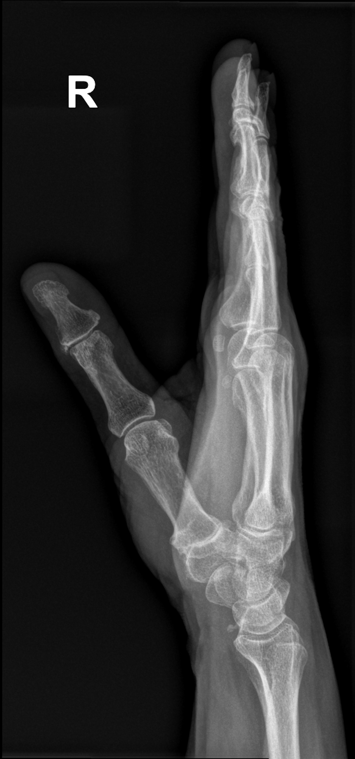
Long description
An AP radiograph of the right thumb of a 66-year-old woman showing trapeziometacarpal osteoarthritis with joint space narrowing, subchondral sclerosis, and osteophytes.

Osteoarthritis is a clinical diagnosis. Imaging should be used to answer a specific question, not to label the disease.
| Test | What it shows or means |
|---|---|
| Clinical assessment | A person aged 45 or over with activity-related joint pain and no or brief morning stiffness needs no imaging to diagnose OA. [1] |
| Plain radiograph | Joint-space narrowing, marginal osteophytes, subchondral sclerosis and subchondral cysts; useful when the diagnosis is uncertain or red flags exist. |
| MRI | Cartilage, meniscus, bone marrow lesions and soft tissue; not a first-line test, but useful for red flags, rapid progression or pre-operative planning. |
| Ultrasound | Synovitis and effusion; can guide injections, especially the hip. [3] |
| Synovial fluid analysis | In OA the fluid is viscous and the white cell count is low; crystals or a high cell count point to crystal or septic arthritis. |
| Blood tests | No blood test diagnoses OA. They exclude alternatives: inflammatory markers, rheumatoid factor and anti-CCP for inflammatory arthritis, urate for gout. |
| Bone mineral density and metabolic tests | Considered when presentation or risk factors suggest secondary OA or osteoporosis. |
Radiographic severity is commonly graded with the Kellgren-Lawrence system, which records joint-space narrowing, osteophytes, subchondral sclerosis and deformity from no radiographic disease to severe disease. A high grade on an X-ray is not the same as a severe patient: images correlate poorly with symptoms, and treatment should follow symptoms and function rather than the film. [1]
The diagnostic approach is deliberately simple. NICE guidance states that OA should be diagnosed clinically without imaging in a person who is 45 or over, has activity-related joint pain and has either no morning joint-related stiffness or morning stiffness that lasts no longer than 30 minutes. [1] This keeps diagnosis practical and avoids unnecessary radiation. Imaging is reserved for atypical features or where an alternative or additional diagnosis is suspected. [1]
Severity in routine practice is judged clinically: pain intensity, limitation of everyday tasks, deformity, gait and the impact on sleep, work and mood. Numerical scoring systems (for example WOMAC, KOOS or HOOS) are useful for measuring change in trials and after arthroplasty, but NICE advises using clinical assessment rather than a numerical severity score to decide when to refer for joint replacement. [1]
Management of OA rests on a small set of principles that apply to every patient, from every guideline:
- The patient is the manager; the clinician is the adviser. Explain the diagnosis, its cause and its course, and correct the common myths (OA is not simply "wearing out", rest does not cure it, and activity does not wear the joint away faster).
- Treat symptoms and function, not the X-ray.
- The core treatments are therapeutic exercise and weight management (if appropriate), along with information and support. [1]
- Start with treatments that have the least systemic exposure or toxicity.
- Use drugs at the lowest effective dose for the shortest possible time. [1]
- Escalate to injections, then joint replacement, only when core and drug treatment are ineffective or unsuitable.
United Kingdom (NICE)
NICE NG226, published in October 2022, is the UK guideline used here. It diagnosed OA clinically when a person is 45 or over with activity-related joint pain and no, or under 30 minutes, of morning stiffness; recommends therapeutic exercise for everyone and weight loss where overweight; offers a topical NSAID first and, if needed, an oral NSAID with a proton pump inhibitor; and refers for arthroplasty when non-surgical care fails.
United States
ACR/Arthritis Foundation 2019 strongly recommends exercise, weight loss for overweight or obese patients with knee or hip OA, self-management programmes, tai chi, topical NSAIDs for knee OA, oral NSAIDs and intra-articular glucocorticoid injection for knee OA. It conditionally recommends acupuncture, topical capsaicin for knee OA, duloxetine, tramadol, balance exercise, yoga and thermal modalities, and recommends against TENS, routine opioids, colchicine, fish oil, hyaluronan, and oral or transdermal opioids. [2][3]
Therapeutic exercise is the central non-surgical treatment for osteoarthritis. Offer exercise tailored to the patient's needs (local muscle strengthening and general aerobic fitness) to everyone with OA, and consider supervised sessions. [1] Pain may initially increase when exercise begins; explain that regular, consistent exercise reduces pain and improves function over time and that long-term adherence magnifies the benefit. [1] Combining exercise with an education or behaviour-change programme in a structured treatment package helps. [1]
The best exercise is the one the patient will actually do. Options with good evidence include walking (often the most accessible), stationary cycling, strengthening with machines, bands or body weight, neuromuscular training for the knee and aquatic exercise. Programmes work better when supervised (often by a physiotherapist) and when combined with self-management support or weight loss. [3] No hierarchy of one exercise over another is implied in current guidance, and there is insufficient evidence to prescribe an exact type, dose or intensity. [3]
Weight management helps whenever the patient is overweight. In OA, any weight loss is likely to be beneficial, but losing 10% of body weight is likely to be better than losing 5%. [1] ACR 2019, summarising a dose-response relationship, notes that loss of at least 5% of body weight can be associated with changes in clinical and mechanistic outcomes, with increasing benefit at 5-10%, 10-20% and above 20% of body weight, and that weight loss works best together with exercise. [3] Refer for dietetic support and use shared goal-setting rather than prescribing a number.
Manual therapy should only be considered for hip or knee OA and only alongside exercise. [1] ACR 2019 advises against adding manual therapy to exercise alone in knee or hip OA. [3]
Adjuncts that may help selected patients: walking aids for lower-limb disease (walking sticks, crutches, frames and rollators), [1] tibiofemoral knee bracing for tibiofemoral disease, hand orthoses for first carpometacarpal OA, and patellofemoral bracing for patellofemoral disease. [3] Devices such as insoles, splints or supports should not be used routinely; reserve them for joint instability or abnormal loading when exercise alone is not enough. [1]
Approaches not to use: acupuncture or dry needling, [1] electrotherapy (TENS, ultrasound, interferential, laser, pulsed short-wave, neuromuscular stimulation), [1] arthroscopic lavage or debridement, [1] intra-articular hyaluronic acid (not offered; NICE advises against it, [1] and OARSI 2019 does not recommend it for hip or polyarticular disease [2]), glucosamine and strong opioids. [1] ACR 2019 additionally lists colchicine, fish oil, lateral or medial wedged insoles, modified shoes, massage, iontophoresis and pulsed vibration therapy as not recommended. [3] Capsaicin is conditionally recommended for knee OA in the ACR guideline but conditionally recommended against for hand disease. [3]
Drug treatment supports exercise and weight loss; it does not replace them. Every agent should be used at the lowest effective dose for the shortest possible time, alongside non-pharmacological care. [1]
Topical NSAIDs are the first drug for many patients. Offer a topical NSAID for knee OA and consider it for OA at other joints. [1] ACR 2019 strongly recommends topical NSAIDs for knee OA and conditionally for hand OA, and recommends considering them before oral NSAIDs. [3] Topical treatment makes particular sense in older patients, in whom systemic NSAID exposure is risky. Practical tips: apply over (not into) the joint, avoid broken skin and the eyes, and avoid heat over the area after application.
Oral NSAIDs are the initial oral drug of choice for OA whatever the joint, but only when topical treatment is ineffective or unsuitable. [1] [3] Choose the lowest-risk agent for the patient, taking into account gastrointestinal, renal, hepatic and cardiovascular toxicity, age, pregnancy, current medication and comorbidities, [1] and always offer a gastroprotective treatment such as a proton pump inhibitor together with the NSAID. [1] Doses should be as low as possible and treatment continued for as short a time as possible. [3] OARSI 2019 declines to recommend any oral NSAID for people with cardiovascular comorbidities or frailty. [2] This is an important example of guideline divergence: NICE leaves the decision to shared assessment of risk, whereas OARSI is more restrictive.
Paracetamol (acetaminophen) receives a weak or absent recommendation. Routine use is not advised because the benefit is small and unsupported by strong evidence. [1] ACR 2019 conditionally recommends it for short-term, episodic use in patients with few alternatives, with regular monitoring for hepatotoxicity and a maximum of 3 g daily in divided doses. [3] NICE limits routine opioid or weak opioid use to infrequent short-term relief when other options are contraindicated, not tolerated or ineffective. [1]
Duloxetine, tramadol and other agents. ACR 2019 conditionally recommends duloxetine and tramadol, and conditionally recommends against non-tramadol opioids; where an opioid is considered at all, tramadol is preferred over others. [3] NICE advises against offering strong opioids. [1] Local and regional opioid policy should guide practice.
Intra-articular injections. Intra-articular hyaluronan is not recommended. [1] [2] Consider intra-articular corticosteroid injection when other pharmacological treatments are ineffective or unsuitable, or to support therapeutic exercise, but explain that relief is short-term, usually 2 to 10 weeks. [1] ACR 2019 strongly recommends intra-articular glucocorticoid for knee and hip OA, considers it for hand OA, and strongly recommends ultrasound guidance for hip injection. [3] OARSI 2019 places intra-articular corticosteroids as level 1B/level 2 options for knee disease depending on comorbidity status, and does not recommend them for hip or polyarticular disease. [2] These differences between guidelines are real and should be explained to the patient. Repeat injections indefinitely are not supported by the evidence; use them sparingly, usually to settle a flare or to enable rehabilitation.
Refer (or discuss referral) when joint symptoms (pain, stiffness, reduced function or progressive deformity) substantially affect quality of life and non-surgical management is ineffective or unsuitable. [1] Use clinical assessment to decide, not a numerical severity score. [1] Do not exclude a patient from referral because of age, sex or gender, smoking, comorbidities or overweight and obesity measured by BMI; unequal exclusion on these grounds worsens outcomes and inequity. [1] Explain that the risk profile of surgery varies with these same factors. [1]
Do not offer arthroscopic lavage or debridement for OA. [1] It does not improve pain or function beyond placebo in this population.
Consider patient-initiated follow-up for most patients and planned follow-up where needs suggest it, taking into account treatments needing monitoring, the patient's ability to seek help, occupation, and symptom severity. [1] Routine imaging is not needed for follow-up or to guide non-surgical management. [1]
- Progressive pain and loss of function, including difficulty with stairs, rising, transfers and walking distance.
- Fixed flexion deformity, varus or valgus angulation and joint instability.
- Falls and reduced activity, leading to deconditioning and loss of independence in older adults.
- Secondary or reactive synovitis and occasional haemarthrosis.
- Side effects of treatment, particularly gastrointestinal bleeding, renal impairment, cardiovascular events and hypertension from NSAIDs; these drive much of the excess morbidity in older adults.
- Complications of surgery in those who proceed to arthroplasty, including infection, thromboembolism, dislocation and revision; these are uncommon but important.
The burden of these complications is why prevention of further joint damage and maintenance of muscle strength matter so much.
OA is a chronic, usually slowly progressive disease. Pain and function fluctuate over months and years; many patients have long stable periods. Structural progression on imaging is not the same as progression of symptoms, and many people with radiographic OA never develop significant symptoms. [1] Once severe fixed deformity and bone-on-bone contact are present, symptoms usually progress. Obesity, malalignment and quadriceps weakness accelerate functional decline; weight loss, exercise and adequate analgesia often produce clinically important improvement even late in the disease.
There is no established population screening programme for OA. Prevention focuses on the modifiable risks listed earlier:
- Maintain a healthy body weight from early adulthood.
- Keep physically active and strengthen the muscles around the joints; activity in general does not damage healthy joints and is protective.
- Prevent and treat joint injuries, including meniscal tears and ligament injuries, and rehabilitate thoroughly.
- Avoid prolonged kneeling, squatting and heavy loading where possible; use ergonomic aids and modify tasks.
- Manage diabetes and other metabolic disease, and keep bone health in mind in older adults.
- Encourage early presentation for joint pain so that core treatment begins before deformity and muscle loss are established.
OA that is diagnosed early is easier to manage; the disease becomes harder to control once deformity and advanced muscle wasting are present.
Pregnancy. Osteoarthritis is uncommon in women of reproductive age. During pregnancy, pain increases in women with existing disease because of weight gain and ligament laxity. Pharmacological treatment should take pregnancy into account as a risk factor, [1] and oral NSAIDs generally need caution, especially later in pregnancy. Use non-drug measures (exercise, physiotherapy, weight management, heat, supports) first and work with the obstetric and rheumatology teams before giving analgesics. Consider relative contraindications carefully and follow local formulary guidance.
Children. Primary OA is not a disease of childhood. Joint pain in a child or young adult should raise the possibility of secondary OA from injury, dysplasia, inflammatory arthritis, metabolic disorder or (in the case of the hip) slipped capital femoral epiphysis or Perthes disease, rather than OA itself. Early referral and investigation of a young patient with "OA" are safer than assuming the adult pattern.
Older adults. This is the most common group. Polypharmacy, frailty, renal impairment, cardiovascular disease and risk of falls shape every decision. Drug choice should emphasise topical over oral agents, the lowest effective doses and close monitoring. Exercise remains beneficial even in frailty, but programmes must be supervised and fall-safe. Consider bone health and function holistically and ensure home and community support.
Red flagWhen the diagnosis is not osteoarthritis
Suspect an alternative or additional diagnosis when the patient has a single hot swollen joint, fever, weight loss, prolonged morning stiffness, recent trauma, rapid progression or deformity, or when routine treatment fails without explanation. These are the atypical features NICE lists, and they warrant imaging and further investigation rather than a label of OA.
- OA can usually be diagnosed clinically without imaging, so a plain radiograph is reserved for red flags or uncertainty. [1]
- Exercise, weight loss and education are the core of treatment and can be delivered by physiotherapists, nurses and community health workers.
- A walking stick, quadriceps strengthening and activity pacing cost little and help a lot.
- Oral NSAIDs are cheap but carry the same risks; use the lowest effective dose for the shortest time and avoid them in renal or cardiovascular disease and in frail patients. [1] [2]
- Topical NSAIDs, if available, are safer for many older patients.
- Joint replacement is effective but requires referral to a centre with surgical capacity; identify severe, end-stage cases early.
- Intra-articular steroid injection, where skills and asepsis are available, can settle a flare and allow rehabilitation.
Approach to suspected osteoarthritis in primary care
- Adult with joint painMost often knee, hip or hand in a person over 45
- Question: Does the patient meet the clinical criteria?
Yes (45 or over, activity-related pain, stiffness under 30 minutes)
- Diagnose osteoarthritis clinicallyExplain, reassure, correct misconceptions; no imaging needed
- Start core treatmentInformation, therapeutic exercise, weight management if overweight
- Question: Are symptoms adequately controlled?
Yes
- Self-management and patient-initiated follow-upReview medicines and exercise plan periodically
No
- Add drug treatmentTopical NSAID first; then consider oral NSAID with PPI if ineffective or unsuitable
- Question: Flare or block to rehabilitation?
Yes
- Consider intra-articular corticosteroid (short-term relief)2 to 10 weeks; ultrasound guidance for the hip
No
- Continue current managementReview if symptoms or function change
- Question: Symptoms still substantially affecting quality of life?
Yes, despite non-surgical care
- Refer for joint replacement assessmentClinical decision; do not exclude because of age, BMI, comorbidity or smoking
No
- Continue and review core and drug treatmentPatient-initiated follow-up
No, or red flags or atypical features present
- Do not label as osteoarthritis yetConsider imaging and tests for inflammatory, crystal or septic arthritis
- Treat or refer for the cause found
- Osteoarthritis is a whole-joint disease of cartilage loss, subchondral bone change, osteophyte formation and low-grade synovial inflammation.
- It affects roughly 302 million people worldwide and is a leading cause of disability in older adults.
- Activity-related pain and brief stiffness under 30 minutes are typical; prolonged morning stiffness suggests inflammatory disease.
- The knees, hips and hands are the common sites; Heberden nodes are at the distal and Bouchard nodes at the proximal interphalangeal joints.
- Diagnosis is clinical in a person aged 45 or over with activity-related joint pain and short-lived morning stiffness.
- Imaging correlates poorly with symptoms; use it for red flags or diagnostic uncertainty, not to label disease.
- Core treatment for everyone is information, therapeutic exercise and weight management.
- Topical NSAIDs come before oral NSAIDs; oral NSAIDs must be combined with a proton pump inhibitor.
- Intra-articular corticosteroids give short (2 to 10 weeks) symptomatic relief; hyaluronic acid is not recommended.
- Joint replacement is indicated for severe symptoms despite non-surgical care and should not be withheld because of age, BMI, comorbidity or smoking.
- Oral or transdermal opioids, glucosamine, TENS, routine hyaluronic acid and arthroscopic lavage or debridement should not be offered.
- Prevention centres on healthy weight, activity, strengthening, injury prevention and early core treatment.
Answer each one in your head first, then open the answer.
A woman has several months of knee pain that is worse on walking, with brief morning stiffness. She is above her ideal body weight. What is the most likely diagnosis, and what is the first-line treatment?
Show answerHide answer
Osteoarthritis of the knee. She meets the clinical criteria: an adult with activity-related joint pain and short-lived morning stiffness. First-line treatment is information and reassurance, a tailored therapeutic exercise programme (especially quadriceps strengthening and aerobic conditioning) and support to lose weight; a topical NSAID can be added if symptoms require it.
Why is intra-articular corticosteroid injection only a short-term treatment in osteoarthritis?
Show answerHide answer
Randomised and clinical experience shows symptomatic benefit that typically lasts from 2 to 10 weeks; injections do not modify the underlying disease. They are best used as a bridge to rehabilitation, to settle a flare, or when oral analgesia is unsuitable.
Which joints are characteristically affected in nodal osteoarthritis of the hand, and where do Heberden and Bouchard nodes appear?
Show answerHide answer
The distal interphalangeal joints, proximal interphalangeal joints and first carpometacarpal (base of thumb) joint. Heberden nodes are at the distal interphalangeal joints and Bouchard nodes at the proximal interphalangeal joints. The metacarpophalangeal joints are relatively spared, which helps distinguish OA from rheumatoid arthritis.
Give two situations in which an X-ray is unnecessary for diagnosing osteoarthritis, and two in which it is appropriate.
Show answerHide answer
Unnecessary: a person aged 45 or over with activity-related joint pain and morning stiffness under 30 minutes, and follow-up of known OA when symptoms are stable. Appropriate: suspected infection, crystal arthritis or tumour; recent trauma or rapid progression; and uncertainty about the diagnosis in a young or atypical patient.
An older man with severe hip osteoarthritis is housebound by pain. He has diabetes, chronic kidney disease and he smokes. Can he be referred for joint replacement?
Show answerHide answer
Yes. Do not exclude patients from referral on grounds of age, smoking, comorbidities or BMI. His risk profile should be discussed and optimised (diabetes control, renal function, smoking cessation), but the decision to refer is clinical and based on symptoms that substantially affect quality of life and failure of non-surgical care.
How do NICE NG226, OARSI 2019 and ACR 2019 differ on oral NSAIDs in a patient with cardiovascular disease?
Show answerHide answer
NICE recommends an oral NSAID be considered when topical treatment fails, taking gastrointestinal, renal, hepatic and cardiovascular risk into account, usually with a proton pump inhibitor. OARSI 2019 does not recommend any oral NSAID for people with cardiovascular comorbidities or frailty. ACR 2019 strongly recommends oral NSAIDs overall but emphasises the lowest effective dose, shortest time and risk mitigation. These differences are genuine and should be shared with the patient.
Musculoskeletal and skin topics such as rheumatoid arthritis, gout, calcium pyrophosphate deposition disease, Paget disease of bone, slipped capital femoral epiphysis, developmental dysplasia of the hip, knee injuries and ligament tears, and obesity and bariatric surgery provide useful adjacent context for osteoarthritis. Some of these topics may be marked as coming soon until published.
- National Institute for Health and Care Excellence (NICE). Osteoarthritis in over 16s: diagnosis and management (NG226). NICE guideline NG226. Published 19 October 2022.. Accessed .
- Bannuru RR, Osani MC, Vaysbrot EE, et al. OARSI guidelines for the non-surgical management of knee, hip, and polyarticular osteoarthritis. Osteoarthritis and Cartilage 2019;27(11):1578-1589. doi:10.1016/j.joca.2019.06.011. Accessed .
- Kolasinski SL, Neogi T, Hochberg MC, et al. 2019 American College of Rheumatology/Arthritis Foundation Guideline for the Management of Osteoarthritis of the Hand, Hip, and Knee. Arthritis Rheumatol 2020;72(2):220-233. doi:10.1002/art.41142. Accessed .
Figure credits
Educational content for medical students and doctors. It is not a substitute for clinical judgement or local guidelines.