Gout and pseudogout
Crystal arthritis from urate and calcium pyrophosphate: diagnosis, flares, urate lowering therapy and pseudogout.
- Gout is crystal arthritis caused by deposits of monosodium urate (MSU) crystals in and around joints, driven by long-standing high serum uric acid.
- Gout declares itself as sudden painful flares, most classically podagra (the first metatarsophalangeal joint), and later as tophi and joint damage if untreated.
- Diagnosis rests on finding needle-shaped, negatively birefringent MSU crystals in synovial fluid; serum uric acid supports but does not prove it.
- Acute flares are treated with NSAIDs, colchicine or glucocorticoids. Long-term, treat to a serum uric acid target below 6 mg/dL using allopurinol first line, with anti-inflammatory prophylaxis for 3 to 6 months. [1]
- Pseudogout (calcium pyrophosphate deposition disease, CPPD) is a similar crystal arthritis from calcium pyrophosphate crystals, which are shorter, often rhomboid, and positively birefringent.
By the end of this page you should be able to:
- Explain urate handling, why some people make crystals and others do not, and how a flare starts and stops.
- Recognise the clinical patterns of acute and chronic gout, including tophi, and the sites they favour.
- Perform or interpret a synovial fluid crystal examination and distinguish MSU from calcium pyrophosphate crystals.
- Choose first-line, second-line and last-line urate-lowering therapy, with starting doses, titration targets and safety cautions.
- Manage a gout flare, including when each drug is contraindicated, and when to aspirate a joint.
- Recognise CPPD (pseudogout), compare it with gout in a table, and know how its treatment differs.
- Apply the current named guidelines (ACR 2020, EULAR 2016, BSR 2017) and discuss where they differ.
DefinitionGout
Gout is a systemic inflammatory arthritis produced by deposits of monosodium urate (MSU) crystals in joints, periarticular tissues, tendons and (at high concentration) the kidneys, driven by chronic hyperuricaemia. The wider disease spectrum runs from asymptomatic hyperuricaemia through acute gouty arthritis, the intercritical (quiescent) period and chronic tophaceous gout. [4][1]
Hyperuricaemia means a serum uric acid above the solubility threshold; in practice a serum uric acid of 6.8 mg/dL or higher. [4] Pathological urate deposition is supersaturation in extracellular fluid: MSU crystallises in cooler, more acidic, less vascular tissue such as the first toe, where urate concentration, pH and water content lower crystal solubility. [4]
Classification by stage is the most useful for a student. [4][1]
- Asymptomatic hyperuricaemia. High serum uric acid without crystals documented. Pharmacologic urate-lowering therapy is generally not started here. [1]
- Acute gouty arthritis. A sudden, intensely painful mono- or oligoarthritis peaking within hours.
- Intercritical gout. The patient is symptom-free between attacks, but low-grade inflammatory activity and stored crystals remain.
- Chronic tophaceous gout. Repeated poorly controlled disease with tophi, radiographic erosions and joint damage.
Pseudogout, properly called calcium pyrophosphate deposition disease (CPPD), is not gout. It is crystal arthritis (and cartilage calcification, chondrocalcinosis) caused by calcium pyrophosphate (CPP) crystals. [5] It can mimic gout acutely and coexists with it. Mixed crystal arthritis is a real diagnostic trap: MSU and CPP crystals can be found together in the same joint. [5][4]
Gout is the most common inflammatory arthritis in many populations. Reported general prevalence is 1 to 4% of the adult population, with higher figures in western countries (3 to 6% in men, 1 to 2% in women) and up to 10% in some countries. Over age 80 years, prevalence is up to 10% in men and 6% in women. Annual incidence is about 2.68 per 1000 persons, with men affected 2 to 6 times as often as women. [4] In the United States, NHANES 2015-16 estimated 9.2 million adults (3.9%) had gout and NHANES 2017-18 estimated 12.1 million (5.1%). [1][8]
Risk rises with age, male sex, menopause, obesity, metabolic syndrome, chronic kidney disease, hypertension, diabetes and the use of loop or thiazide diuretics, low-dose aspirin, cyclosporine and transplantation. [4][1] Gout also co-occurs with osteoarthritis: damaged cartilage seems to permit urate crystal nucleation. [4]
Purine overproduction accounts for about 10% of gout and is due to rare enzyme defects (hypoxanthine-guanine phosphoribosyltransferase deficiency, phosphoribosyl pyrophosphate synthetase superactivity) or high cellular turnover. [4] Most gout (about 90%) is from under-excretion by the kidney and intestine. [4]
CPPD becomes common with age, often on a background of osteoarthritis, and is associated with hyperparathyroidism, haemochromatosis and hypomagnesaemia. Recent data suggest its prevalence in developed countries is growing, partly through longer life, more osteoarthritis and possibly magnesium-depleting drugs (loop and thiazide diuretics, proton pump inhibitors). [5]
Uric acid is the end product of purine metabolism in humans, who lack uricase. It circulates mainly as urate. Rising serum urate, sustained over time, leads to formation and deposition of MSU crystals in cooler peripheral tissues. Crystal deposition alone is usually silent until tissue-resident immune cells encounter free crystal surfaces. [4][6]
A gout flare is a short, explosive inflammatory burst, not slow cartilage wear. MSU crystals are phagocytosed by macrophages and monocytes, damage lysosomes, release pro-inflammatory cytokines and activate the NLRP3 inflammasome, which releases interleukin-1beta. [6] Reported activation mechanisms include lysosomal disruption with cathepsin release and altered ion flux: monosodium urate crystals, on reaching lysosomes, raise intracellular sodium, causing water influx and cellular swelling, which lowers intracellular potassium concentration below the threshold that licenses inflammasome assembly. [6] Neutrophils are recruited rapidly, which explains the dramatic onset and the pain. [4]
Flares are self-limited because well-differentiated macrophages later clear crystals and debris, and anti-inflammatory cytokines such as transforming growth factor beta terminate signalling; urate levels actually fall slightly during an acute attack. [4] Repeated inflammation produces chronic synovitis, tophi (aggregations of crystals with a surrounding granuloma), cartilage damage and the classic overhanging-edge erosions. [4]
Genetic variation matters. URAT1 and other renal urate transporters (SLC2A9, ABCG2) determine how much urate is excreted; common variants explain why hyperuricaemia clusters in families. [4] Secondary causes include major cell turnover, malignancy, chemotherapy and obesity-driven endogenous production. [4]
Figure 1Gout.IMG 0364
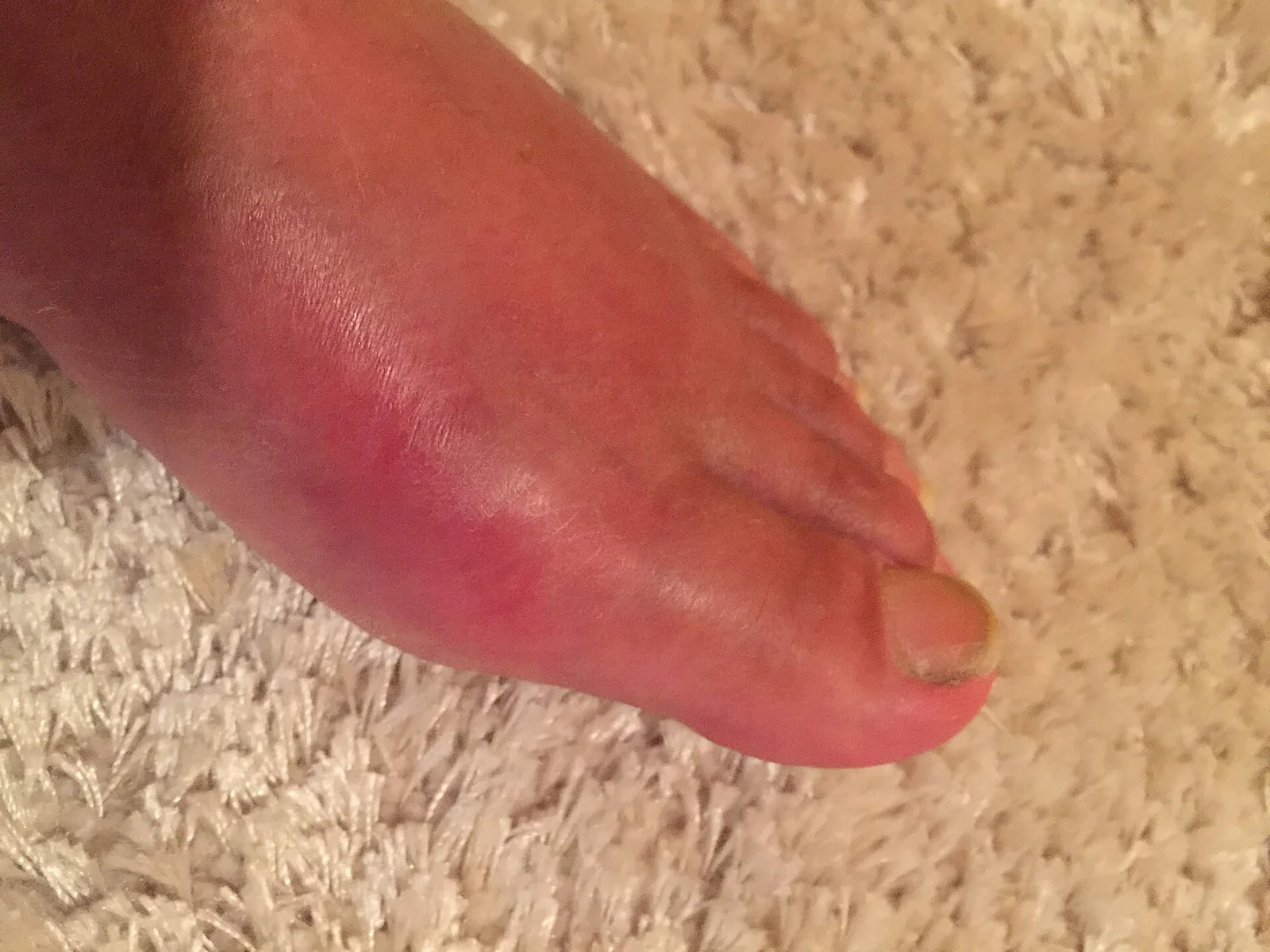
Long description
A clinical photograph of gout presenting in the metatarsophalangeal joint of the big toe, with redness of the skin overlying the joint.

An acute gout flare is a step-change in pain. The patient often wakes at night; the joint is, in the patient's words, "the worst pain I have ever had". It begins within hours and peaks within a day, with striking erythema and swelling, often of the first metatarsophalangeal joint (podagra). Lower-limb joints (ankle, mid-foot, knee) are the usual early targets. [4]
Other presentations:
- Monoarticular flare is classic; polyarticular flares occur, more often in postmenopausal women or long-standing disease. [4]
- Systemic features such as fever and malaise can mimic septic arthritis, so both can coexist in the same joint. [4]
- Intercritical flares may be followed by months or years of freedom, then recur.
- Chronic tophaceous gout shows persistent lumps, bony deformity and mechanical pain.
- Extra-articular crystal deposition includes olecranon bursitis, Achilles tenosynovitis and, clinically important, renal colic from uric acid stones. Patients on loop or thiazide diuretics may have diuretic-precipitated gouty bursitis. [4]
- Acutely: a hot, red, exquisitely tender joint with shiny overlying skin and swelling; range of motion is lost.
- Chronic gout: palpable tophi (chalky white material under skin, common at helix of ear, olecranon, fingers and toes), joint deformity and bony erosions with overhanging edges on examination/imaging.
- Tophi can be mistaken for rheumatoid nodules, Heberden/Bouchard nodes, lipomas or xanthomas; a needle aspirate showing MSU crystals confirms them. [4]
Examine for comorbidity as part of the joint consultation: blood pressure, body mass index, signs of chronic kidney disease, hypertension and dyslipidaemia, because these drive treatment choices. [1][4]
The gold standard is polarised light microscopy of synovial fluid or tophus aspirate showing MSU crystals. [4][5] In practice, diagnosis is often clinical with a compatible history plus high serum uric acid, but aspiration should be performed whenever septic arthritis is possible or the presentation is atypical. [4]
Synovial fluid is examined as soon as possible (ideally within 6 hours, or kept at 4 degrees Celsius for up to 24 hours). Crystals are visible with ordinary light at 600x to 1000x magnification; compensated polarised light improves detection and lets you read birefringence. [4]
| Test | What it measures or shows | Key number or reading |
|---|---|---|
| Serum uric acid (SUA) | Baseline and target | Solubility/threshold about 6.8 mg/dL (405 micromol/L). Hyperuricaemia usually uses this. [4] |
| SUA target on ULT | Cure-oriented goal | Below 6 mg/dL (360 micromol/L); below 5 mg/dL (300 micromol/L) in severe/tophaceous gout. [2] |
| SUA during a flare | Not a diagnostic test | Often falls during an acute flare; a normal value does not exclude gout. [4] |
| Annual risk from hyperuricaemia | Context only | With SUA 7.0 to 8.9 mg/dL about 0.5% develop gout per year; above 9 mg/dL about 4.9% per year (about 22% over 5 years). [7] [4] |
| Synovial fluid crystal microscopy | Gold standard | MSU crystals: needle-shaped, strongly negatively birefringent (yellow when parallel to the compensator axis). CPP crystals: often short or rhomboid, weakly positively birefringent (blue). [4] |
| Synovial fluid leukocyte count | Exclude sepsis | Can exceed 50,000 cells per microlitre in an acute flare; gout usually has normal glucose, unlike septic arthritis. [4] |
| Synovial fluid culture and Gram stain | Exclude sepsis | Always send when septic arthritis is in the differential; co-infection happens. [4] |
| Renal function (eGFR, creatinine) | Dosing safety | CKD stage 3 or worse is an indication to start ULT; it also limits colchicine and NSAID use. [1] |
| 24-hour urinary uric acid | Production vs excretion | Above 800 mg per 24 h suggests overproduction and favours a xanthine oxidase inhibitor, not a uricosuric, plus stone precautions. [4] |
| Plain radiograph (CR) | Chronic damage | In 10 or more years, or high burden; sensitivity about 31% for gout, specificity about 93%. Tophi, punched-out erosions with overhanging edges, preserved joint space early. [4] |
| Ultrasound (US) | Crystal deposits | Double contour sign on hyaline cartilage; hyperechoic tophus aggregates; more sensitive than plain radiograph for early deposits and erosions. [4][5] |
| Dual-energy CT (DECT) | Urate mapping | Colour-codes urate deposits, including subclinical ones; highly specific but less available. [4] |
| Advanced imaging (CT, MRI) | All presentations, staging | Best for unusual sites and for quantifying tophi; not first-line for a simple flare. [4] |
| Condition | What separates it |
|---|---|
| Septic arthritis | Fever/systemic illness; crystals do not exclude infection; Gram stain and culture; therapeutic aspiration and antibiotics needed. [4] |
| Pseudogout (CPPD) | See table below; crystals are CPPD, not MSU. [5] |
| Rheumatoid arthritis | Prolonged morning stiffness, symmetric small-joint swelling, rheumatoid factor/anti-CCP, not single explosive self-limited flares. [4] |
| Osteoarthritis | Age-related, mechanical, bony nodes, no crystals unless mixed CPPD; gout worsens at OA joints. [4] |
| Psoriatic or reactive arthritis | Enthesitis, dactylitis, skin/nail disease or genitourinary/gut infection link; different crystal result. [4] |
| Gout with mixed crystals | Both MSU and CPP crystals can coexist in the same fluid; report both. [5] |
| Feature | Gout | Pseudogout (CPPD) |
|---|---|---|
| Crystal | Monosodium urate (MSU) | Calcium pyrophosphate (CPP) |
| Shape | Needle-like | Short, rhomboid or parallelepiped |
| Birefringence | Strongly negative | Weakly positive |
| Colour with compensator | Yellow when parallel to axis | Blue when parallel to axis |
| Typical joint | First MTP, ankle, knee, mid-foot | Knee, wrist, shoulder (often knees) |
| Cartilage calcification | Not typical | Chondrocalcinosis common |
| Risk setting | Hyperuricaemia, male, CKD, diuretics | Age, OA, hyperparathyroidism, haemochromatosis, hypomagnesaemia |
| Acute treatment | NSAIDs, colchicine, steroids, IL-1 blockade | NSAIDs, colchicine, steroids; no ULT; treat underlying metabolic cause if any |
| Chronic strategy | Urate lowering to target | No crystal-dissolving drug; manage symptoms and OA |
Approach to a suspected gout flare
- Acute hot swollen jointPain peaking within hours, redness, tenderness; lower limb common
- Is septic arthritis possible?Fever, systemic illness, immunocompromise, atypical joint or first episode
- Question: Aspirate the joint (synovial fluid)
Crystals found
- MSU crystalsNeedle-shaped, negatively birefringent: acute gout
- CPP crystalsShort, positively birefringent: pseudogout
- Both presentMixed crystal arthritis: treat and investigate both
No crystals
- Consider septic arthritis, trauma, OA, RA, reactive or psoriatic arthritisCulture and treat if infection cannot be excluded
- Treat the flare earlyNSAID at full dose, or colchicine 1.8 mg (1.2 mg then 0.6 mg after 1 hour), or prednisone 30 mg daily for 7 days, or intra-articular steroid. Combine only in severe disease.
- Review comorbidity before choosing the drugCKD limits NSAIDs and colchicine; diabetes or hypertension make steroids unattractive; check drug interactions
- When ULT is indicated, start low and go slowAllopurinol 100 mg (50 mg if CKD stage 3 or worse) with titration toward SU below 6 mg/dL; prophylaxis colchicine for 3 to 6 months
The named guidelines used here are the ACR 2020 gout guideline, the EULAR 2016 updated recommendations (published 2017), and the BSR 2017 guideline. [1][2][3] They agree on treat-to-target and allopurinol first line, and differ in detail: for example, on whether colchicine should be used within 12 (EULAR) or 24 (ACR) hours of flare onset, and on how aggressively to lower urate in severe gout. [4] A patient leaflet and lifestyle advice accompany drug therapy in all three.
Start treatment early; all flare drugs work much better in the first 12 to 24 hours. [4] Choose one, or combine in severe disease. [4]
- NSAIDs (or COX-2 inhibitors) at full analgesic dose, with proton pump inhibitor cover when indicated. Efficacy is well accepted although placebo-controlled trials are lacking. [4]
- Colchicine. When taken within 12 hours of onset, 1.8 mg total (1.2 mg then 0.6 mg one hour later) is as effective as older high-dose regimens. [4] It has a narrow therapeutic window; diarrhoea, nausea or vomiting are usually the first toxic signs and should prompt dose reduction or stopping. [4] Renal dosing differs by source: Ragab 2017 (Ref 4) advises 0.5 to 0.6 mg daily for eGFR 30 to 60 and 0.5 to 0.6 mg every 2 to 3 days for eGFR 15 to 30, with avoidance in stage 5 CKD; the UK SmPC contraindicates severe renal impairment and advises caution and dose reduction in moderate impairment; the US product label does not require adjustment in mild to moderate impairment and uses reduced starting doses with monitoring in severe impairment and dialysis. [4][9] Reduce in hepatic impairment. It interacts with CYP3A4 and P-glycoprotein inhibitors (macrolides, azole antifungals, cyclosporin, diltiazem, verapamil), which raises toxicity; muscle and nerve toxicity can occur with long-term low-dose use in CKD. [4][1]
- Glucocorticoids. Oral prednisone 30 mg daily for 7 days is effective and is recommended by ACR and EULAR as a first-line option, especially in CKD or where NSAIDs/colchicine are unsuitable; intra-articular steroid injection is highly effective for mono- or oligo-articular flares. [2][4] Steroids can worsen hypertension and diabetes. [4] [1]
- IL-1 blockers are reserved for patients with frequent flares who cannot tolerate colchicine, NSAIDs or steroids. [2] They are not first-line.
- During the flare: keep the joint rested, use ice, analgesia and fluids; aspiration rules out sepsis. [4]
Pharmacologic urate-lowering therapy is strongly recommended for: any subcutaneous tophus, radiographic damage attributable to gout, or frequent flares defined as 2 or more per year. It is conditionally recommended for more than 1 prior flare with infrequent flares (fewer than 2 per year), and conditionally recommended against after a single uncomplicated flare, except when CKD stage 3 or worse, serum uric acid above 9 mg/dL, or urolithiasis is present. [1]
Choice of agent. Allopurinol is the preferred first-line agent, including in moderate-to-severe CKD, because of efficacy at appropriate doses, safety and cost. [1] Start at 100 mg daily or less, and 50 mg daily or less in CKD stage 3 or worse, then titrate. [1] It is metabolised to oxypurinol and can reach a maximum FDA-approved dose of 800 mg daily. [1] Testing for HLA-B*5801 is conditionally recommended before starting allopurinol in patients of Southeast Asian descent (Han Chinese, Korean, Thai) and in African American patients, in whom prevalence is 7.4% and 3.8% respectively versus 0.7% in white and Hispanic patients, because of the risk of allopurinol hypersensitivity syndrome. [1]
Febuxostat is an alternative xanthine oxidase inhibitor. Start at 40 mg daily or less and titrate. [1] Switch patients with a cardiovascular history or a new cardiovascular event to an alternative agent where possible, because of the FDA boxed warning and the CARES trial signal (cardiovascular death and all-cause mortality). [1] The absolute cardiovascular risk attributable to febuxostat remains uncertain. [1]
Uricosurics (probenecid) are an option when a second drug is needed; start at 500 mg once to twice daily. [1] Alkalinising urine is not recommended, but adequate hydration and counselling about stones are. [1] Switching to a second xanthine oxidase inhibitor is conditionally preferred over adding a uricosuric when the first has failed. [1]
Treat to target. Use serial serum uric acid measurements to guide dose titration rather than fixed doses, toward a serum uric acid below 6 mg/dL, and below 5 mg/dL in severe or tophaceous disease. [1][2] This nurse- or pharmacist-led augmented care improves adherence and outcomes. [1] Continuing therapy indefinitely is conditionally recommended over stopping, even in clinical remission. [1]
Pegloticase is strongly recommended against as first line, and strongly recommended (over continuing current therapy) for refractory disease after xanthine oxidase inhibitors, uricosurics and other options have failed, in patients with frequent flares or non-resolving tophi; it is strongly recommended against where flares are infrequent and there are no tophi. [1]
Prophylaxis against flares on starting ULT (colchicine, NSAID or prednisone/prednisolone, chosen by patient factors) is strongly recommended for 3 to 6 months, with reassessment. [1] Starting ULT during a flare (rather than waiting) is conditionally recommended. [1]
For asymptomatic hyperuricaemia (serum uric acid 6.8 mg/dL or higher with no prior flare or tophus), pharmacologic ULT is conditionally recommended against, even when MSU deposition is seen on imaging, because the absolute risk of progression is low. [1] (Urate 9 mg/dL over 5 years progresses to gout in only about 20% of people. [1]) Lifestyle advice still applies. [1]
- Educate the patient and provide a written flare plan; self-treatment of early flares is encouraged. [2][4]
- Reduce purine-rich foods (especially animal and seafood sources), limit alcohol (beer worst, spirits less, wine least), and favour weight loss, exercise and waist reduction. [4]
- Favour high vitamin C and low-fat dairy, vegetables and plant oils; these do not need restriction. [4]
- For medication-related hyperuricaemia, when feasible switch hydrochlorothiazide to another antihypertensive, prefer losartan, do not stop low-dose aspirin taken for a correct indication, and do not add fenofibrate for gout. [1]
- Treat comorbidities (hypertension, diabetes, hyperlipidaemia, chronic kidney disease, obesity) aggressively; they matter as much as the gout. [1][4]
- Septic arthritis in a gouty joint (co-infection occurs; aspirate and culture). [4]
- Renal colic and uric acid stones from high urinary urate; stone prophylaxis with hydration and stone-directed therapy. [4]
- Joint destruction, tophi and deformity from uncontrolled disease. [4]
- Erosions with characteristic overhanging edges on radiograph. [4]
- Allopurinol hypersensitivity syndrome (serious and potentially fatal) - mitigated by low starting dose and HLA-B*5801 screening in Asian and African American patients. [1]
- Colchicine toxicity, especially with renal failure or interacting drugs. [4][1]
- Cardiovascular implications of febuxostat (boxed warning). [1]
With consistent treat-to-target therapy the outlook is excellent: flares stop, tophi dissolve, radiographic progression slows. [1][2] About 13% of patients in stable remission whose serum uric acid stayed below 7 mg/dL without therapy had no flares over 5 years, which argues for continuing therapy in most patients. [1] Flare frequency and tophus burden are the clinically important outcomes that guide intensity of therapy. [1][2]
There is no population screening test for gout. Prevention means identifying hyperuricaemia risk factors: family history, renal impairment, medication review, diet, alcohol, obesity and metabolic syndrome. [4][1] Patients with gout should be monitored for CKD and cardiovascular risk. [1][4] Low vitamin C and low-fat dairy intake, weight control and avoiding excess beer and spirits modify risk. [4]
Pregnancy. Gout in pregnancy is uncommon and needs specialist care; decisions about flares and urate-lowering therapy should be made jointly with obstetrics and rheumatology, following local guidance. [1]
Children and adolescents. Gout is rare in children and, when it occurs, suggests an inborn error of purine metabolism; tophi in a child or young adult should prompt a search for familial disease, and renal/genetic evaluation. [4]
Older adults. Chronic kidney disease, polypharmacy, diuretic use and under-nutrition are common; colchicine and NSAID doses must be reduced, and drug interactions checked; tophi may be long-standing. [1][4]
Renal impairment. Allopurinol remains first line but starts lower and titrates cautiously; colchicine and NSAIDs are restricted; febuxostat has boxed CV concerns. [1][4]
Where joint aspiration is unavailable, use clinical features plus serum uric acid, and treat the flare empirically while considering sepsis. Crystal confirmation and radiographs may be unavailable; an ultrasound (double contour sign, tophus aggregates) and basic biochemistry often suffice. Colchicine is inexpensive but has a narrow safety window; low-dose regimens are safer and effective. [1][4]
- Gout is inflammatory crystal arthritis from MSU deposition driven by chronic hyperuricaemia, with stages from asymptomatic hyperuricaemia to chronic tophaceous gout.
- Male sex, age, obesity, CKD, diuretics and purine-rich diet raise risk; most gout is from under-excretion, not overproduction.
- Hyperuricaemia thresholds start at 6.8 mg/dL; crystals form in cooler, more acidic, poorly vascular peripheral joints.
- A flare is a neutrophilic burst from lysosomal damage and NLRP3/IL-1 activation by MSU crystals; it is self-limited but best treated early.
- Podagra is classic; tophi, erosions and joint damage occur if untreated; septic arthritis and gout can coexist.
- Diagnosis rests on polarised light microscopy of synovial fluid: needle-shaped, negatively birefringent MSU crystals.
- Serum uric acid supports but does not prove gout; it drops during flares; high urinary uric acid above 800 mg/24 h suggests overproduction.
- Treat flares early with an NSAID, low-dose colchicine (1.8 mg total) or a steroid (prednisone 30 mg for 7 days); add IL-1 blockade for refractory cases.
- Long-term treat to a serum uric acid below 6 mg/dL (below 5 mg/dL in severe disease) with allopurinol first line, starting low and titrating, plus 3 to 6 months of prophylaxis.
- Allopurinol, including in CKD; screen for HLA-B*5801 in Southeast Asian and African American patients; febuxostat has cardiovascular cautions; pegloticase is last resort.
- Pseudogout (CPPD) is a separate crystal arthritis from positively birefringent CPP crystals, managed without urate lowering; look for OA, hyperparathyroidism, haemochromatosis, hypomagnesaemia.
- Follow ACR 2020, EULAR 2016 and BSR 2017 principles: treat to target, shared decisions, patient education and comorbidity care.
Answer each one in your head first, then open the answer.
A 54-year-old man wakes at 03:00 with a red, hot, exquisitely tender first MTP joint. Synovial fluid shows needle-shaped crystals that are strongly negatively birefringent. What is the diagnosis and first management step?
Show answerHide answer
Why can serum uric acid be normal during an acute gout flare, and what is the gold-standard diagnostic test?
Show answerHide answer
Uric acid falls slightly during an acute attack. The gold standard is polarised light microscopy of synovial fluid showing MSU crystals (needle-shaped, strongly negatively birefringent). [4]
A patient with CKD stage 3 wants urate-lowering therapy. Which drug is preferred, how is it started and what is the renal dosing caution for colchicine?
Show answerHide answer
Synovial fluid shows short, rhomboid crystals that are weakly positively birefringent. Which condition is this and what underlying causes should you seek?
How do the named guidelines differ on serum uric acid targets?
A tophus is found in a 22-year-old. What should you consider and how will you confirm it?
Show answerHide answer
Consider familial or early-onset gout (inborn errors of purine metabolism); confirm by aspirating the tophus and demonstrating MSU crystals. [4]
Plain text placeholders until topics are verified: rheumatoid arthritis; osteoarthritis; septic arthritis; urinary stone disease; chronic kidney disease; metabolic syndrome; hyperparathyroidism; haemochromatosis.
- FitzGerald JD, Dalbeth N, Mikuls T, et al. 2020 American College of Rheumatology Guideline for the Management of Gout. Arthritis Care Res (Hoboken) 2020;72(6):744-760. Accessed .
- Richette P, Doherty M, Pascual E, et al. 2016 updated EULAR evidence-based recommendations for the management of gout. Ann Rheum Dis 2017;76(1):29-42. Accessed .
- Hui M, Carr A, Cameron S, et al. The British Society for Rheumatology Guideline for the Management of Gout. Rheumatology (Oxford) 2017;56(7):e1-e20. Accessed .
- Ragab G, Elshahaly M, Bardin T. Gout: An old disease in new perspective - A review. J Adv Res 2017;8(5):495-511. Accessed .
- Wu Y, Chen K, Terkeltaub R. Systematic review and quality analysis of emerging diagnostic measures for calcium pyrophosphate crystal deposition disease. RMD Open 2016;2(2):e000339. Accessed .
- Kelley N, Jeltema D, Duan Y, He Y. The NLRP3 Inflammasome: An Overview of Mechanisms of Activation and Regulation. Int J Mol Sci 2019;20(13):3328. Accessed .
- Campion EW, Glynn RJ, DeLabry LO. Asymptomatic hyperuricemia. Risks and consequences in the Normative Aging Study. Am J Med 1987;82(3):421-426. Accessed .
- Yokose C, McCormick N, Lu N, et al. Trends in Prevalence of Gout Among US Asian Adults, 2011-2018. JAMA Netw Open 2023;6(4):e239501. Accessed .
- Wockhardt UK Ltd. Colchicine 500 micrograms Tablets. Summary of product characteristics. emc (medicines.org.uk), updated 31 July 2025. Accessed .
Figure credits
Educational content for medical students and doctors. It is not a substitute for clinical judgement or local guidelines.