Spondyloarthritis and ankylosing spondylitis
Chronic inflammatory spondyloarthritides: classification, HLA-B27 biology, clinical features, BASDAI cut-offs, NSAID strategy, TNF and IL-17 inhibitors per.
- Spondyloarthritis is a family of interrelated inflammatory diseases whose hallmark is enthesitis and axial or peripheral arthritis in a seronegative (rheumatoid-factor-negative) host; ankylosing spondylitis is the form with radiographic sacroiliitis and spinal involvement.
- It usually starts as inflammatory back pain in a person younger than 45 years, with stiffness that improves with movement and worsens with rest.
- HLA-B27 is strongly associated with the disease, but it is neither required for the diagnosis nor sufficient on its own.
- Plain films of the sacroiliac joints diagnose ankylosing spondylitis; magnetic resonance imaging detects the earlier, non-radiographic stage.
- Non-steroidal anti-inflammatory drugs are first-line treatment; tumour necrosis factor inhibitors are the usual first biologic, followed by interleukin-17 inhibitors when a first tumour necrosis factor inhibitor fails [2] [3].
By the end of this page you should be able to:
- Classify the spondyloarthritides by their predominant axial or peripheral pattern and name the members of the family.
- Use the modified New York criteria and the ASAS axial classification criteria, including their imaging and clinical arms, with their stated sensitivities and specificities.
- Explain inflammatory back pain, list the ASAS definition, and distinguish it from mechanical back pain.
- Perform and interpret the Schober test and other spinal mobility measures.
- List the typical laboratory, radiographic, and magnetic resonance findings and the cut-offs that separate ankylosing spondylitis from non-radiographic axial disease.
- Outline stepwise pharmacological management, with the relevant doses, according to the 2019 ACR/SAA/SPARTAN and NICE NG65 recommendations.
- Choose an appropriate biologic for a patient with axial disease, including comorbid iritis or inflammatory bowel disease, and know the ACR 2019 sequencing rules.
- Recognise extra-articular disease, complications, and the factors that determine prognosis.
DefinitionSpondyloarthritis
A group of inflammatory rheumatic diseases that are seronegative (rheumatoid factor negative) and share clinical features: enthesitis, axial or peripheral arthritis, and characteristic extra-articular disease (anterior uveitis, psoriasis, inflammatory bowel disease), together with a strong association with the HLA-B27 allele. "Axial spondyloarthritis" denotes predominant spine and sacroiliac joint disease; "peripheral spondyloarthritis" denotes predominant joint, tendon, and digit disease.
Spondyloarthritis may be predominantly axial or peripheral, and the two patterns often coexist. Under the NICE NG65 framework the family includes ankylosing spondylitis (radiographic axial spondyloarthritis), non-radiographic axial spondyloarthritis, psoriatic arthritis, reactive arthritis, and enteropathic spondyloarthritis [3]. Axial disease usually begins in late adolescence or early adulthood and, when it is allowed to progress, produces sacroiliitis and spinal new bone formation; peripheral disease preferentially involves the lower limbs and the insertions of tendons and ligaments.
In axial disease the radiographic stage, historically called ankylosing spondylitis, is reached when plain films show sacroiliitis meeting the modified New York (mNY) criteria: bilateral grade 2 to 4 sacroiliitis, or unilateral grade 3 to 4 sacroiliitis [3]. Patients with the same clinical picture but without those radiographic changes are classified as having non-radiographic axial spondyloarthritis. In cohort studies the two stages share similar age at onset, prevalence of HLA-B27, peripheral and extra-articular manifestations, and disease activity [1]. The prevalence of non-radiographic disease is comparable with that of ankylosing spondylitis at roughly 0.3 to 0.6% of the population [1]; about 12% of patients with non-radiographic disease progress to ankylosing spondylitis over 2 years, with elevated C-reactive protein (CRP) and active sacroiliitis on magnetic resonance imaging (MRI) as the strongest predictors [1]. [5]
The ASAS classification criteria are the most widely used set for axial disease. In a patient with chronic back pain beginning before age 45, the criteria are fulfilled if there is either:
- Imaging arm: definite radiographic sacroiliitis (modified New York grade) or active sacroiliitis on MRI plus at least one other spondyloarthritis feature, or
- Clinical arm: HLA-B27 positivity plus at least two other spondyloarthritis features (inflammatory back pain, arthritis, enthesitis, uveitis, dactylitis, psoriasis, inflammatory bowel disease, good response to non-steroidal anti-inflammatory drugs (NSAIDs), elevated CRP, family history of spondyloarthritis).
The criteria have a sensitivity of 82.9% and a specificity of 84.4% overall, with 66.2% sensitivity and 97.5% specificity for the imaging arm alone [1]. The original European Spondyloarthropathy Study Group (ESSG) and Amor criteria remain valid historical alternatives, but the ASAS criteria better capture early disease because they incorporate MRI. Classification criteria are not diagnostic criteria; they should be applied to patients in whom the disease is already plausible, not used as a checklist on otherwise unexplained back pain [1].
For non-specialist recognition, NICE NG65 offers a referral algorithm. A person with low back pain that began before 45 years and has lasted longer than 3 months should be referred to rheumatology if at least four of the following additional criteria are present: back pain beginning before 35 years, waking in the second half of the night, buttock pain, improvement with movement, improvement within 48 hours of NSAIDs, a first-degree relative with spondyloarthritis, current or past arthritis, current or past enthesitis, or current or past psoriasis. If exactly three criteria are present, HLA-B27 testing is recommended, with referral if it is positive [3].
Ankylosing spondylitis affects roughly 1 in 200 people, and axial spondyloarthritis overall affects about 1 in 100 [5]. A population survey in the United States estimated axial disease at 0.9 to 1.4% of adults [5]. In Europe and Asia the number of ankylosing spondylitis cases is estimated at 1.30 to 1.56 million and 4.63 to 4.98 million respectively [5]. Disease onset is usually before age 45, often in the late teens or twenties, and men are affected roughly twice as often as women in radiographic disease, whereas non-radiographic disease affects men and women in roughly equal proportions, with some sources describing a slight female preponderance [4] [5].
HLA-B27 is present in the general population in up to 8% [5], but in ankylosing spondylitis it is found in 85 to 95% of patients of white and Han Chinese ancestry, over 86% of Hispanic patients, and roughly 50 to 84% of patients in the Middle East, North Africa, and Black American populations [5]. Only about 5% of HLA-B27-positive carriers develop spondyloarthritis [5], which is why screening carriers in the general population is not useful. Overall, HLA-B27 is found in about 90% of patients of European ancestry with ankylosing spondylitis, 60% with psoriatic arthritis, 70% with reactive arthritis, and 70% with the spondylitis associated with inflammatory bowel disease [6].
A family history carries substantial risk. Genetic factors account for over 90% of the population variance in ankylosing spondylitis, although HLA-B27 itself contributes only about 20% of that heritable risk [4] [5], and twin concordance is 63% in monozygotic twins and between 23% and 27% in dizygotic twins (27% in Hwang 2021, 23% in Zhu 2019) [4] [5]. The relative risk of disease is about 94, 25, and 4 for first-, second-, and third-degree relatives respectively [4]. In an HLA-B27-positive population the risk of disease increases from about 1 to 2% to about 15 to 20% when a first-degree relative is affected [4].
Genetic background dominates. HLA-B27 contributes roughly 20% of heritable risk and defines many of the mechanistic hypotheses discussed below. Over 100 non-HLA loci are now associated with disease, including endoplasmic reticulum aminopeptidase 1 (ERAP1), interleukin-23 receptor (IL-23R), RUNX3, and T-box transcription factor (TBX21), several of which are shared with psoriasis, Crohn disease, and ulcerative colitis, supporting the idea of a common immunological substrate across the family [5]. [4]
Non-genetic influences matter too, even though they are less well quantified. Asymptomatic intestinal inflammation, usually involving the terminal ileum, occurs in 57 to 70% of patients with ankylosing spondylitis, suggesting that gut mucosal immune activation is not an occasional accompaniment but a routine feature. Subclinical bowel inflammation is seen in up to half of patients with ankylosing spondylitis in other series [6]. In Western populations 5 to 10% of patients with ankylosing spondylitis present with inflammatory bowel disease, whereas in East Asian cohorts the figure has been as low as 0.4% [4]. In a Swedish case-control study, respiratory tract infections and tonsillitis in childhood increased later risk, while appendicitis was associated with a decreased risk, and mechanically demanding occupations were associated with more radiographic progression. [5]
Enthesitis is the primary lesion. At tendon and ligament insertions, chronic inflammation brings CD4-positive and CD8-positive T lymphocytes and macrophages, with tumour necrosis factor alpha (TNF-alpha) and transforming growth factor beta (TGF-beta) driving the triad of inflammation, fibrosis, and ossification that eventually produces syndesmophytes and, in late disease, ankylosis. This is why every clinically useful therapy either suppresses inflammation or targets the cytokines produced at the enthesis.
HLA-B27 contributes through several incompletely understood routes [5]:
- Arthritogenic peptide presentation: an HLA-B27-bound self or microbial peptide may be recognised by CD8-positive T cells and trigger autoimmunity.
- Protein misfolding: HLA-B27 heavy chains fold slowly, accumulate in the endoplasmic reticulum, induce cellular stress, and activate the interleukin-23 and interleukin-17 axis.
- Homodimer formation: free HLA-B27 heavy-chain homodimers on the cell surface are recognised by killer immunoglobulin-like receptors on natural killer and CD4-positive T cells, promoting survival and differentiation of T helper 17 (Th17) cells.
- Microbial interactions: commensal gut bacteria are required for disease expression in HLA-B27-transgenic animal models; dysbiosis is present in patients.
- Altered intracellular killing of arthritogenic bacteria, particularly in reactive arthritis.
HLA-B27 is therefore neither necessary nor sufficient: 10% or more of ankylosing spondylitis cases occur without it. Its classical association is with anterior uveitis: patients who are HLA-B27 positive have earlier-onset disease and a higher prevalence of uveitis [4]. [5]
The interleukin-23 and interleukin-17 axis is the other dominant pathway. Genome-wide studies linked IL-23R to disease risk, and patients show raised levels of interleukin-23 and interleukin-17 with Th17 cells present in facet joints. Differentiated T cells release interleukin-17 (activating osteoclasts and favouring erosion) and interleukin-22 (stimulating osteoproliferation), which helps to explain the paradoxical coexistence of erosion and new bone formation in the same spine. TNF-alpha blockade and interleukin-17 blockade both produce clinical and imaging responses because they interrupt this cascade at different points [4].
The hallmark is inflammatory back pain: chronic pain in the lumbar region or buttocks lasting more than 3 months, with onset before age 45 and an insidious course. Characteristically the pain improves with movement and exercise, does not improve with rest, wakes the patient in the second half of the night, and improves within 48 hours of taking NSAIDs. Morning stiffness lasts longer than 30 minutes. Alternating buttock pain is typical. A widely used simple rule is that back pain lasting at least 3 months combined with two of four features (morning stiffness of at least 30 minutes, improvement with exercise but not rest, alternating buttock pain, awakening in the second half of the night) separates inflammatory from mechanical pain with a sensitivity of 70.3% and specificity of 81.2% [6]. The ASAS expert definition of inflammatory back pain refines these questions for use in classification; it is intended for patients in whom axial spondyloarthritis is already plausible rather than as a self-diagnosis tool [1].
Because the pain is often worse at night, patients may leave the bed and walk around for relief. As disease progresses, patients notice loss of height, difficulty looking over the shoulder when driving, and a stooped posture. Fatigue is common. The pattern differs from mechanical back pain, where pain worsens with activity and with specific movements, is relieved by rest, and does not wake the patient at night.
Peripheral manifestations are common: an asymmetric oligoarthritis of the lower limbs (knees, ankles), usually affecting one or a few joints, is typical. Enthesitis, especially of the Achilles tendon and plantar fascia, and dactylitis ("sausage digits") are characteristic and often more disabling than the arthritis itself. Hip arthritis occurs in about 50% of patients and is associated with more severe disease overall [6]. Shoulder involvement and small-joint arthritis are less common.
Extra-articular disease is what usually brings the patient to a different specialty first:
- Anterior uveitis is the commonest extra-articular feature, present across the spondyloarthritis family. It presents as a painful red eye with photophobia, lacrimation, and blurred vision, can threaten vision, and always warrants same-day ophthalmological assessment [3] [6].
- Inflammatory bowel disease: Both Crohn disease and ulcerative colitis occur in roughly 5 to 10% of patients with ankylosing spondylitis in Western cohorts, up to 50% of which have subclinical bowel inflammation; flares of enteropathic peripheral arthritis track bowel activity, whereas axial disease does not [4] [6].
- Psoriasis is present in many patients and supports a diagnosis of psoriatic spondyloarthritis when found.
- Cardiac involvement (conduction abnormality, aortic regurgitation) and an upper-lobe-predominant interstitial lung pattern are uncommon but recognised complications.
- Osteoporosis and vertebral fragility fractures occur earlier and more often than in the general population, particularly in longstanding ankylosing spondylitis.
There is a recognisable sexual difference in presentation: men have earlier onset, worse radiographic damage, and more restricted spinal mobility, whereas women more often have peripheral arthritis, dactylitis, and enthesitis, and higher BASDAI scores with a lower response to tumour necrosis factor inhibitors [5].
Inspection should note loss of lumbar lordosis, a forward-stooped posture, a flattened sacral curve, and, in long-standing disease, a compensatory hyperextension of the neck. Palpation is unremarkable in early disease; the important findings are measures of spinal mobility and chest expansion.
Spinal mobility. The Schober test should increase by more than 5 cm when the patient bends forward to touch the toes; a change of less than 5 cm indicates restriction of lumbar flexion [6]. Chest wall expansion at the xiphisternal junction should be greater than 2.5 cm between maximal expiration and inspiration [6]. Occiput-to-wall distance is tested with the patient standing against a wall; the patient with normal spinal extension can touch the wall (0 cm distance). Lateral flexion should normally be greater than 10 cm between the starting and end positions of the third finger above the floor [6]. These measurements are combined in the Bath Ankylosing Spondylitis Metrology Index (BASMI), which scores cervical rotation, tragus-to-wall distance, lumbar flexion, lateral lumbar flexion, and intermalleolar distance and is used to follow spinal mobility [1].
Examination of the peripheral joints looks for asymmetric oligoarthritis of the large joints, heel enthesitis, Achilles tendon thickening, tender plantar fascia insertion, and dactylitis. Skin examination should include the scalp, retroauricular region, pinna, nails, and gluteal cleft for psoriasis and nail pitting, dystrophy, or onycholysis; nails are abnormal in 60 to 80% of patients with psoriatic arthritis, particularly when distal interphalangeal joints are involved [6]. Eye redness and anterior chamber activity are assessed in chronic uveitis. Palpation of the sacroiliac joints may reveal tenderness, though this is neither sensitive nor specific.
Cardiovascular and respiratory examination should include auscultation for aortic regurgitation and measurement of chest expansion; kyphotic deformity may cause a restrictive ventilatory defect. Neurological examination is mandatory when pain, weakness, or sphincter change develops in a rigid spine, because of the risk of fracture and cauda equina syndrome.
The differential is broad and one of the reasons for diagnostic delay. Mechanical low back pain is the most common alternative: it worsens with movement, improves with rest, lacks morning stiffness longer than 30 minutes, and does not wake the patient. Osteoarthritis of the spine produces stiffness in older patients and degenerative radiographic changes rather than sacroiliitis. Diffuse idiopathic skeletal hyperostosis (DISH, Forestier disease) creates flowing ossification of the anterior longitudinal ligament with preserved disc height and only minimal or absent sacroiliac change. Fibromyalgia can coexist with spondyloarthritis and amplify symptoms.
Rheumatoid arthritis usually affects middle-aged women with symmetrical small-joint polyarthritis, rheumatoid factor and anti-citrullinated protein antibody positivity, and neither sacroiliitis nor the enthesitic pattern of disease. Psoriatic arthritis produces oligoarthritis, distal interphalangeal involvement, dactylitis, nail changes, and often a family or personal history of psoriasis. Reactive arthritis follows genitourinary or gastrointestinal infection and presents with asymmetric large-joint oligoarthritis, often with conjunctivitis and urethritis. Enteropathic arthritis parallels inflammatory bowel disease; its peripheral form waxes and wanes with bowel activity, whereas the axial form does not. Infection (septic arthritis, brucellosis, tuberculosis of the spine) and spinal tumour or metastasis must be excluded in older or systemically unwell patients.
| Condition | Age and pattern | Key separating feature |
|---|---|---|
| Ankylosing spondylitis | Younger than 45; chronic inflammatory back pain | Sacroiliitis on plain film; HLA-B27 in 85-95% of white and Asian patients |
| Non-radiographic axial SpA | Same clinical pattern | No modified New York sacroiliitis; inflammation on MRI may be present |
| Mechanical back pain | Any age | Pain with exertion, relieved by rest; no morning stiffness beyond 30 min |
| Osteoarthritis of spine | Older adults | Degenerative change; sacroiliac joints spared |
| DISH | Older adults, often metabolic disease | Flowing anterior ossification; disc spaces preserved; no true sacroiliitis |
| Rheumatoid arthritis | Middle age, female predominance | Symmetrical small-joint polyarthritis; rheumatoid factor positive |
| Psoriatic arthritis | Middle age | DIP joint arthritis, nail change, current or past psoriasis |
| Reactive arthritis | Young adults, after infection | Asymmetric lower-limb oligoarthritis, conjunctivitis, urethritis |
| Enteropathic arthritis | Any age with IBD | Peripheral arthritis parallels bowel disease; axial disease independent |
| Spinal fracture or tumour | Older, often focal pain | Focal spinal tenderness, neurological deficit, weight loss |
Figure 1Bamboo spine ankylosing spondylitis
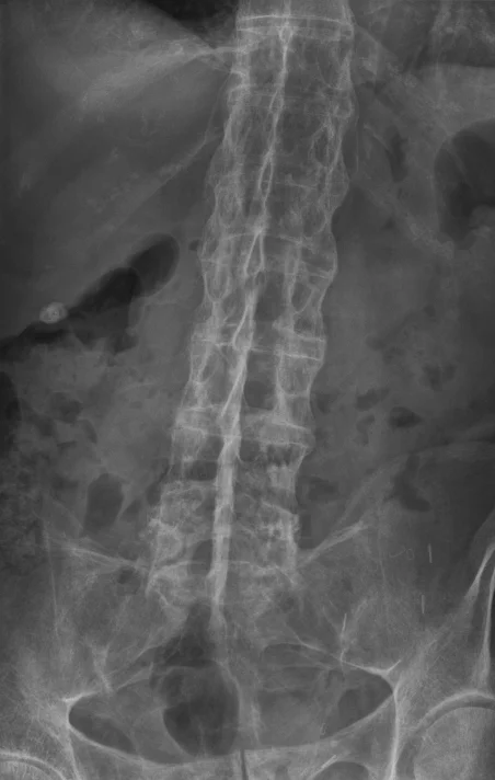
Long description
An X-ray showing bamboo spine in a patient with ankylosing spondylitis.

No single test is diagnostic. CRP and erythrocyte sedimentation rate (ESR) are often raised, but a normal result does not exclude spondyloarthritis [3].
- HLA-B27: increases the likelihood of disease in the right clinical setting; a negative result never excludes the diagnosis [3].
- Radiography: plain film of the sacroiliac joints is the first imaging study in suspected axial disease; ankylosing spondylitis is diagnosed when sacroiliitis meets the modified New York grade (bilateral 2 to 4, or unilateral 3 to 4). Spine films show shiny corners (Romanus lesions), squaring of vertebral bodies, syndesmophytes, and eventually a "bamboo spine" [3] [6].
- MRI: when plain films are normal, or in a patient whose skeleton is not fully mature, an unenhanced inflammatory back pain MRI with STIR and T1-weighted whole-spine sagittal and coronal-oblique sacroiliac sequences is requested. Active sacroiliitis is interpreted using the ASAS/OMERACT criteria (definitions of bone marrow oedema). If MRI is negative but clinical suspicion persists, the result does not exclude the disease, and a follow-up MRI, specialist musculoskeletal radiology review, or HLA-B27 testing can be used [3].
- Other imaging: ultrasound and MRI are used for peripheral joints and enthesitis; scintigraphy is not recommended [3].
- Laboratory safety checks: before starting a biologic, screen for tuberculosis and hepatitis B and C, check full blood count and renal and hepatic function, and update immunisations.
| Test or measure | What it shows | Quoted cut-off or interpretation |
|---|---|---|
| Modified Schober test | Lumbar flexion by tape measure | Increase greater than 5 cm normal; less than 5 cm suggests restricted flexion [6] |
| Chest wall expansion | Costovertebral function | Greater than 2.5 cm normal |
| Occiput-to-wall distance | Spinal extension | 0 cm normal; any distance abnormal |
| Lateral flexion | Lateral spinal mobility | Greater than 10 cm normal |
| ESR / CRP | Non-specific inflammation | Often elevated; normal results do not exclude disease |
| HLA-B27 | Genetic susceptibility | Present in 85-95% of white and Asian patients; negative in at least 10% of patients |
| Plain film sacroiliac joints | Structural sacroiliitis | Modified New York grade: bilateral 2-4 or unilateral 3-4 |
| MRI sacroiliac joints / spine | Active and structural change | ASAS/OMERACT MRI criteria for active sacroiliitis |
| BASDAI | Patient-reported disease activity | Scale 0-10; a score of 4 or more supports eligibility for biologic therapy under NICE |
| Spinal VAS | Axial pain severity | 4 cm or more on a 10 cm visual analogue scale |
| BASFI | Function | Higher scores indicate worse function; used to monitor change over time |
| ASDAS | Composite disease activity | Less than 2.1 low disease activity in the trial definitions used; higher values indicate active disease |
Staging is mostly clinical and radiographic: early inflammatory back pain and normal or equivocal films (non-radiographic axial disease), definite modified New York sacroiliitis (radiographic ankylosing spondylitis), and late ankylosis with syndesmophytes and spinal fusion. Severity measures rather than stages guide treatment:
- BASDAI (Bath Ankylosing Spondylitis Disease Activity Index) is a 0-to-10 score of pain, stiffness, fatigue, and axial discomfort.
- BASFI (Bath Ankylosing Spondylitis Functional Index) measures everyday function.
- ASDAS combines patient measures with CRP; it is the preferred outcome measure in treat-to-target studies because it includes an objective marker.
- BASMI measures spinal mobility as described above.
Under NICE NG65, active ankylosing spondylitis is assumed to qualify for the biologic medicines considered in the technology appraisals when the BASDAI score is 4 or more and the spinal visual analogue scale is 4 cm or more and NSAIDs have not controlled the disease or are not tolerated [3].
The goals are to control pain and stiffness, preserve spinal mobility and function, prevent spinal complications, and treat extra-articular disease. Because structural damage is only partly reversible, early diagnosis matters more here than in almost any other arthritis.
Every patient should be referred to a specialist physiotherapist for a structured, individualised exercise programme covering stretching, strengthening, postural and extension exercises, spinal range-of-motion work in the lumbar, thoracic and cervical spine, deep breathing, and aerobic conditioning [3] [7]. Hydrotherapy is a useful adjunct. Patients should sleep on a firm mattress with a thin or no pillow, avoid prolonged flexed postures such as low chairs and heavy lifting, and perform daily home exercises; people who exercise 200 minutes per week report less pain and disability than those who exercise less [6]. Smoking cessation should be advised because smoking is associated with more radiographic progression. Surgery is reserved for advanced hip arthritis (total hip arthroplasty), severe kyphosis (corrective osteotomy only in selected cases), and spinal deformity that is severe or progressing despite optimal non-surgical management [2]. Spinal manipulation is strongly discouraged in patients with spinal fusion or advanced spinal osteoporosis [2].
NSAIDs are the cornerstone of treatment (strongly recommended over no NSAID) [2] [3]. Both NICE NG65 and the 2019 ACR/SAA/SPARTAN guideline, like the ASAS-EULAR 2016 management recommendations before them, strongly recommend an NSAID over no NSAID [1] [2] [3]. NICE advises the lowest effective dose with appropriate clinical assessment, monitoring of risk factors, and gastroprotective treatment where indicated, while the ACR conditionally favours continuous over on-demand use in active disease and on-demand use once disease is stable [2] [3]. No single NSAID is favoured over another, including in patients with inflammatory bowel disease, where the risk of worsening bowel symptoms has not been shown to differ between agents [2] [3]. If pain is not controlled at the maximum tolerated dose after 2 to 4 weeks, switching to another NSAID is appropriate [3]. Concomitant glucocorticoid use is not a substitute: systemic glucocorticoids are strongly recommended against, although local injected glucocorticoids can be useful for isolated sacroiliitis, enthesitis, or peripheral arthritis, with avoidance of peri-tendon injection at the Achilles, patellar, and quadriceps tendons [2].
When disease remains active despite NSAIDs, conventional synthetic disease-modifying drugs (sulfasalazine, methotrexate, leflunomide) are not effective for axial disease and are reserved for prominent peripheral arthritis or when biologics are unavailable [1] [2]. Sulfasalazine is recommended only for persistent peripheral arthritis when TNF inhibitors are contraindicated [2]. Methotrexate at conventional doses is not supported by evidence for axial disease, although it may be used for peripheral involvement. Both NICE and the ACR place biologics at the next step.
The biologic sequence is where the 2019 ACR/SAA/SPARTAN guidance is most explicit [2]:
- Tumour necrosis factor inhibitors (TNFi) are recommended over secukinumab or ixekizumab as the first biologic and over tofacitinib. [2]
- TNFi, secukinumab, and ixekizumab are all recommended over no biologic, and all three are favoured over tofacitinib.
- Secukinumab or ixekizumab is recommended over tofacitinib.
- In patients with active disease despite a first TNFi, secukinumab or ixekizumab is recommended over a second TNFi when the first was a primary non-response; a different TNFi is recommended over a different biologic class when the first produced a secondary non-response.
- Switching to the biosimilar of the first TNFi, or adding low-dose methotrexate to a TNFi, is not recommended. [2]
- Biologics should not be discontinued or tapered as a standard approach, nor should a strict treat-to-target strategy with an ASDAS below 1.3 or 2.1 be mandated over physician assessment.
Tumour necrosis factor inhibitors approved for axial disease are infliximab, etanercept, adalimumab, golimumab, and certolizumab pegol. Typical adult doses are:
| Drug | Route | Typical adult dose |
|---|---|---|
| Adalimumab | Subcutaneous | 40 mg every 2 weeks [8] |
| Certolizumab pegol | Subcutaneous | 200 mg every 2 weeks (or 400 mg every 4 weeks), with standard loading as per label [8] |
| Etanercept | Subcutaneous | 50 mg once weekly, or 25 mg twice weekly [8] |
| Golimumab | Subcutaneous | 50 mg once monthly [8] |
| Infliximab | Intravenous | 5 mg/kg, starting at weeks 0, 2, and 6, then as per label [8] |
| Secukinumab | Subcutaneous | 150 mg monthly (after loading) [8] |
| Ixekizumab | Subcutaneous | 160 mg at week 0, then 80 mg every 4 weeks [9] (80 mg every 4 weeks, with optional 160 mg loading, in some national labels) [8] |
| Tofacitinib | Oral | 5 mg twice daily (in selected guidelines) [8] |
Secukinumab loading for axial disease in many trials uses 10 mg/kg intravenously at baseline, weeks 2 and 4, then 150 mg subcutaneously every 4 weeks; maintenance can be increased to 300 mg monthly [7] [8]. Infliximab was effective at 5 mg/kg intravenously in pivotal trials at weeks 0, 2, 6, 8, 12, and 18 [7]. Adalimumab at 40 mg subcutaneously every other week produced remission in 58% of patients versus 21% on placebo at 12 weeks [7].
No single TNFi is preferred for the typical patient, but comorbidities matter:
- Recurrent anterior uveitis or active inflammatory bowel disease: TNF monoclonal antibodies (infliximab, adalimumab, certolizumab, golimumab) are favoured over etanercept and over the interleukin-17 inhibitors, which are not effective in inflammatory bowel disease and may worsen it [2] [7].
- High tuberculosis risk or recurrent infection: other agents should be considered before infliximab, and sulfasalazine preferred when other biologics are unsafe [2].
- Congestive heart failure or demyelinating disease: interleukin-17 inhibitors are safer than TNF inhibitors, which can worsen these conditions.
- Patient preference: route, frequency of dosing, and prior tuberculosis risk all guide the choice of a individual TNFi [2].
- Biosimilar or mandated switching: originator TNFi is strongly preferred over mandated biosimilar switching in patients already stable.
Under NICE, the biologic options available for active ankylosing spondylitis with a BASDAI of 4 or more and spinal visual analogue scale of 4 cm or more are the five TNF inhibitors listed above [3]. Secukinumab, ixekizumab, tofacitinib, upadacitinib, and bimekizumab are also options if TNF inhibitors are unsuitable or insufficient [3]. For non-radiographic disease NICE restricts TNF inhibitors to those with objective signs of inflammation (elevated CRP or active sacroiliitis on MRI), matching European regulatory approvals. In the United States, the FDA did not initially approve TNF inhibitors for non-radiographic axial spondyloarthritis over concerns about specificity of the ASAS criteria and the natural history of the disease, but approved certolizumab pegol for this indication in March 2019 as the first and only TNF inhibitor for non-radiographic disease [1] [8].
Monitoring is deliberately simple: validated disease-activity measures (BASDAI or ASDAS) and CRP/ESR at regular intervals, with MRI of the spine or pelvis reserved for unclear disease activity on treatment, and no routine serial spine radiographs [2]. Treatment is reviewed continuously and continued only if there is clear evidence of response [3]. Annual or problem-triggered uveitis assessment, cardiovascular risk discussion, fall counselling, and patient education all form part of routine care [2].
Iritis is treated by an ophthalmologist, with at-home topical glucocorticoid drops for patients with recurrent episodes, and TNF monoclonal antibodies are preferred when iritis is recurrent [2]. For isolated active sacroiliitis or active peripheral arthritis despite NSAIDs, intra-articular or local parenteral glucocorticoid injections are conditionally recommended, with repeating of radiographs avoided on a routine basis [2]. Total hip arthroplasty is strongly recommended over no surgery for advanced hip arthritis [2].
Complications span skeletal, extra-articular, and iatrogenic domains:
- Persistent inflammatory pain and disability are the commonest morbidity, particularly in patients with late diagnosis.
- Vertebral fragility fracture, which may occur after minor trauma in a rigid, osteoporotic spine, and is a medical emergency because of the risk of spinal cord injury. Fractures occur in about 4.6% of patients at a mean of roughly 25 years after disease onset and are more frequent as disease duration increases [6].
- Atlanto-axial subluxation and, rarely, cauda equina syndrome.
- Aortic regurgitation and conduction disease, occurring in a minority.
- Restrictive pulmonary function from reduced chest wall expansion, with severe pulmonary involvement in about 1% of patients, and upper-lobe interstitial change. [6]
- Osteoporosis: ACR conditionally recommends bone mineral density testing with dual x-ray absorptiometry, repeating every 2 years on the NICE schedule, with particular attention to the hips because spinal values can be falsely elevated by syndesmophytes [2] [3].
- Postoperative and rehabilitation complications, especially atrial fibrillation after spinal or joint surgery in other contexts; after surgery for ankylosis, careful airway management is required.
- Cardiovascular comorbidity is increased and should be discussed with all patients.
- Drug-related harms: infusion and injection-site reactions, serious infection including tuberculosis reactivation, demyelinating disease, and a potentially increased skin cancer risk with TNF inhibitors, which patients should be told about [3].
Most patients retain reasonable function, but outcomes vary widely. Individual factors that predict more radiographic progression include male sex, hip arthritis, persistent elevation of inflammatory markers, and smoking. Long-standing disease carries increased cardiovascular mortality. From a treatment perspective, roughly 12% of patients progress from non-radiographic to radiographic disease over 2 years, the best predictor being active inflammation on imaging with raised CRP [1]; the proportion in remission at 12 weeks after starting biologic therapy depends on the drug, 58.2% remission reported at 12 weeks with adalimumab versus 20.6% with placebo in one trial [7]. Early treatment, sustained exercise, and smoking cessation improve the outlook.
There is no primary prevention, because the genetic contribution is large and no modifiable trigger is certain. Screening that is supported includes:
- Targeted case-finding in patients presenting with acute anterior uveitis, using the NICE questions about joint problems and low back pain starting before age 45 and lasting more than 3 months [3].
- Careful attention to skin and nails in psoriasis clinics.
- Osteoporosis assessment every 2 years in axial disease, with hip as well as spinal measurements [3].
- Fall evaluation and counselling in all patients [2].
- Tuberculosis and hepatitis screening before any biologic.
- Family history taking and HLA-B27 awareness, with the caveat that population screening is not useful because only 5% of carriers develop disease.
Pregnancy. Women often have stable or improved axial symptoms during pregnancy, with flares post-partum; many of the NSAIDs and biologics used in disease management have specific reproductive cautions, and decisions about drug continuation, including interleukin-17 inhibitors, are best made with rheumatology, obstetrics, and gastroenterology or ophthalmology as indicated. Methotrexate, leflunomide, and live vaccines are avoided; sulphasalazine and certolizumab pegol have more favourable reproductive profiles but each decision is individual [3].
Children and adolescents. Juvenile spondyloarthritis may present with enthesitis, dactylitis, or lower-limb oligoarthritis before axial symptoms appear. Diagnosis may be missed if the physician waits for radiographic sacroiliitis; HLA-B27 is again common but not diagnostic. NICE NG65 covers those aged 16 and over, and transition arrangements should be planned [3]. In younger adolescents, plain films of the sacroiliac joints may not show changes because the skeleton is not mature; MRI should be used instead [3].
Older adults. Disease burden is dominated by spinal stiffness, osteoporosis, and fracture risk. Regular osteoporosis assessment and avoidance of spinal manipulation are important, and drug safety (renal and gastrointestinal) guides NSAID choice and dosing [2].
Immunocompromised patients. Latent tuberculosis and hepatitis B must be treated before biologic therapy, and in patients at high infection risk, sulfasalazine is preferred over TNF and interleukin-17 inhibitors [2].
- A careful history of inflammatory back pain plus a plain film of the sacroiliac joints makes the diagnosis of ankylosing spondylitis; MRI is helpful but not essential, and scintigraphy is not recommended.
- If imaging is limited, clinical recognition, HLA-B27 testing where affordable, and referral to a rheumatologist are reasonable, with repeated reassessment if suspicion remains.
- The mainstay of treatment is an NSAID, structured exercise, and patient education; physiotherapy can be delivered by a trained therapist or by a home programme.
- Tuberculosis and hepatitis screening is essential before any biologic; where biologics are unavailable, conventional disease-modifying drugs and local glucocorticoid injections for peripheral or sacroiliac disease can be useful.
- Send every unexpected surgical or post-traumatic spinal specimen for histology in rigid spines and remember the high risk of fracture after apparently minor trauma.
Approach to suspected axial spondyloarthritis in a young adult with chronic back pain
- Chronic back or buttock painAge at onset under 45 years, duration longer than 3 months
- Is it inflammatory back pain?Morning stiffness over 30 min; improves with exercise; worse with rest; nocturnal pain; NSAID responsiveness
- Question: At least 4 NICE referral criteria or 3 plus HLA-B27 positive?
Yes
- Refer to rheumatologyX-ray of sacroiliac joints first
- Question: Sacroiliitis modified New York grade?
Yes
- Diagnose ankylosing spondylitis (radiographic axial SpA)NSAIDs, structured exercise, and assess for biologic therapy
No or skeleton immature
- Inflammatory-back-pain MRI (STIR/T1)Apply ASAS/OMERACT criteria
- Question: MRI positive for active sacroiliitis?
Yes
- Diagnose non-radiographic axial SpA
No, suspicion remains
- Reassess clinically; HLA-B27; consider follow-up MRI or specialist radiology reviewDo not exclude disease on one negative test
No
- Manage as mechanical back pain unless red flags or uveitis, psoriasis, IBD, infection, or family historyReassess if new specific features develop
- Question: Peripheral disease, psoriasis, or IBD suspected?
Yes
- Assess with ultrasound/MRI of affected joints, hands/feet X-ray, CASPAR criteria for psoriatic arthritisRefer promptly if arthritis, dactylitis, or enthesitis is present
Uveitis present
- Same-day ophthalmology referral
- ManagementNSAID lowest effective dose, structured physiotherapy; if active disease and BASDAI 4 or more with spinal VAS 4 cm or more, discuss TNFi (preferred first biologic), then IL-17 inhibitor, then other agents
- Ankylosing spondylitis is the radiographic stage of axial spondyloarthritis: definite modified New York sacroiliitis plus clinical features.
- Always separate inflammatory back pain from mechanical pain by its age of onset (under 45), nocturnal awakening, relief with exercise, and NSAID responsiveness.
- HLA-B27 is present in 85 to 95% of white and Asian patients but also in up to 8% of the general population; only about 5% of carriers develop disease.
- Non-radiographic axial spondyloarthritis is not a milder early label: its clinical features and response to treatment parallel radiographic disease, although progression to ankylosis occurs in only about 12% over 2 years.
- The modified Schober test should increase by more than 5 cm, chest expansion by more than 2.5 cm, and lateral flexion by more than 10 cm; smaller values point to restricted mobility.
- ESR and CRP are normal in many patients; normal inflammatory markers never exclude the diagnosis.
- Plain films diagnose ankylosing spondylitis; MRI diagnoses the non-radiographic stage and is interpreted with the ASAS/OMERACT criteria.
- NSAIDs are the first-line drug, with gastroprotection where needed, and one NSAID is not preferred over another.
- Systemic glucocorticoids do not treat axial disease; local injection helps isolated sacroiliitis or peripheral arthritis.
- Conventional DMARDs (sulfasalazine, methotrexate) do not treat axial disease but help persistent peripheral arthritis.
- TNF inhibitors are the recommended first biologic, with IL-17 inhibitors used after primary non-response, and tofacitinib or JAK inhibitors reserved for further treatment lines.
- No single TNFi is preferred for the typical patient; comorbid recurrent uveitis and inflammatory bowel disease favour TNF monoclonal antibodies, and demyelinating or cardiac disease favours IL-17 inhibitors.
- Extra-articular uveitis occurs in 25 to 35% of patients and always needs same-day ophthalmological care.
- Subclinical intestinal inflammation is present in up to half of patients and inflammatory bowel disease in 5 to 10% in Western cohorts.
- Osteoporosis screening every 2 years, fall counselling, smoking cessation, and avoidance of high-risk spinal sports are part of long-term management.
- Screening biologic candidates for tuberculosis and hepatitis B is mandatory.
Answer each one in your head first, then open the answer.
A 24-year-old man has 6 months of low back pain that started insidiously, wakes him in the second half of the night, is worse after sitting, and improves after a morning run. He takes indomethacin with good relief. His ESR and CRP are normal. What is the most important diagnostic concept, and what test is most likely to clinch the diagnosis if his plain film is normal?
Show answerHide answer
Inflammatory back pain in axial spondyloarthritis: normal inflammatory markers do not exclude the disease. If the plain film of the sacroiliac joints is normal, the next step is an unenhanced inflammatory-back-pain MRI interpreted with the ASAS/OMERACT criteria; the absence of sacroiliitis does not rule out non-radiographic axial disease if the clinical picture is supportive.
What is the modified New York radiographic criterion that separates ankylosing spondylitis from non-radiographic axial spondyloarthritis?
Show answerHide answer
Bilateral grade 2 to 4 sacroiliitis, or unilateral grade 3 to 4 sacroiliitis, on plain film. The modified Schober test should increase by more than 5 cm; chest expansion should exceed 2.5 cm.
In a patient with ankylosing spondylitis that remains active despite two NSAID courses, state the 2019 ACR/SAA/SPARTAN first-line biologic choice and the drug class used after primary non-response to it.
Show answerHide answer
A tumour necrosis factor inhibitor is recommended as the first biologic over secukinumab or ixekizumab and over tofacitinib. After primary non-response to the first TNF inhibitor, secukinumab or ixekizumab is preferred over a second TNF inhibitor; after secondary non-response, a different TNF inhibitor is preferred first. [2]
A 32-year-old woman with active axial spondyloarthritis also has recurrent anterior uveitis and active Crohn disease. Which biologic class is preferred and which should be avoided?
Show answerHide answer
A TNF monoclonal antibody (infliximab, adalimumab, certolizumab or golimumab) is preferred for both recurrent uveitis and inflammatory bowel disease. Interleukin-17 inhibitors, which are not effective in inflammatory bowel disease and may worsen it, should be avoided; etanercept is less effective for these comorbidities than the monoclonal antibodies.
State the NICE NG65 BASDAI and spinal visual analogue scale thresholds that support use of the biologic medicines for axial spondyloarthritis.
Show answerHide answer
A BASDAI score of 4 units or more and a spinal visual analogue scale of 4 cm or more, with NSAIDs not controlling the disease well enough or not tolerated.
A rheumatology clinic has a patient with non-radiographic axial spondyloarthritis whose plain films are normal and whose MRI is negative, but who is HLA-B27 positive with two other spondyloarthritis features. According to the ASAS criteria, which arm is satisfied, and what is the reported specificity of the imaging arm alone?
Show answerHide answer
The clinical arm is satisfied because HLA-B27 positivity plus at least two other features is required. The imaging arm alone, by contrast, has a reported sensitivity of 66.2% and specificity of 97.5%, while the criteria overall have sensitivity 82.9% and specificity 84.4%.
Ankylosing spondylitis and spondyloarthritis are linked in practice to low back pain, mechanical back pain and disc disease, rheumatoid arthritis, inflammatory bowel disease, psoriasis, anterior uveitis, osteoporosis, spinal fracture, and the biologic disease-modifying antirheumatic drugs.
- Proft F, Poddubnyy D. Ankylosing spondylitis and axial spondyloarthritis: recent insights and impact of new classification criteria. Therapeutic Advances in Musculoskeletal Disease 2018;10(5-6):129-139. Accessed .
- Ward MM, Deodhar A, Gensler LS, et al. 2019 Update of the American College of Rheumatology/Spondylitis Association of America/Spondyloarthritis Research and Treatment Network Recommendations for the Treatment of Ankylosing Spondylitis and Nonradiographic Axial Spondyloarthritis. Arthritis & Rheumatology 2019;71(10):1599-1613. Accessed .
- National Institute for Health and Care Excellence (NICE). Spondyloarthritis in over 16s: diagnosis and management. NICE guideline NG65. Accessed .
- Zhu W, He X, Cheng K, et al. Ankylosing spondylitis: etiology, pathogenesis, and treatments. Bone Research 2019;7:22. Accessed .
- Hwang MC, Ridley L, Reveille JD. Ankylosing spondylitis risk factors: a systematic literature review. Clinical Rheumatology 2021;40(8):3079-3093. Accessed .
- Harper BE, Reveille JD. Spondyloarthritis: Clinical Suspicion, Diagnosis, and Sports. Current Sports Medicine Reports 2009;8(1):29-34. Accessed .
- Progress in targeted therapy for ankylosing spondylitis review group. Progress in targeted therapy for ankylosing spondylitis: A review. Review article, PMC11608699. Accessed .
- Michielsens CAJ, den Broeder AA, et al. Dose reduction and withdrawal strategy for TNF-inhibitors in psoriatic arthritis and axial spondyloarthritis: design of a pragmatic open-label, randomised, non-inferiority trial. Trials 2020;21:90. Accessed .
- Eli Lilly and Company (FDA label). Taltz (ixekizumab) prescribing information. US prescribing information for Taltz (ixekizumab), 2024. Accessed .
Figure credits
Educational content for medical students and doctors. It is not a substitute for clinical judgement or local guidelines.