Cataracts and age-related macular degeneration
Cataracts and AMD: from classification and risk factors to surgery and anti-VEGF therapy.
- A cataract is a cloudy lens: age breaks lens proteins into clumps, and the only cure is surgery, which is safe and usually restores vision.
- Age-related macular degeneration (AMD) damages the macula at the centre of the retina and is a leading cause of central vision loss in people over 55.
- Cataract is by far the commonest removable cause of blindness worldwide; AMD is rarer but accounts for much severe vision loss in high-income countries.
- Dry AMD is managed with slowing strategies and, in intermediate disease, AREDS 2 supplements; wet (exudative) AMD needs prompt anti-VEGF injections.
- Many people have both conditions; cataract surgery in an eye with macular disease needs individual counselling about what vision can realistically improve.
- Guidelines differ between health systems, so check the current local protocol for referral thresholds, screening intervals and drug choice.
By the end of this page you should be able to:
- Classify a cataract by where it sits in the lens and an eye with AMD by severity stage.
- Explain how lens ageing and drusen/VEGF biology produce the clinical picture in each condition.
- Plan the investigation of an older adult with central visual loss and choose the right imaging.
- Advise on cataract surgery, AREDS 2 supplements and anti-VEGF therapy, including when to refer urgently.
- Recognise low-vision support, driving rules and safety considerations for both conditions.
- Summarise UK (NICE), US (AAO) and low-resource approaches and where they differ.
A cataract is a cloudy area in the lens of the eye, the transparent structure behind the iris that focuses light onto the retina. Age-related (senile) cataract is by far the commonest form in adults: the proteins of the lens fibres break down and clump together from about the fifth decade, so the lens slowly loses transparency.[5] Cataracts in adults can be classified by where the opacity sits in the lens:
- Nuclear sclerotic cataract: hardening and yellowing of the central (nuclear) fibres. It tends to blur distance vision more than near and often causes myopic shift (better reading without glasses).[2]
- Cortical cataract: spoke-like opacities in the outer cortex; glare and contrast problems often dominate and monocular diplopia can occur.[2]
- Posterior subcapsular (PSC) cataract: a plaque just inside the posterior lens capsule. It disproportionately affects younger patients, those on corticosteroids and those with diabetes or uveitis, and produces disabling glare and near blur early.[2][5]
- Secondary cataract: after eye injury, surgery (especially vitrectomy or trabeculectomy), inflammation, radiation, or long-term systemic or topical corticosteroids.[2][5]
- Congenital/infantile cataract: presents in children; causes and management differ and are outside this adult guidance.[2]
A cataract in one eye does not spread to the other, though both eyes are commonly affected.[5]
AMD is a progressive disorder of the macula, the central part of the retina responsible for sharp (central) vision. It occurs without any other obvious cause in people aged 50 years and over, with extracellular drusen accumulating between the retinal pigment epithelium and Bruch membrane and progressive degeneration of photoreceptors and adjacent tissues.[1][6]
NICE classifies AMD as follows (drusen sizes refer to the standard grading of the Age-Related Eye Disease Study).[1]
| Stage | Defining findings |
|---|---|
| Normal eyes | No AMD; small (hard) drusen less than 63 micrometres only [1] |
| Early AMD (low risk) | Medium drusen 63 micrometres or more and less than 125 micrometres, or pigmentary abnormalities [1] |
| Early/intermediate AMD (higher risk) | Large drusen 125 micrometres or more, reticular drusen, or medium drusen with pigmentary abnormalities [1] |
| High-risk early AMD | Large drusen or reticular drusen with pigmentary abnormalities; vitelliform lesion without significant visual loss (best-corrected acuity better than 6/18); or atrophy under 175 micrometres not involving the fovea [1] |
| Late AMD (dry) | Geographic atrophy without neovascular AMD [1] |
| Late AMD (dry, significant visual loss) | Significant visual loss (6/18 or worse) with dense/confluent drusen, advanced pigmentary change or atrophy, or a vitelliform lesion [1] |
| Late AMD (wet active) | Classic, occult or mixed choroidal neovascularisation (CNV), retinal angiomatous proliferation (RAP), or polypoidal choroidal vasculopathy (PCV) |
| Late AMD (wet inactive) | Fibrous scar, sub-foveal atrophy/fibrosis, cystic degeneration unresponsive to treatment; not to be called 'dry AMD' |
The AAO Preferred Practice Pattern (2024, updated 2026) and the JAMA review use a simpler clinical ladder that is worth knowing: early AMD = medium drusen (over 63, up to 125 micrometres) without pigmentary change; intermediate AMD = large drusen (over 125 micrometres) and/or any pigmentary abnormality; late (advanced) AMD = geographic atrophy or neovascular (exudative) AMD. JAMA itself notes that there is no evidence-based treatment for early AMD.[3][6]
By mechanism, neovascular ('wet') AMD develops when new vessels grow under or into the retina in response to vascular endothelial growth factor (VEGF-A), leaking fluid or blood and producing sub-retinal or intra-retinal fluid and fibrosis. Geographic atrophy ('dry' late AMD) is confluent loss of photoreceptors and retinal pigment epithelium.[6]
Cataract. Cataract is the leading cause of blindness worldwide. The 2020 Global Burden of Disease meta-analysis estimated that of 43.3 million blind people and 295 million with moderate to severe vision impairment, 17.0 million (39.6%) blind and 83.5 million (28.3%) with moderate to severe impairment were due to cataract. The burden is concentrated in older adults and in low- and middle-income countries, where cataract caused 62.9% of blindness in South Asia and 47.9% in South-East Asia and Oceania.[7] The majority are age-related nuclear cataracts, and most people are affected by the sixth decade or later.[7] In the US, more than half of people aged 80 years or older have a cataract or have had cataract surgery, and cataract surgery is one of the commonest operations performed.[5]
AMD. AMD is a leading cause of severe vision loss in people over 55 worldwide. It affected an estimated 196 million people in 2020 and is projected to affect 288 million by 2040. About 20 million people in the US live with AMD, of whom 1.5 million have late-stage disease. Pooled prevalence of any AMD is about 8.69% in people aged 45 to 85 years, with higher prevalence in people of European (12.33%), Hispanic (10.43%), African (7.53%) and Asian (7.38%) origin. Yet only 10 to 15% of people with AMD progress to the late stages that threaten central vision. The incidence of late-stage AMD rises sharply with age: about 0.3 per 1000 person-years at 55 to 59 years, 5.7 per 1000 at 75 to 79 years and 36.7 per 1000 at 90 years or older. In the US the annual incidence of late-stage AMD in White people aged 50 or older was about 3.5 per 1000 in a 2015 report. Roughly 71% of late-stage AMD risk is genetic (heritability).[6]
- Age: the dominant factor.[5]
- Diabetes and other metabolic disease (oxidative stress and lens changes; diabetic cataracts progress faster).[5][7]
- Smoking (dose-related damage to lens; effect strongest with heavy smoking over many pack-years).[5][2]
- Alcohol (modestly increased extraction rates at daily intake of at least one alcoholic drink).[2]
- High body mass index and the metabolic syndrome.[2]
- Steroid use (systemic, inhaled, topical, intraocular): especially posterior subcapsular cataract.[5][2]
- Sunlight/UV exposure, particularly at younger ages, and radiation to the head/neck.[5]
- Eye surgery/trauma, uveitis, glaucoma (trabeculectomy increases cataract risk), vitrectomy (intraocular gas accelerates nuclear sclerosis).[2]
- Systemic disease and congenital infection (important in children).[2]
- Family history of cataract.[5]
Risk factors for advanced AMD given by NICE include older age, AMD in the other eye, family history, smoking, hypertension, a body-mass index of 30 or higher, a diet low in omega-3 and omega-6 fatty acids, vitamins, carotenoids and minerals, and a diet high in fat, plus lack of exercise.[1] [2][3]
- Smoking is the most consistently reported and most important modifiable risk factor. In prospective cohort data, smokers of at least 20 to 25 cigarettes a day had roughly double the AMD incidence of non-smokers.[3][6]
- Genetics: heritability of late-stage AMD is about 71%; variants at CFH and ARMS2-HTRA1 dominate. Routine genetic testing is not currently recommended.[6][3]
- Diet and lifestyle: a Mediterranean-style pattern (vegetables, fruit, whole grains, fatty fish) is associated with lower AMD risk; late-stage risk is also linked to drusen abundance and pigmentary changes.[6]
- Aspirin: there is no need to stop a prescribed long-term aspirin on account of AMD; continue as directed by the physician.[3]
- Northern European ancestry raises risk.[3]
- Other associations under study include physical activity, body size, hypertension and complement pathway variants; diet supplements show benefit only in intermediate/late disease (see Treatment).[6]
The lens is avascular and depends on antioxidants in the aqueous to keep its long-lived crystallins soluble. From about age 40, oxidative and photo-oxidative stress (including UV light) progressively cross-links and aggregates lens proteins, and a mid-life 'barrier' reduces antioxidant movement to the lens centre, so opacification accelerates.[2] Added modifiers include diabetes (osmotic/sorbitol stress), steroid exposure, inflammation and intraocular surgery. The clinical type of cataract depends on which fibre population is hit (nuclear, cortical or subcapsular), which is why the visual consequences and risk groups differ between them.[2]
AMD is a multifactorial disease of ageing superimposed on genetic susceptibility and environment. Ageing changes choroidal (choriocapillaris) blood flow, thickens Bruch membrane and reduces photoreceptor density, while oxidative stress and chronic complement-driven inflammation deposit lipid-, mineral- and protein-rich material (drusen) in the outer retina. Drusen size and pigmentary change mark progression. In dry (atrophic) AMD these changes culminate in confluent photoreceptor and retinal pigment epithelium atrophy (geographic atrophy). In wet (exudative) AMD, local hypoxia and inflammation drive VEGF-A over-expression, which promotes choroidal neovascularisation that leaks fluid and blood, producing rapid visual decline.[6]
Figure 1Macular Soft Drusen
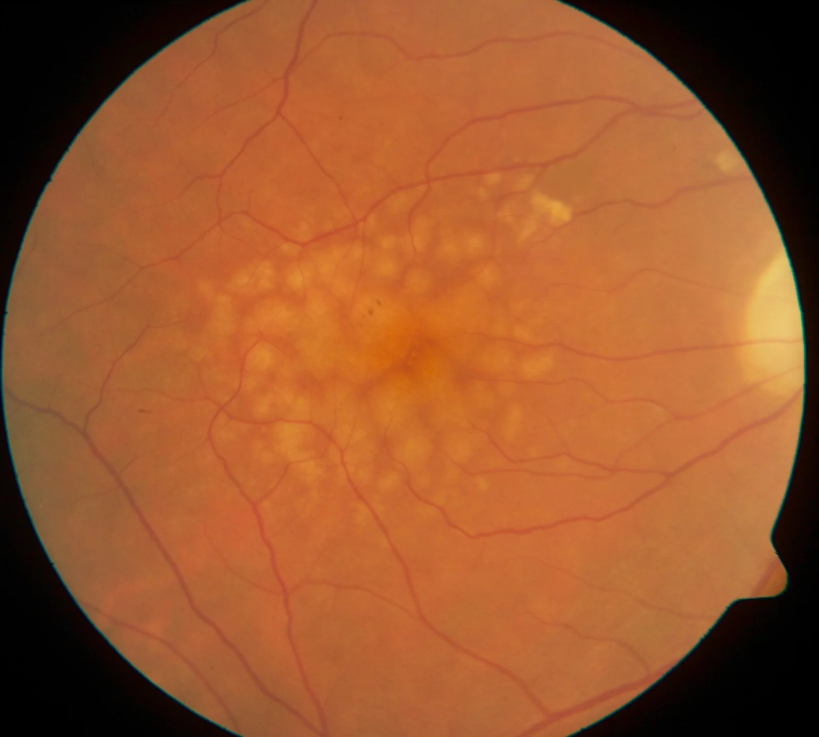
Long description
Fundus image of macular soft drusen in the right eye of a 70 year old male.

Patients usually describe painless, slowly worsening blur, often starting in one eye. Features include misty or filmy vision, faded colours (often noticed on TV/fabric), glare and dazzle (oncoming headlights, bright sun), trouble with night driving, monocular double vision and frequent changes of glasses prescription. Examination shows reduced best-corrected visual acuity (often correctable only partly), a dull or absent red reflex, and, on slit-lamp examination, the visible opacity; direct ophthalmoscopy reveals the opacity silhouetted against the fundus red reflex. In advanced (mature) cataract the fundus cannot be seen; secondary changes include phacomorphic (angle-closure) glaucoma from swelling of the lens and phacolytic glaucoma from leaking lens protein.[2][5] With bilateral dense cataract patients may have marked visual disability even when each eye's acuity does not look very low.[2]
Early and intermediate AMD may cause no symptoms, but patients often notice poor dark adaptation, difficulty in low-contrast or dim settings, intermittent blur and, later, metamorphopsia (straight lines wavy) or a blurred/dark patch in central vision. Wet AMD can present suddenly with distortion and rapid visual loss in one eye, sometimes asymptomatic until the fellow eye is engaged. Functional consequences include loss of reading, driving, face recognition and, in late disease, 'invisibility' of the clock hands while the rim is seen. Charles Bonnet syndrome (release visual hallucinations) is common in significant vision loss and should be explained. Depression and multimorbidity are frequent and should be anticipated.[1][6]
Examination centers on dilated fundus (slit-lamp biomicroscopy) looking for drusen and pigment change, OCT of the macula, and self-monitoring devices. Visual acuity is measured with Snellen or, in trials, ETDRS charts; a drop of 15 or more ETDRS letters is 'moderate visual loss' and 30 or more 'severe'.[6]
Amsler grid: a high-contrast grid of black lines on a white background, viewed monocularly at reading distance. Patients report distortion or missing squares. Its sensitivity for detecting new wet AMD is only about 33.7% (specificity about 81.4%), so a normal grid does not exclude disease and clear patient education on symptom thresholds matters.[6]
The following is a practical, low-threshold summary of what each test establishes and the cut-offs that matter.[1][6]
| Investigation | What it establishes | Key numbers/notes |
|---|---|---|
| Visual acuity (Snellen/ETDRS) | Baseline function; referral decisions | Cataract: referral not based on acuity alone. AMD: NICE anti-VEGF window best-corrected VA 6/12 to 6/96 (NICE 1.5.6). Loss of 3 or more lines (15 or more ETDRS letters) = moderate visual loss; 6 or more lines = severe [1] |
| Slit-lamp fundus biomicroscopy | Cataract type and maturity; drusen and pigment change in AMD | First-line for both; for early AMD confirm on biomicroscopy alone |
| Optical coherence tomography (OCT) | AMD subtypes, activity, treatment response | Sensitivity 91.7% for exudative AMD. Dry: atrophy; wet: intraretinal/subretinal fluid [6] |
| Fundus fluorescein angiography (FFA) | Neovascularisation | Gold standard for exudative AMD; offer if OCT does not exclude neovascular disease; risks include allergy/anaphylaxis |
| Colour fundus photography | Documentation, staging, GA follow-up | Accepted for geographic atrophy area in trials |
| Fundus autofluorescence | Geographic atrophy mapping and progression | Used to monitor GA lesion area |
| Optical coherence tomography angiography | Non-invasive flow imaging | Promising replacement for FFA; mainly research use currently |
| Amsler grid | Self-monitoring of distortion | Sensitivity 33.7%, specificity 81.4% |
| Biometry/keratometry | Cataract surgery planning | Optical biometry for axial length; SRK/T, Haigis, Hoffer Q, Barrett Universal II by axial length range |
NICE further specifies the anti-VEGF selection criteria that count as a clinical 'threshold': ranibizumab within its licence is recommended for wet AMD when, in the treated eye, best-corrected visual acuity is between 6/12 and 6/96, there is no permanent structural damage to the central fovea, lesion size is 12 disc areas or less in greatest linear dimension, and there is evidence of recent presumed disease progression; in eyes with acuity worse than 6/96, anti-VEGF should be considered only if a benefit to overall visual function is expected (for example, the better-seeing eye).[1] Treatment for wet AMD should be offered as soon as possible and within 14 days of referral to the macula service, with referral made urgently and normally within one working day.[1] [3][6]
Conditions that look like, or accompany, cataract or AMD, and the feature that separates them.[5][6][1]
| Condition | Typical presentation | Feature that separates it |
|---|---|---|
| Age-related cataract | Painless blur, glare, faded colour, reduced acuity | Clouded lens on slit lamp; red reflex lost; surgery curative |
| Wet AMD | Sudden distortion, central scotoma, rapid decline in one eye | OCT shows intraretinal/subretinal fluid; FFA shows neovascular leakage |
| Dry AMD (geographic atrophy) | Slow central visual decline, difficulty in low light | Atrophy of photoreceptors/RPE on OCT and fundus autofluorescence |
| Open-angle glaucoma | Peripheral field loss first, often asymptomatic | Optic disc cupping, raised IOP; central acuity preserved until late |
| Diabetic retinopathy/maculopathy | Blur, distortion in person with diabetes | Haemorrhage/exudate; OCT shows macular oedema (also cataract risk raised) |
| Corneal opacity/scar | Blur, glare; acuity often poor | Corneal opacity seen on exam; lens clear |
| Optic neuropathy (e.g. giant cell arteritis) | Sudden monocular visual loss in the elderly | Pale or swollen optic disc; systemic symptoms; constitutes an emergency |
| Central serous chorioretinopathy | Distortion, blur in younger adults | Fluid under retina on OCT, often self-limiting |
| Macular hole | Central metamorphopsia, scotoma | OCT shows full-thickness retinal defect |
| 'Dry eye'/refractive error | Fluctuating blur, irritation | Surface staining/tear film problems; clear ocular media |
Cataract and AMD commonly coexist in the same older eye; neither condition should be assumed to be the only cause of symptoms without examination.
Cataract: following NICE NG77 (2017, last reviewed 2025) and AAO PPP (Cataract in the adult eye, 2021)
Non-surgical measures first: ensure spectacles/contact lenses are up to date, adjust lighting, use anti-glare sunglasses and magnifiers, and treat any coexisting disease. Early cataracts do not require urgent surgery.[2][5]
Who needs surgery: the decision should be based on discussion of how the cataract affects quality of life and function (reading, driving, work, recognising faces), whether one or both eyes are involved, and the person's willingness; NICE says do not restrict access to cataract surgery on the basis of visual acuity alone. Surgery is the only cure.[2]
Timing: a dense cataract that is left to mature carries a higher surgical complication rate, so delay should not be indefinite; bilateral, simultaneous cataract surgery can be considered for low-risk patients or those needing general anaesthesia with added risk.[2]
Surgical technique (standard): phacoemulsification with foldable intraocular lens implant under topical or sub-Tenon anaesthesia (peribulbar if both of the first-line options are contraindicated; retrobulbar avoided). Multifocal intraocular lenses should not be offered. Bio-metry uses optical biometry for axial length, keratometry for corneal curvature, and standard formulas chosen by axial length (Haigis/Hoffer Q for under 22 mm, Barrett Universal II (or SRK/T) for 22 to 26 mm, Haigis/SRK/T over 26 mm). Wrong-lens safety relies on the WHO checklist (modified), biometry verification and lens reconciliation in theatre. Intracameral cefuroxime is used to prevent endophthalmitis; topical steroids +/- NSAIDs prevent inflammation and cystoid macular oedema (NSAID+steroid for high-risk groups such as diabetes/uveitis). Enhanced recovery includes no in-person first-day review after uncomplicated surgery.[2]
AAO concurrence: the AAO Preferred Practice Pattern (cataract in the adult eye, 2021) supports day-case small-incision phacoemulsification with a foldable lens, intracameral antibiotics and systematic wrong-lens checks, and regards symptomatic cataract as a surgical disorder with minimal benefit from nutritional supplements.[2]
Early AMD: no pharmacological treatment; NICE recommends only healthy lifestyle and counselling, and no routine hospital monitoring.[1][3]
Intermediate AMD (or late AMD in one eye): offer AREDS 2 supplements daily (vitamin C 500 mg, vitamin E 400 IU, lutein 10 mg, zeaxanthin 2 mg, zinc 80 mg, copper 2 mg) to slow progression; note that retinal risk reduction exists only for intermediate/late disease, and that early AMD does not benefit. Patients should keep regular ophthalmic review and supplement use should be discussed with a clinician (possible steroid/supplement interactions). The *original* AREDS formula, which contains beta carotene, should not be given to people who smoke or have stopped smoking (lung cancer signal); the AREDS 2 formula replaces beta carotene with lutein/zeaxanthin. Dietary advice stresses vegetables, fruit and fatty fish (Mediterranean-style), and not exceeding supplement doses.[4][6][1]
Wet (exudative) AMD: anti-VEGF intravitreal injections are the first-line, most effective treatment and should be started as soon as possible, within 14 days of referral. NICE has no clinically significant difference in effectiveness or safety between aflibercept, bevacizumab and ranibizumab, so comparable regimens are chosen partly by net cost. Bevacizumab has no UK marketing authorisation for this indication and its use is off-label, prescriber's responsibility. Pegaptanib and photodynamic therapy alone are not recommended; PDT as an adjunct only in a trial; intravitreal corticosteroids as adjunct are not recommended. Referral should be urgent (within one working day) and treatment selection should follow OCT and, where needed, FFA.[1][3]
Dosing from licensed product information and major trials:
- Ranibizumab: 0.5 mg intravitreal injection, typically monthly in pivotal trials; real-world regimens (treat-and-extend, pro re nata) vary with disease activity. In trials, monthly ranibizumab reduced visual loss (less than 15 letters) in about 94.6% of patients at 12 months versus 62.2% with sham.[6]
- Aflibercept: 2 mg intravitreal injection monthly for the first 12 weeks, then 2 mg every 8 weeks; some patients need monthly maintenance, and up to 12-weekly intervals may be used after a year of effective therapy.[8]
- Bevacizumab: mentioned in NICE as an off-label alternative on cost grounds; dose decision is the prescriber's.[1]
- More recently, faricimab (bispecific anti-VEGF/angiopoietin-2) and biosimilars allow longer intervals between injections for selected patients; brolucizumab, pegcetacoplan and avacincaptad pegol address selected neovascular/geographic atrophy subgroups and are introduced under specialist care. Where quoted ranges are uncertain, follow the current SPC.[6][3]
Switching between anti-VEGF agents has limited clinical benefit but may be done for practical/preference reasons; anti-VEGF can be stopped when the eye develops late inactive AMD with no prospect of functional improvement, or continued observationally when disease is stable.[1]
Geographic atrophy (late dry): no proven treatment that reverses vision in routine practice; two complement inhibitors were approved in the US in 2023 for slowing GA progression (pegcetacoplan, avacincaptad pegol), with uncertain benefit on visual function and high rates of new neovascularisation; NICE/AAO position needs checking locally before use. Referral for certification of sight impairment, low-vision services, research participation.[6][1]
Support for visual loss in AMD: certification of visual impairment; low-vision aids, eccentric viewing training, group rehabilitation; screen for depression and multimorbidity; advise on driving standards and DVLA/local equivalents.[1]
Monitoring: wet AMD on treatment has ongoing OCT of both eyes; early/dry AMD is not routinely monitored in hospital but patients self-monitor and keep regular optometric sight tests. Self-monitoring symptoms to report promptly: blurred/grey patch, straight lines distorted, objects smaller than normal.[1]
Adult with central visual symptoms or known cataract/AMD
- History and symptomsDuration, one or both eyes, glare, distortion, night vision, smoking, diabetes, steroids
- Measure acuity (Snellen/ETDRS); dilated exam; OCT if central lossDon't rely on acuity alone for cataract; Amsler and home monitoring for AMD
- Question: What is the main abnormality?
Clouded lens (cataract)
- Conservative measuresUpdate spectacles, lighting, anti-glare; treat co-morbidity
- Refer for surgery if quality of life affectedDo not restrict by visual acuity; NICE NG77
- Pre-operative biometry and wrong-lens checksOptical biometry, anaesthesia, IOL, intracameral cefuroxime
Drusen/pigment change, no fluid (dry AMD)
- Stage the disease (early/intermediate/late)OCT, autofluorescence, fundus photos
- Lifestyle + AREDS 2 if intermediate or late in one eye10 mg lutein, 2 mg zeaxanthin, no beta carotene if smoker
- Certify/low-vision services if impaired; routine optometric testsNICE: do not monitor early/dry AMD in hospital routine
Fluid/haemorrhage (wet active AMD)
- Urgent referral within 1 working dayOffer OCT, FFA if OCT inconclusive
- Start anti-VEGF within 14 days of referralRanibizumab 0.5 mg or aflibercept 2 mg per schedule; bevacizumab off-label
- Monitor both eyes; reassess by OCT according to disease activitySwitch/stop if stable or inactive late AMD
Other cause
- Manage differentialsGlaucoma, diabetic eye disease, corneal disease, optic neuropathy
- Progressive visual loss until the lens is opaque; falls, fractures and social isolation.
- Secondary glaucomas: phacomorphic (swelling lens) and phacolytic (leaking lens protein) are sight-threatening.
- Surgery-related: posterior capsule rupture, endophthalmitis (prevented with intracameral cefuroxime), cystoid macular oedema, raised intraocular pressure, floppy iris syndrome (alpha-blockers), residual refractive error, retinal detachment (higher risk in high myopia).
- Cataract surgery can accelerate AMD progression (conflicting observational evidence), so the macula should be assessed and patients counselled pre-operatively.[2]
- Conversion from dry to wet AMD, or enlargement of geographic atrophy causing central scotoma.
- Depression and loss of independence; many patients have multimorbidity.[1]
- Therapy complications: intravitreal injections carry a low but finite risk of endophthalmitis (0.06 to 0.16% per injection in trials), retinal detachment and pressure elevation; systemic anti-VEGF (intravenous) carries vascular risks, but intravitreal use was not associated with major cardiovascular events in trials. Anticoagulation does not need to be stopped for injections.[6]
- Repeated anti-VEGF is associated with macular atrophy in long-term studies.[6]
- Cataract: highly treatable. Surgery restores vision in about 9 out of 10 people and improves function; delaying very dense (mature) cataract raises operative risk.[5][2]
- Early/intermediate AMD: many patients never reach late disease; AREDS 2 plus lifestyle slows progression in intermediate disease.[6][4]
- Late wet AMD: with anti-VEGF, about 95% lose fewer than 15 letters at 12 months versus about 62% without treatment; long-term SEVEN-UP data show 34% of treated eyes lose 15 letters or more and 43% remain stable or improve over ~7 years, with macular atrophy in nearly all eyes.[6]
- Late dry AMD (geographic atrophy): roughly half of affected patients have moderate vision loss within 2 years, and a quarter severe loss on standard charts.[6]
- Cataract: sunglasses/hat UV protection, protective eyewear in industry/sport, smoking cessation, good glycaemic control, limit excessive alcohol, balanced diet; dilated eye exam every 1 to 2 years from age 60. No tablet prevents cataract.[5]
- AMD: stop smoking, Mediterranean-style diet (vegetables, fruit, fatty fish), regular physical activity, control of vascular risk, AREDS 2 only at intermediate/late stage, prompt reporting of new distortion central patch/dimmer vision. Prompt treatment of wet AMD matters: delays beyond ~7 weeks reduce visual gain from 38% to about 20% of patients.[3][6]
- Screening: NICE does not recommend routine hospital monitoring of early/dry AMD, but public screening rests on community optometrists and self-monitoring; cataract detection is opportunistic via optometrists/GPs using vision testing and red-reflex/slit-lamp checks.[1][5]
- Pregnancy/breastfeeding: cataract surgery, when needed, is usually planned outside pregnancy; anti-VEGF agents have not been studied in pregnancy and should be used only if benefit justifies risk (aflibercept warning); intravitreal injection technique and informed consent apply to all patients.[8][1]
- Older adults: very common; correction of hearing/sensory, fall risk and transport planning matter. Coexisting AMD and glaucoma are common.
- Children: cataract and AMD are diseases of adulthood. A cataract in a child is usually congenital, traumatic or steroid-related and managed in paediatric ophthalmology.
- Immunocompromised / steroid users: high risk of posterior subcapsular cataract and infection; prophylaxis and monitoring should be heightened.
- A direct ophthalmoscope and a good slit lamp (where available), plus a portable Amsler grid and measurement of visual acuity, diagnose most cataracts; OCT/FFA may not be available and wet AMD can be presumed clinically, but anti-VEGF is costly.
- Cataract surgery is the first investment: it is one of the most cost-effective interventions in medicine and should be made available before sophisticated AMD biologics.
- Where anti-VEGF is not available, referral to a higher-level service and counselling about expected outcomes and visual rehabilitation are the realistic options. Community low-vision services can be organised locally (magnifiers, lighting, eccentric viewing training) without expensive technology.
- Screening rests on community optometrists and trained primary workers checking acuity and red reflex in older adults.
- Cataract is opacification of the lens; the only cure is phacoemulsification with intraocular lens implant, a safe, day-case procedure.
- Types are nuclear sclerotic, cortical and posterior subcapsular, plus secondary forms; posterior subcapsular cataract affects steroid users, diabetics and young patients early. [2]
- Do not refer cataract for surgery on the basis of acuity alone; refer on functional impact and the patient's wishes.
- Guide biometry and IOL choice by axial length; use intracameral cefuroxime; avoid retrobulbar anaesthesia.
- AMD affects the macula; late (advanced) AMD is geographic atrophy ('dry') or exudative neovascular AMD ('wet').
- Non-neovascular AMD makes up about 80% of patients, but neovascular AMD causes most severe vision loss. [3]
- Early AMD has no evidence-based treatment; intermediate AMD needs lifestyle advice and AREDS 2 supplements; wet active AMD needs prompt anti-VEGF.
- AREDS 2 dose: vitamin C 500 mg, vitamin E 400 IU, lutein 10 mg, zeaxanthin 2 mg, zinc 80 mg, copper 2 mg daily; do not use beta-carotene-containing AREDS in smokers.
- Wet AMD referral should be made within one working day; treatment started within 14 days.
- Ranibizumab (0.5 mg) and aflibercept (2 mg, monthly times three then 8-weekly) are licensed anti-VEGF options; bevacizumab is off-label in the UK.
- An Amsler grid is a useful self-monitor (sensitivity about 33.7%); OCT is far more sensitive (91.7%) and should be used when wet AMD is suspected.
- Certification, low-vision aids, driving advice and depression screening are part of care.
- NICE (NG77, NG82), the AAO Preferred Practice Patterns (cataract 2021; AMD 2024, updated 2026) and NEI guidance informed this page.
Answer each one in your head first, then open the answer.
A 62-year-old smoker with intermediate AMD in both eyes asks what he can do. What is appropriate advice?
Show answerHide answer
Advise smoking cessation, dietary advice (vegetables, fruit, fatty fish) and, if everyday foods are insufficient, AREDS 2 supplementation daily (vitamin C 500 mg, vitamin E 400 IU, lutein 10 mg, zeaxanthin 2 mg, zinc 80 mg, copper 2 mg). Do not use the older AREDS formula containing beta carotene in a smoker. Book regular eye review with self-monitoring for distortion and prompt reporting.
Why can a posterior subcapsular cataract disable a driver even when visual acuity is only mildly reduced?
Show answerHide answer
PSC opacities sit just inside the posterior capsule and produce intense glare and light scatter, so oncoming headlights and bright sunlight blow out contrast and night vision out of proportion to the letters on the chart. The patient may still read much of the chart yet be unable to drive at night.
A fundus photo shows medium-sized drusen with no pigment change. Which AMD stage is this and what treatment is indicated?
Show answerHide answer
Early (low-risk) AMD. There is no evidence-based pharmacological treatment: counsel, address lifestyle risk (especially smoking and diet), and arrange routine follow-up; do not give AREDS 2 yet.
An 80-year-old woman with a mature cataract in one eye has late dry AMD in the other. How should you counsel before cataract surgery?
Show answerHide answer
Surgery should restore the structural clarity blocked by the cataract, but the macular disease limits central vision. Check the macula by OCT before surgery, set realistic expectations about reading and face recognition, and discuss low-vision aids and lighting. Surgery does not treat AMD.
What findings on OCT specifically distinguish active wet AMD from dry AMD, and what must happen next?
Show answerHide answer
Intraretinal and/or subretinal fluid (and sometimes haemorrhage or fibrosis) indicate exudative activity. Reassure the patient and make an urgent referral (within one working day) to macular services for consideration of anti-VEGF, which should be offered within 14 days of referral.
A patient self-monitoring with an Amsler grid rings with new distortion but normal VA. How do you interpret this?
Show answerHide answer
Amsler changes can precede acuity loss and are the kind of change patients are taught to report promptly, but the grid's sensitivity is only about 33.7%. Arrange prompt slit-lamp/OCT assessment rather than reassuring on the grid or acuity alone.
Cataract reduces the view of the macula, so significant AMD can be masked; OCT should be obtained whenever visual acuity is worse than the cataract appears to explain, before surgery. Cataract surgery does not by itself treat macular disease, and observational evidence suggests it can sometimes be associated with faster AMD progression, so patients with coexisting disease need a realistic discussion of which problems surgery will and will not solve. Good communication about spectacles and low-vision aids after surgery improves satisfaction.
Plain-text (no separate topic files yet): glaucoma; diabetic retinopathy and diabetic macular oedema; visual field testing; retinal detachment; vision rehabilitation and low-vision services; neuro-ophthalmology (optic neuropathy); smoking cessation.
- National Institute for Health and Care Excellence. Age-related macular degeneration (NICE guideline NG82): recommendations. NICE guideline NG82, published 23 January 2018 (last updated context page). Accessed as recommendations page.. Accessed .
- National Institute for Health and Care Excellence. Cataracts in adults: management (NICE guideline NG77): recommendations and context. NICE guideline NG77, published 26 October 2017, last reviewed 20 May 2025 (IOL selection update pending).. Accessed .
- American Academy of Ophthalmology Preferred Practice Pattern Retina/Vitreous Committee. Age-Related Macular Degeneration Preferred Practice Pattern (2024, updated 2026). American Academy of Ophthalmology, 2024 (updates December 2025 and June 2026).. Accessed .
- National Eye Institute. AREDS 2 supplements for age-related macular degeneration (AMD). NEI patient/professional information, last updated 22 June 2021.. Accessed .
- National Eye Institute. Cataracts. NEI patient/professional information, last updated 19 August 2026.. Accessed .
- Fleckenstein M, Schmitz-Valckenberg S, Chakravarthy U. Age-related macular degeneration: a review. JAMA 2024;331(2):147-157. doi:10.1001/jama.2023.26074 (author manuscript PMC12935482). Accessed .
- Vision Loss Expert Group of the Global Burden of Disease Study et al. Global estimates on the number of people blind or visually impaired by cataract: a meta-analysis from 2000 to 2020. Eye (Lond) 2024;38(11):2156-2172. doi:10.1038/s41433-024-02961-1. Accessed .
- Regeneron Pharmaceuticals (FDA label). EYLEA (aflibercept) injection, for intravitreal use. FDA prescribing information, revised 10/2024 (DailyMed, updated 07/2026).. Accessed .
Figure credits
Educational content for medical students and doctors. It is not a substitute for clinical judgement or local guidelines.