Retinal vascular occlusion
Sudden, painless loss of vision in one eye from retinal artery embolism or venous thrombosis, including the CRAO emergency.
- Retinal vascular occlusion is a sudden blockage of the blood vessels supplying the retina. An artery is usually blocked by an embolus (central or branch retinal artery occlusion) and is an ophthalmic emergency. A vein is usually blocked by thrombus (central or branch retinal vein occlusion).
- Central retinal artery occlusion (CRAO) causes sudden, painless, severe loss of vision in one eye, often with a "cherry-red spot" and box-carring of vessels. Treat it as a stroke: urgent referral to a stroke centre or emergency department, with brain and vascular work-up. Brain imaging and thrombolysis are time-critical. [2]
- Retinal vein occlusions cause painless loss that can be mild or severe. The main threat to central vision is macular oedema; intravitreal anti-VEGF therapy is first line, with monthly injections until the vision is stable.
- All patients need a systemic vascular work-up (blood pressure, glucose, lipids, carotid imaging, cardiac assessment), and in older patients assessment for giant cell arteritis.
- Arteritic CRAO and ischaemic central retinal vein occlusion carry a risk of neovascular glaucoma and need close ophthalmic follow-up. [1][3][5]
By the end of this page you should be able to:
- Define retinal vascular occlusion and classify arterial (CRAO, BRAO, OAO) and venous (CRVO, HRVO, BRVO) forms.
- Recognise CRAO from its history and fundus findings and state why it is treated as a stroke.
- Distinguish ischaemic from non-ischaemic central retinal vein occlusion and state the capillary non-perfusion cut-off.
- Plan ocular and systemic investigations, including the tests shown in the investigation table.
- State evidence-based treatment steps and doses from the AHA scientific statement (2021), AAO Preferred Practice Patterns (arterial 2024; vein 2024/updated 2026), and Royal College of Ophthalmologists guideline (2022) for management of retinal vein occlusion. [2][3][4][5]
- Counsel a patient on prognosis, complications, secondary prevention, and follow-up.
DefinitionRetinal vascular occlusion
A sudden or subacute blockage of the retinal arterial supply or venous drainage, leading to retinal ischaemia, leakage and, in the case of venous occlusion, macular oedema and neovascularisation. "Retinal vascular occlusion" covers both arterial occlusion (a "retinal stroke") and venous occlusion (thrombosis of a retinal vein).
- Central retinal artery occlusion (CRAO): obstruction of the central retinal artery, usually by embolus. [1]
- Branch retinal artery occlusion (BRAO): obstruction of a branch artery, causing a sectoral visual field defect.
- Ophthalmic artery occlusion (OAO): less common and more devastating; may combine retinal and choroidal ischaemia.
- Amaurosis fugax: a transient retinal ischaemic attack lasting minutes, with normal fundus by the time of examination.
CRAO can be non-arteritic or arteritic (usually giant cell arteritis), and permanent, transient, or with cilioretinal sparing. On fundoscopy a patent cilioretinal artery, present in about one third of eyes, may spare central vision. [1]
- Central retinal vein occlusion (CRVO): obstruction at or just behind the lamina cribrosa, affecting all four quadrants.
- Hemi-retinal vein occlusion (HRVO): obstruction of one trunk before the central vein, affecting a hemisphere.
- Branch retinal vein occlusion (BRVO): obstruction at an arteriovenous crossing, affecting one quadrant.
Both CRVO and BRVO are divided into ischaemic and non-ischaemic forms on the basis of capillary non-perfusion on fluorescein angiography. The historical definition of ischaemic CRVO is more than 10 disc areas of capillary non-perfusion on seven-field angiography; with wide-field imaging, more than 10 disc areas of non-perfusion in the posterior pole, or an ischaemic index above 45%, is used. [5]
HRVO behaves clinically like CRVO, including a higher risk of anterior segment neovascularisation than BRVO. [4]
A hyperviscous state (sickle cell disease, myeloproliferative disorder, Waldenstrom macroglobulinaemia) or external compression can mimic or cause both types, and a combined retinal artery and vein occlusion occasionally occurs. Combined occlusion carries a high risk of iris neovascularisation and needs urgent pan-retinal photocoagulation if it develops. [3]
CRAO is rare but serious. Incidence is estimated at 1 in 100,000 people per year and 1 in 10,000 outpatient visits, with an average age in the early sixties and a higher incidence in men. [1] Overall spontaneous functional visual recovery without treatment is only 17.7%. [1] Arteritic CRAO from giant cell arteritis accounts for about 4% of cases and is more likely in people over 70 years. [1]
CRAO occurs as three broad clinical patterns: non-arteritic permanent CRAO is the most common (about two thirds of cases), transient CRAO accounts for 15% to 17%, and CRAO with cilioretinal sparing accounts for the remainder. [1] In fewer than 10% of patients a retinal embolus is visible. [1]
RVO is the second most common retinal vascular disorder after diabetic retinopathy. A global meta-analysis estimated the 2015 prevalence in people aged 30 to 89 years as 0.77% for any RVO, 0.64% for BRVO, and 0.13% for CRVO, or roughly 28.06 million, 23.38 million, and 4.67 million people respectively. [6] BRVO is therefore far more common than CRVO, but CRVO is more likely to be blinding. The pooled five-year cumulative incidence is 0.86% and the ten-year cumulative incidence is 1.63%. [6] [5]
Prevalence rises steeply with age, from about 0.26% in the 30–39-year group to 3.4% in the 80–89-year group for any RVO. [6] Hypertension is the strongest risk factor (meta-odds ratio 2.82), followed by prior myocardial infarction (2.23) and stroke (2.07). [6] Women and men are affected equally. [6]
Over 90% of CRAOs are non-arteritic. Risk factors mirror those of atherosclerotic cerebrovascular disease: hypertension, diabetes, dyslipidaemia, coronary artery disease, carotid disease, smoking, family history, and transient ischaemic attacks. [1] The embolus is usually cholesterol, platelet-fibrin, or calcific material and impacts where the artery is narrowest as it pierces the dural sheath of the optic nerve. [1]
Arteritic CRAO is due to giant cell arteritis and is an emergency for both eyes. [1][3]
RVO is not, in most people, a consequence of systemic venous thrombosis. The underlying problem is at the retinal arteriovenous crossing, where the artery shares a sheath with the vein, or at the lamina cribrosa in CRVO, and compression by an atherosclerotic artery injures the vein wall and promotes local thrombosis. [5]
Common associations and risk factors include:
- Hypertension (the dominant association, see above). [6]
- Diabetes is no more common than in the general population, but testing at diagnosis detects undiagnosed disease. [5]
- Hyperlipidaemia, heart attack, stroke, raised creatinine, and intraocular pressure/glaucoma. [5][6]
- Hyperviscosity and thrombophilia (sickle cell disease, myeloproliferative disorders, antiphospholipid syndrome) in younger patients. [5]
- RVO has been reported in people taking oestrogen-containing preparations; the decision to continue is made case by case. [5]
Routine testing for anti-phospholipid antibodies is not recommended in a retinal vein occlusion occurring in isolation without other features of the anti-phospholipid syndrome. In isolated RVO there is no high-quality evidence supporting anticoagulation or antiplatelet drugs, and finding a thrombophilic abnormality does not change management or predict prognosis. [5]
RVO is a disease of any age: prevalence is 0.26% in people aged 30–39 years and 0.44% in 40–49-year-olds. [5]
The central retinal artery is the first branch of the ophthalmic artery (itself a branch of the internal carotid artery) and supplies the inner retinal layers. The outer retina, including the photoreceptors, is supplied by the choroid. When the central artery blocks, the inner retina infarcts but the fovea is fed by the choroid, producing the pale swollen retina with a red central spot. A cilioretinal artery, present in about one third of eyes, can spare central vision and leave acuity at 20/50 or better. [1]
Experimental work suggests no detectable damage within 97 minutes, but occlusion lasting 240 minutes or longer causes irreversible damage; the exact reversible window in humans is thought to lie between 4.5 and 12 hours. [1]
A thrombosed vein raises capillary pressure, producing leakage, retinal haemorrhage and oedema. Retinal ischaemia releases vascular endothelial growth factor (VEGF), which increases vascular permeability and drives macular oedema, and later new vessel formation on the iris, angle or retina. This is why ischemia, not just oedema, determines prognosis. [5]
Figure 1Fondo de ojo Ostrucción Vena central
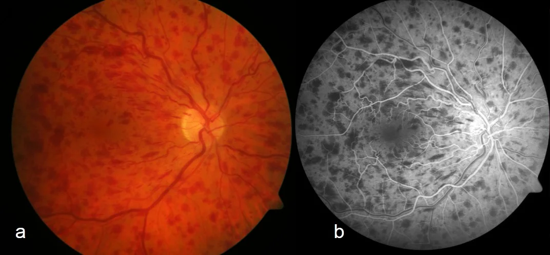
Long description
A two-panel figure whose left panel is a color fundus photograph of the posterior pole in central retinal vein occlusion showing intraretinal haemorrhages, paired with a fluorescein angiogram showing blockage of choroidal fluorescence by those haemorrhages.

Figure 2Cherry red spot in patient with central retinal artery occlusion (CRAO)
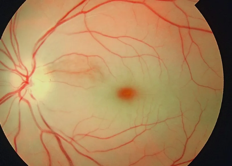
Long description
Fundus photograph of a patient with central retinal artery occlusion showing a cherry red spot and retinal swelling.

- CRAO: sudden, painless, profound monocular visual loss, often described as a curtain or like a switch. It may follow episodes of amaurosis fugax. Ask about vascular risk factors and neurological symptoms. [1]
- Transient CRAO: visual loss lasts minutes to hours and the fundus may be normal when you see the patient. [1]
- RVO: painless visual loss of variable degree; many BRVOs are found on routine examination. Central metamorphopsia, micropsia, or scotoma point to macular oedema. [4][5]
- Visual acuity: usually very poor (under 20/200) unless the attack is transient or cilioretinal sparing. [1]
- Relative afferent pupillary defect.
- Fundus: retinal arterial whitening (oedema) with a cherry-red spot at the fovea, thinned vessels with box-carring, possible disc oedema and splinter haemorrhages; emboli are seen in fewer than 10% of cases. [1]
- Chronic stage: pale disc, vascular attenuation, retinal pigment epithelial mottling. [1]
- Arteritic CRAO: suspect giant cell arteritis in patients over 70 years with headache, jaw claudication, scalp tenderness, polymyalgia rheumatica, and raised inflammatory markers. Fundus fluorescein angiography may show choroidal filling defects, and the fellow, asymptomatic eye may have cotton-wool spots. [1][3]
- CRVO: disc oedema, dilated and tortuous veins in all quadrants, widespread deep and superficial haemorrhages, cotton-wool spots, and macular oedema. After the acute phase: disc and retinal collaterals, telangiectatic capillaries, and persistent venous change. [5] [3]
- BRVO: identical changes, but confined to the sector drained by the occluded branch (most often superotemporal), with nicking at the responsible arteriovenous crossing. [5]
- Ischaemic CRVO: more than 10 disc areas of capillary non-perfusion; higher risk of iris or angle new vessels and neovascular glaucoma. [5]
- Associated glaucoma is common in CRVO and must be controlled alongside the retinal condition. [4]
| Condition | Key distinguishing feature |
|---|---|
| CRAO | Sudden severe loss, RAPD, cherry-red spot, box-carring, "ischaemic" inner retina; high stroke risk. [1][3] |
| BRAO | Sectoral field defect matching a branch, otherwise as CRAO in the affected sector. |
| Amaurosis fugax (transient RAO) | Lasts minutes, fundus normal later, signals carotid disease. [1] |
| Non-arteritic anterior ischaemic optic neuropathy | Disc swelling, altitudinal field defect, not a cherry-red spot. |
| Retinal detachment | Photopsia, floaters, visual field curtain moving; not sudden central only. |
| Vitreous haemorrhage | Floaters, absent red reflex; possible proliferative disease. |
| Acute angle-closure glaucoma | Pain, haloes, shallow anterior chamber, high pressure. |
| Central retinal vein occlusion | Venous dilation, widespread haemorrhages and oedema, not severe acute arterial ischaemia. [5] |
| Branch retinal vein occlusion | Sectoral haemorrhages and venous change, often asymptomatic. [5] |
- Visual acuity and refraction; test for a relative afferent pupillary defect.
- Dilated fundus examination with slit-lamp biomicroscopy is the bedside diagnosis.
- Optical coherence tomography (OCT) of the macula in CRAO shows hyperreflectivity of the inner retina; in RVO it measures central subfield thickness and monitors macular oedema. [1][5]
- Fluorescein angiography (FFA) shows delayed retinal arterial filling in CRAO; in RVO it quantifies capillary non-perfusion (the ischaemic CRVO cut-off is over 10 disc areas in the posterior pole). [5]
- OCT angiography demonstrates capillary perfusion deficits in CRAO and RVO. [1]
- Intraocular pressure and gonioscopy to detect iris and angle neovascularisation in ischaemic CRVO. [5]
| Test | What it shows or measures | Key cut-off or meaning |
|---|---|---|
| Dilated fundus exam | Arterial whitening, cherry-red spot, box-carring (CRAO); venous dilation/haemorrhage (RVO) | Bedside diagnostic pattern. |
| OCT macula | Inner retinal hyperreflectivity (acute CRAO); macular oedema (RVO) | Repeat to confirm response. |
| FFA | Delayed arterial filling (CRAO); capillary non-perfusion (RVO) | Ischaemic CRVO: over 10 disc areas of non-perfusion in the posterior pole. [5] |
| VA | Baseline and follow-up vision | Guides treatment decisions. |
| OCTA | Capillary perfusion deficit | Confirms ischaemia. |
| IOP, gonioscopy | Iris/angle new vessels | Detects NVG risk in ischaemic CRVO. |
The aim is both to identify a source of embolus (CRAO) and to control modifiable risk factors (all RVO).
- Blood pressure, fasting glucose or HbA1c, fasting lipids, full blood count, ESR and CRP. [1][5]
- Carotid duplex ultrasound and echocardiography to look for embolic sources. [1][3]
- ECG for atrial fibrillation or other cardiac disease.
- In patients under 50 years with no vascular risk factors, a hypercoagulable and vasculitic screen (protein C and S, factor V Leiden, antiphospholipid antibodies, antinuclear and related antibodies, angiotensin-converting enzyme) and blood film for myeloproliferative or sickle cell disease. [1]
- In patients over 50 years, or any patient with symptoms, a work-up for giant cell arteritis (ESR, CRP, temporal artery studies) because arteritic CRAO needs emergency steroid treatment. [1][3]
- Testing for anti-phospholipid antibodies in isolated RVO without other features of the anti-phospholipid syndrome is not recommended. [5]
| Component | When to use | Purpose |
|---|---|---|
| Glaucoma/IOP | All ischaemic CRVO; BRAO/CRAO with NV risk | Detect neovascularisation. |
| Duplex carotid ultrasound | All non-arteritic CRAO, RAO | Carotid atherosclerosis or stenosis. |
| Echocardiogram | All non-arteritic CRAO, RAO | Cardiac embolic source. |
| Hypercoagulable/vasculitic screen | Under 50 years, no risk factors | Thrombophilia/vasculitis. [1] |
| GCA blood tests | Over 50 years with suspicious features | Arteritic CRAO. [1][3] |
| HbA1c (NICE type 2 target 48 mmol/mol (6.5%)) | At diagnosis of RVO | Detect or monitor diabetes. [5] |
There is no single laboratory criterion for CRAO. The diagnosis is clinical, supported by imaging, and is classified as non-arteritic versus arteritic and permanent versus transient or cilioretinal-sparing. [1]
For RVO, the degree of ischaemia measured on fluorescein angiography is the main severity marker and drives treatment intensity and follow-up. Ischaemic CRVO is defined as over 10 disc areas of non-perfusion in the posterior pole. [5] Macular ischaemia on OCT/FFA/OCTA is distinct from global ischaemia and predicts limited response to treatment. [5]
Suspected retinal vascular occlusion presenting to eye casualty
- Sudden, painless monocular visual loss
- Question: Fundus pattern?
Retinal arterial whitening, cherry-red spot, box-carring (arterial)
- Central retinal artery occlusion (CRAO)Treat as a stroke
- If symptom onset under 4.5 hDiscuss IV alteplase (0.9 mg/kg, maximum 90 mg) with the stroke team, if eligible
- If onset over 4.5 hAntiplatelet therapy and referral to acute stroke unit for secondary prevention
- Assess for giant cell arteritisUrgent IV methylprednisolone if suspected
Venous dilation, haemorrhage, oedema (venous)
- Central or branch retinal vein occlusionRefer to eye centre within 2 to 4 weeks
- Measure perfusion (FFA/OCTA)Ischaemic CRVO = over 10 disc areas of non-perfusion in the posterior pole
- Macular oedema present? Start monthly anti-VEGFranibizumab 0.5 mg, aflibercept 2 mg, bevacizumab 1.25 mg (off-label), or faricimab 6 mg; continue until vision is stable
- Neovascularisation? PRP/anti-VEGFMonitor monthly for 6 months, then less often
CRAO is the ocular analogue of cerebral stroke. There is no consensus on treatment and no guideline-endorsed single protocol, so care is provided by the stroke team. [1] The 2021 American Heart Association scientific statement on CRAO places it within the stroke care pathway, and the AAO 2024 Preferred Practice Pattern recommends immediate referral of acute symptomatic arterial occlusion to the nearest stroke centre. [2][3]
Acute management by interval since symptom onset
- Under 4.5 hours: intravenous alteplase 0.9 mg/kg (maximum 90 mg, with a 10% intravenous bolus and the remainder over 1 hour) can be considered in patients who meet stroke criteria, in collaboration with the stroke team. A patient-level meta-analysis of intravenous fibrinolysis for CRAO reported better functional visual outcomes with treatment within 4.5 hours. [1][2]
- Over 4.5 hours: start antiplatelet therapy and arrange review by the acute stroke unit for vascular work-up and secondary prevention. [1]
- The AAO 2024 Preferred Practice Pattern states that there is insufficient evidence to support intra-arterial or intravenous thrombolysis in CRAO. This differs from the fibrinolysis position in the stroke literature summarised by the review above; guidelines differ, and the decision rests with the stroke team. [3][1]
Principles running through all time windows
- Immediate referral to a stroke centre or emergency department for brain imaging and vascular assessment, because ocular ischaemia signals cerebral and cardiac disease. [1][3]
- Telemedicine or telestroke can be used to confirm the diagnosis when no eye specialist is present on site, as recommended by the AHA statement. [1]
- Non-invasive adjuncts (ocular massage, intravenous acetazolamide 500 mg, mannitol 1.5 to 2 g/kg, topical glaucoma drops, anterior chamber paracentesis, carbogen inhalation, hyperbaric oxygen) are often attempted, but all of these therapies, singly or combined, do not alter the final clinical outcome. [1] A meta-analysis found hyperbaric oxygen did not improve final visual outcomes. [1]
- Giant cell arteritis: seek it actively in patients over 50 years; if suspected or diagnosed, give intravenous methylprednisolone and arrange urgent specialist care, because it threatens the fellow eye. [1][3]
- Subacute phase: watch for ocular neovascularisation, reported after CRAO with a prevalence of 2.5% to 31.6% (mean onset 8.5 weeks); apply pan-retinal photocoagulation if it develops. [1]
- Secondary prevention: a multidisciplinary team (ophthalmology, neurology, primary care) should manage hypertension, lipids, diabetes, obesity, sleep apnoea, diet, exercise, and smoking cessation. In patients without contraindication an initial 21 days of dual antiplatelet therapy is reasonable, followed by aspirin 81 mg daily or clopidogrel 75 mg daily long term, mirroring the recommendation for transient ischaemic attack or minor stroke. [1][2]
- Risk context: patients with symptomatic arterial occlusion have the highest stroke risk in the 2 weeks before and up to 1 month after the event. [3] In one series 64% of patients had at least one previously undiagnosed vascular risk factor (hyperlipidaemia in 36%). [1]
The RCOphth RVO guideline (2022 executive summary), AAO Preferred Practice Pattern (2024, updated 2026), and AAO real-world data agree on the structure below. [4][5]
- Refer suspected RVO from primary care or optometry to the eye centre within 2 to 4 weeks, and initiate treatment within 1 to 2 weeks of assessment. [5]
- Delaying anti-VEGF treatment by up to 6 months produces lower visual gains than immediate treatment, so start promptly once macular oedema is confirmed. [5]
- Intravitreal anti-VEGF agents are first line for macular oedema in both CRVO and BRVO. [4][7] [5]
- Licensed options and doses: ranibizumab 0.5 mg, aflibercept 2 mg, bevacizumab 1.25 mg (off-label), and faricimab 6 mg, which is also approved for macular oedema associated with RVO. [7]
- Regimen: monthly injection until maximum stable visual acuity is achieved, then an individualised PRN or treat-and-extend regimen with 4 to 8-weekly monitoring. [5]
- Just over one third of patients need only three injections and another third need six consecutive injections to reach maximum vision. [5]
- The eye treated should be reassessed after three loading injections: if vision and OCT central subfield thickness do not improve after three, and not at all after six, further treatment is not recommended. [5]
- Eyes presenting with visual acuity below 6/96 were excluded from trials but may still benefit if significant oedema is present; stop after three injections if oedema resolves without visual gain. [5]
- A licensed dexamethasone intravitreal implant is an alternative, chosen by the clinician, with discussion of treatment frequency, intraocular pressure rise, and cataract formation. [5][4]
- There is no benefit from combining anti-VEGF or steroid with grid/pan-retinal laser for macular oedema in CRVO. [5]
- Eyes with over 10 disc areas of posterior pole non-perfusion should not be excluded from intravitreal therapy. [5]
- Stop-start criteria for poor presenting vision are as above. [5]
- Monitor 1 to 2-monthly for neovascularisation during the first year after stopping anti-VEGF. [5]
- CRVO: monitor monthly for iris and angle new vessels. At the earliest sign, start anti-VEGF, followed by sufficient pan-retinal photocoagulation, either the same day (before anti-VEGF) or within 1 to 2 weeks. [5]
- Established neovascular glaucoma: control intraocular pressure (topical agents, surgery, or cyclodestruction) and aim for regression of iris and angle vessels. [5]
- BRVO: screen at 3 to 4-monthly intervals when one or more quadrants are ischaemic. Sector (not full) pan-retinal photocoagulation is applied once disc or retinal neovascularisation appears; a quadrant typically needs at least 500 shots of 500 microns, one shot width apart, creating mild grey-white lesions. [5] Waiting for vitreous haemorrhage before lasering may not worsen prognosis, according to available evidence. [5]
- Laser photocoagulation (grid/sector) is considered only when macular oedema has been present for at least 3 months, visual acuity is 6/12 or worse, there is no significant macular haemorrhage, and fluorescein angiography shows perfusion of the macula without foveal blood. Few patients in routine practice meet all these criteria. [5]
- Routine fluorescein angiography is not usually required before peripheral laser, because the ischaemic sector is visible clinically. [5]
- Monthly review in the first 6 months, then every 3 months for 1 year. [5]
- In uncomplicated cases, patients are usually discharged after 3 years. [5]
- Spontaneous resolution of macular oedema with the development of optociliary collaterals predicts a good outcome. [5]
- Test blood pressure, glucose (HbA1c) and lipids, and screen for previously unknown disease, in partnership with the primary care physician. [4][5]
- Treat hypertension, diabetes, and dyslipidaemia, encourage smoking cessation, and advise regular follow-up, because these are the important modifiable risk factors. [4]
- Anticoagulation or antiplatelet drugs have no evidence base in isolated RVO, and thrombophilia testing is not recommended when no other clinical features of the anti-phospholipid syndrome exist. [5]
- Patients with any RVO have an increased risk of cardiovascular events and all-cause mortality, but there is no evidence that a different therapeutic approach to medical risk factors is warranted beyond standard primary prevention. [4]
- In arterial occlusion, the key test is prompt fundus examination; where angiography and OCT are unavailable, assume recent onset severe loss with a cherry-red spot is CRAO and refer as a stroke.
- In venous occlusion, where OCT is unavailable, monitor vision and clinically visible macular oedema, and refer or treat according to local laser and anti-VEGF access.
- Where eye-centre treatment is limited, tele-ophthalmology and shared stroke-care pathways become the safety net. [1][3]
- CRAO and RAO: permanent severe visual loss; ocular neovascularisation and neovascular glaucoma; future stroke and cardiovascular events. [1][3]
- RVO: macular oedema and foveal ischaemia; epiretinal membrane; neovascularisation leading to vitreous haemorrhage and tractional retinal detachment; neovascular glaucoma in ischaemic CRVO. [4][5]
- Systemic: patients with symptomatic arterial occlusion have excess stroke risk around the event; RVO is associated with cardiovascular events and all-cause mortality. [3][4]
- CRAO: poor untreated (only 17.7% recover useful vision). Transient CRAO has the best outcome and outcome varies with the duration of the transient event. Cilioretinal sparing can leave vision at 20/20. Arteritic CRAO worsens rapidly but may be limited by urgent steroid treatment. [1]
- BRVO/CRVO: prognosis depends on the site, degree of ischaemia, and macular oedema. Distal occlusions with less ischaemia do better. [4]
- BRVO: only 18% to 41% of eyes improve spontaneously without reaching 6/12 vision on average, so early treatment is reasonable when vision is significantly reduced. [5]
- Non-ischaemic CRVO: up to 30% convert to the ischaemic form over three years. Anti-VEGF does not completely prevent worsening of non-perfusion. [5]
- Collaterals and spontaneous resolution of oedema in CRVO indicate a good outcome and may allow discharge after 6 months. [5]
- There is no recommended population screening for these conditions in the cited guidelines; early detection depends on symptom awareness and access to eye care.
- Primary prevention: control hypertension, diabetes, and dyslipidaemia, stop smoking, and treat carotid and cardiac disease. [4][6]
- Secondary prevention after CRAO: vascular risk-factor control and antiplatelet therapy as described above. [1]
- After RVO: standard vascular risk-factor management jointly with primary care, because the eye and systemic circulations share risk factors. [4][5]
- Older adults. Suspect giant cell arteritis in every CRAO occurring after 70 years of age, and in patients over 50 with systemic symptoms, and treat urgently. [1][3]
- Children and young adults. CRAO is rare; look for embolic sources and thrombophilia. RVO occurs in young people (prevalence 0.26% at 30 to 39 years and 0.44% at 40 to 49 years), and at least one in five young RVO patients develop severe neovascular complications, so follow-up is careful. [1][5]
- Pregnancy. CRAO and RVO have been reported in pregnancy (hypercoagulability, pre-eclampsia). Management is by a multidisciplinary team; therapeutic decisions, including whether to treat, should follow local obstetric and ophthalmology protocols. The cited guidelines do not give pregnancy-specific drug regimens.
- Immunocompromised or dual pathology is handled according to the underlying condition and the same vascular principles; recent retinal vein occlusion does not itself warrant anticoagulation. [5]
- Retinal vascular occlusion is either arterial (embolic stroke of the eye) or venous (thrombosis of a retinal vein), and the two need different, urgent strategies. [1][5]
- CRAO is an emergency: painless sudden severe monocular visual loss with a cherry-red spot and box-carring; refer same day to a stroke service. [1][2][3]
- IV alteplase within 4.5 hours may be offered by the stroke team; beyond this, antiplatelet therapy and vascular work-up take priority. [1]
- Sources differ on whether IV or intra-arterial thrombolysis is supported in CRAO; the AAO states insufficient evidence, while stroke-pathway reviews support the 4.5-hour window. [1][3]
- Giant cell arteritis must be excluded (or treated) in anyone over 50 with CRAO. [1][3]
- CRAO recovery is poor without treatment (17.7%), and ocular neovascularisation is seen in 2.5% to 31.6% of cases. [1]
- RVO is common (0.77% any RVO, 0.64% BRVO, 0.13% CRVO), mainly age- and hypertension-driven. [6]
- Macular oedema is the main cause of visual loss in RVO; anti-VEGF therapy is first line, given monthly until vision stabilises. [4][7]
- Standard licensed anti-VEGF doses for RVO are ranibizumab 0.5 mg, aflibercept 2 mg, bevacizumab 1.25 mg off-label, and faricimab 6 mg. [7]
- Ischaemic CRVO threshold: over 10 disc areas of posterior pole non-perfusion, with higher neovascular glaucoma risk. [5]
- Do not anticoagulate isolated RVO; manage shared cardiovascular risk factors instead. [5]
- Neovascular complications need timely photocoagulation, particularly in ischaemic CRVO and ischaemic BRVO. [5]
- Follow-up after RVO is monthly for six months, then three-monthly for a year. [5]
Answer each one in your head first, then open the answer.
A 72-year-old woman with headache and jaw claudication loses vision suddenly in one eye. Fundoscopy shows CRAO. What is the most likely cause and what must happen next?
Why does a preserved cilioretinal artery produce a cherry-red spot with good central acuity in CRAO?
Show answerHide answer
The retina around the fovea is ischaemic (inner layers), while the fovea retains choroidal supply via the spared cilioretinal artery; the contrast makes the fovea look bright red. Acuity can stay 20/20 or better. [1]
A patient has CRVO with macular oedema and vision of 6/24. What is first-line treatment and how is it dosed?
Fluorescein angiography shows 14 disc areas of posterior pole non-perfusion in CRVO. How does this change management and follow-up?
Show answerHide answer
This is ischaemic CRVO. The eye still benefits from anti-VEGF, and monthly monitoring for iris and angle new vessels is needed, with anti-VEGF followed by pan-retinal photocoagulation at the first sign of neovascularisation. [5]
A 55-year-old man presents with BRVO and mild macular oedema of 2 weeks' duration. Should laser or anti-VEGF be given immediately?
Show answerHide answer
Anti-VEGF can be started promptly because early treatment improves gain, whereas laser for macular oedema is only considered after 3 months, with vision 6/12 or worse and good macular perfusion on angiography. [5]
When is thrombophilia testing appropriate in RVO?
Show answerHide answer
Not in isolated RVO; only when other features of the anti-phospholipid syndrome are present. Anticoagulation is not supported in uncomplicated RVO. [5]
Classify the following as ischaemic or not: BRVO with one quadrant of capillary closure and no neovascularisation yet.
Show answerHide answer
Sector capillary closure is present, so this is an ischaemic BRVO at risk of neovascularisation. Follow-up every 3 to 4 months and apply sector pan-retinal photocoagulation once new vessels appear. [5]
- Stroke and transient ischaemic attack (neurology)
- Giant cell arteritis (rheumatology)
- Open-angle and neovascular glaucoma (ophthalmology)
- Diabetic retinopathy (ophthalmology)
- Hypertension and atherosclerosis (cardiovascular)
- Macular oedema (ophthalmology)
- Madike R, Cugati S, Chen C. A review of the management of central retinal artery occlusion. Taiwan Journal of Ophthalmology 2022;12(3):273–281. Accessed .
- Mac Grory B, Schrag M, Biousse V, et al. Management of Central Retinal Artery Occlusion: A Scientific Statement From the American Heart Association. Stroke 2021;52:e282–e294. doi:10.1161/STR.0000000000000366. Accessed .
- Flaxel CJ, Kovach JL, Bailey ST, et al. Retinal and Ophthalmic Artery Occlusions Preferred Practice Pattern 2024. American Academy of Ophthalmology 2024. Accessed .
- Kovach JL, Bailey ST, Kim SJ, et al. Retinal Vein Occlusions Preferred Practice Pattern 2024 (updated 2026). American Academy of Ophthalmology 2024/2026. Accessed .
- Nicholson L, Talks SJ, Amoaku W, Talks K, Sivaprasad S. Retinal vein occlusion (RVO) guideline: executive summary. Eye (London) 2022;36:909–912 (RCOphth). Accessed .
- Song P, Xu Y, Zha M, Zhang Y, Rudan I. Global epidemiology of retinal vein occlusion. Journal of Global Health 2019;9:010427. Accessed .
- Koksaldi S, Karti O, Saatci AO. Anti-vascular endothelial growth factor therapies in ophthalmology. Medical Hypothesis, Discovery and Innovation in Ophthalmology 2025;14:107–135. Accessed .
Figure credits
Educational content for medical students and doctors. It is not a substitute for clinical judgement or local guidelines.