Ischaemic stroke and transient ischaemic attack
How blocked brain arteries cause stroke or TIA, how to recognise, image and treat them fast, and how to prevent recurrence.
- An ischaemic stroke is death of brain tissue from sudden blockage of an artery supplying it; a transient ischaemic attack (TIA) is the same process with symptoms resolving within 24 hours.
- Roughly 85% of strokes are ischaemic and the remainder haemorrhagic; TIA is a warning event with a high early risk of stroke, and much of that risk is in the first days. [1]
- The commonest artery involved is the middle cerebral artery; face and arm are usually weaker than the leg, and speech or attention can be affected.
- Treatments that reopen the artery (thrombolysis within 4.5 hours, thrombectomy in selected patients up to 24 hours) are strongly time-dependent; "time is brain".
- After the emergency phase, preventing another stroke means blood pressure below 130/80 mmHg, a statin with LDL below 1.8 mmol/L, single antiplatelet therapy, and fast action on atrial fibrillation and carotid stenosis.
By the end of this page you should be able to:
- Define ischaemic stroke and TIA and classify them (occlusive versus embolic, lacunar, large-vessel, cardioembolic, cryptogenic).
- Explain how symptoms map to vascular territories, and recognise the common stroke syndromes.
- Choose and time the first investigations, including the head CT and vascular imaging.
- Apply the thrombolysis and thrombectomy windows, list the main contraindications to alteplase, and manage the first 24 hours.
- Plan secondary prevention after an ischaemic stroke or TIA, including blood pressure, lipids, antiplatelet therapy and the TIA pathway.
- Use the ABCD2 score for what it is (and is not) good for, and counsel on prognosis.
DefinitionIschaemic stroke and transient ischaemic attack
An ischaemic stroke is a sudden focal deficit of brain, spinal cord or retinal function from arterial occlusion and infarction. A TIA is the same mechanism with symptoms lasting less than 24 hours and no permanent tissue damage on imaging. The distinction cannot be made reliably while the patient is still symptomatic.
TIA is therefore a tissue-and-time-based idea, but in practice clinicians use a clinical definition so they do not wait for imaging before acting: transient focal brain or eye symptoms, presumed vascular, lasting under 24 hours [3]. Whether symptoms fully settle within an hour or take several hours, the early risk of a full stroke is high and identical pathways apply.
Pathophysiologically, ischaemic stroke is one of two broad mechanisms:
- Thrombotic: a clot forms on an atherosclerotic plaque in a cerebral or carotid artery, grows and finally occludes it.
- Embolic: a clot formed elsewhere (most often the left atrium in atrial fibrillation, or a carotid plaque) travels and lodges in a cerebral artery. A cardioembolic stroke arises from the heart; atheroembolic stroke from a large artery; lacunar stroke from blockage of a single small penetrating vessel, often from lipohyalinosis due to hypertension or diabetes. A sizeable minority of strokes remain cryptogenic (no cause found after standard work-up).
Stroke can also be partial anterior circulation stroke, posterior circulation stroke or (rarely) retinal. A radiologically confirmed infarct in a patient with TIA symptoms upgrades the diagnosis to "stroke with resolved symptoms".
Globally, stroke is the second most frequent cause of death and a major source of adult disability. In the United Kingdom, where the numbers below come from [1]: first-ever stroke affects about 230 people per 100,000 each year, the median age is 77 years (interquartile range 67 to 85), and a quarter occur in people of working age. A stroke admission carries a roughly 13.6% chance of death within 30 days. The United Kingdom holds about 1.2 million stroke survivors, and about 85% of all strokes are ischaemic rather than haemorrhagic.
Recurrence is the more relevant figure for a person who has just survived: ESO data put recurrent stroke at 9 to 15% within one year [4], while UK registry data give a 26% chance within 5 years and 39% by 10 years [1]. Risk is highest in the first hours and days, which shapes every recommendation below.
Modifiable vascular risk factors dominate, and most are shared with coronary heart disease. Hypertension is the most important single one: each 20 mmHg rise in systolic pressure roughly doubles stroke risk. Others: [4]
- Atrial fibrillation (cardioembolic risk rises steeply with age and is the most common cause in older adults).
- Diabetes mellitus, dyslipidaemia, smoking, excess alcohol.
- Large-artery atherosclerosis, carotid stenosis, patent foramen ovale.
- Hypercoagulability, sickle cell disease, vasculitis in younger patients.
- Recent pregnancy, particular in the post-partum period, and the oral contraceptive pill in combination with smoking.
Risk factors can be thought of in two groups: those that predispose to atherosclerosis and hypertension (most patients) and those that point to a specific mechanism (cardioembolic, dissection in young adults, hypercoagulable).
The brain has almost no energy reserve, so when an artery occludes a core of cells dies within minutes unless blood flow is restored. Around the core is the ischaemic penumbra: tissue that is electrically silent but still alive, kept going by collateral vessels [6]. The penumbra is the target of every acute treatment, and it shrinks hour by hour as collaterals fail, which is why speed matters so much.
If flow is restored early, the penumbra is saved and the patient recovers function. If not, the infarct grows into the penumbra and the deficit becomes permanent. Collateral quality (and blood pressure) explains why two patients with the same blocked artery can have very different severity.
Hypertension after stroke is common (raised blood pressure is seen in up to 80% of acute stroke) [6]. It may be a protective response to maintain perfusion through damaged autoregulation. That is why blood pressure is generally not lowered aggressively in acute ischaemic stroke without reperfusion therapy, except at extreme levels.
Stroke is usually sudden: a deficit present at its maximum within seconds to minutes, often on waking. Key questions:
- "When were you last completely well?" Clock time sets the thrombolysis window.
- "What exactly happened?" Sudden versus stuttering onset; the latter suggests crescendo TIA or capsular warning syndrome.
- "Did you lose vision in one eye?" (Amaurosis fugax points to carotid or ophthalmic disease.)
- Risk factors, medicines (especially anticoagulants), recent trauma or procedures.
- "My arm went dead" or "my face drooped" (contralateral weakness).
- Difficulty getting words out, or not understanding what others say (dysphasia).
- "The room spun" alone is rarely stroke; vertigo with another posterior sign raises concern.
- "A curtain came down over one eye" (amaurosis fugax, seconds to minutes).
- Headache is uncommon in bland ischaemic stroke; a severe "thunderclap" headache suggests haemorrhage.
Use the National Institutes of Health Stroke Scale (NIHSS) to quantify deficit and route decisions. Bedside findings by territory:
| Artery / division | Typical deficits |
|---|---|
| Internal carotid | Combined anterior and middle cerebral artery syndromes; amaurosis fugax or branch retinal occlusion |
| Anterior cerebral | Contralateral leg numbness and weakness; transcortical motor aphasia (left); neglect (right) |
| Middle cerebral, superior division | Contralateral face and arm weakness more than leg; lower-field homonymous hemianopia; Broca (expressive) aphasia if dominant; neglect if non-dominant |
| Middle cerebral, inferior division | Contralateral upper-field homonymous hemianopia; Wernicke (receptive) aphasia if dominant; constructional apraxia if non-dominant |
| Posterior cerebral | Contralateral homonymous hemianopia; alexia without agraphia if the splenium is involved |
| Lacunar (small perforator) | Pure motor hemiparesis, pure sensory loss, sensorimotor stroke, ataxic hemiparesis, dysarthria-clumsy hand |
| Vertebrobasilar | Ataxia, vertigo, dysarthria, diplopia, "locked-in" syndrome; Horner syndrome and crossed sensory loss in lateral medullary (Wallenberg) stroke |
Other key signs: gaze or head deviation toward the side of a large MCA infarct, visual neglect, global aphasia with dense hemiplegia in dominant MCA occlusion, and the "cortical hand" pattern of isolated hand weakness.
The biggest mimic trap is to call a seizure, migraine, syncope or sepsis a stroke. The rule: sudden focal deficit = stroke until proven otherwise, but always check blood glucose and oxygen saturation and re-examine.
| Condition | Feature that separates it |
|---|---|
| Intracranial haemorrhage | Sudden severe headache, vomiting, hypertension, coma; only imaging separates it |
| Seizure with post-ictal (Todd's) paresis | Positive phenomena before the weakness; short, resolving; often known epilepsy |
| Migraine with aura | Spreading visual/sensory symptoms over minutes; headache; younger patient |
| Hypoglycaemia | Low capillary glucose; global or fluctuating rather than focal |
| Brain tumour / abscess | Subacute, progressive; fits or infection |
| Multiple sclerosis / demyelination | Relapsing, longer evolution, younger |
| Functional neurological disorder | Inconsistent, non-anatomical, variable with distraction |
| Transient global amnesia | Pure memory loss, no focal deficit, patient remains alert and interactive |
Strokes also need separating from each other: a cardioembolic stroke is more sudden and severe, often with AF and a known source; lacunar stroke is pure motor or pure sensory without cortical signs; and TIA may leave a small infarct on MRI ("tissue-positive TIA").
Figure 1Langstreckiger Verschluss der Carotis interna rechts 84W - MR T2 und MRA MIP - 001
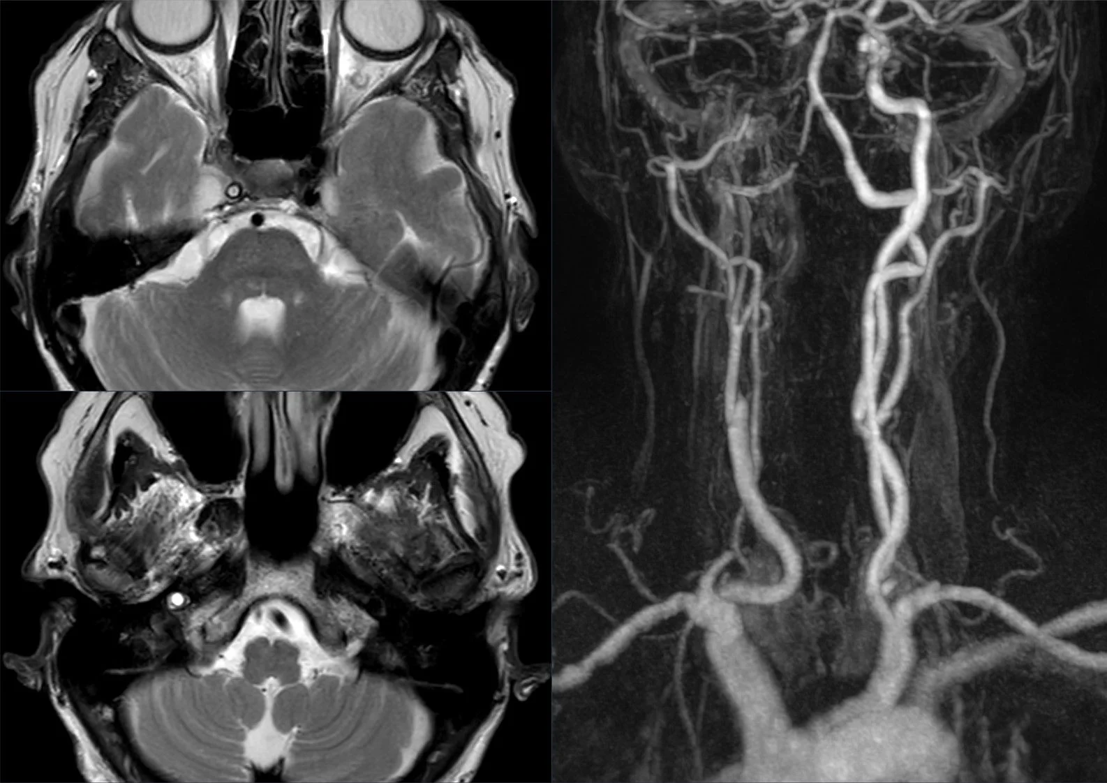
Long description
A long-segment (langstreckiger) occlusion of the right internal carotid artery extending intracranially, with absent flow void on the T2-weighted sequence and a corresponding MRA MIP.

Figure 2MRI perfusion in cerebral ischemia - (A) Improved collateral supply (B) Post-reperfusion hyperperfusion (C) Persistent hypoperfusion
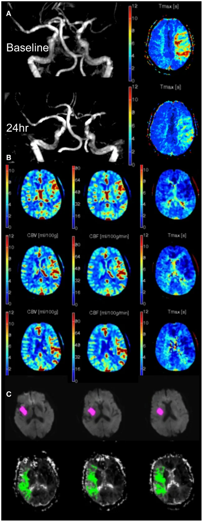
Long description
A composite open-access figure whose panel A is an MR angiography and Tmax map in a patient with a left middle cerebral artery occlusion at 5 and 24 hours.

Figure 3DWE MRI of cerebral infarction
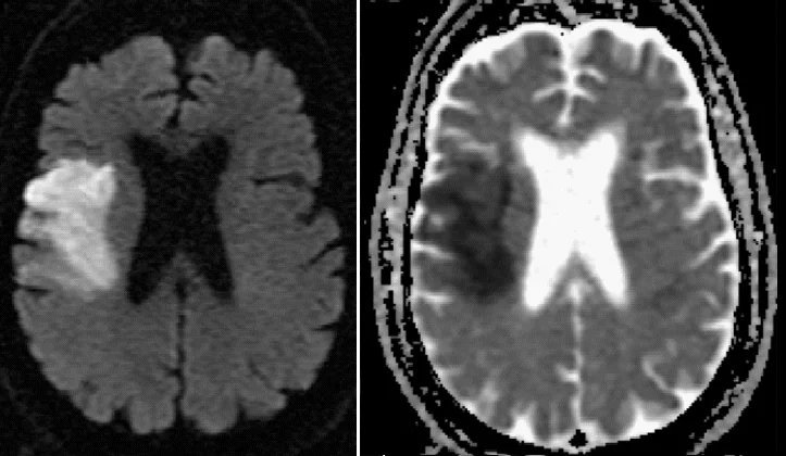
Long description
Axial brain MRI with DWI on the left showing a hyperintense (bright) region in the right middle lobe area and a corresponding hypointense region on the right ADC (or similar) view, indicating infarcted tissue.

Figure 4INFARCT
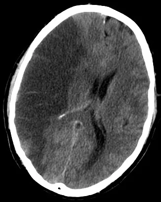
Long description
A CT scan of the brain showing a right middle cerebral artery infarct.

- Non-contrast CT of the head - immediate if any of the following apply: candidate for thrombolysis or thrombectomy; on anticoagulants; known bleeding tendency; depressed consciousness (Glasgow Coma Scale below 13); progressive or fluctuating symptoms; neck stiffness, fever or papilloedema; severe headache at onset [1]. Otherwise as soon as possible and within 24 hours of onset.
- CT angiography after the non-contrast scan if thrombectomy might be indicated, to look for large-vessel occlusion; add CT perfusion if thrombectomy may be considered beyond 6 hours of onset [1].
- MRI (DWI, FLAIR, blood-sensitive sequences) for suspected TIA, or when the diagnosis or vascular territory is uncertain. If MRI is done after a TIA, it should be performed on the same day as specialist assessment [1].
- Blood tests: full blood count, glucose, renal and liver function, coagulation if on anticoagulants, lipids later, HbA1c, troponin if chest pain or cardiac suspicion.
- ECG and cardiac monitoring: look for atrial fibrillation (acute, subclinical or paroxysmal; more monitoring finds more).
- Carotid (and intracranial) imaging: urgent carotid imaging in anyone who after specialist assessment is a candidate for carotid endarterectomy [1].
- Non-contrast CT's job in the first hours is only to exclude haemorrhage (and show major early ischaemia). A normal CT does not exclude ischaemic stroke.
- CTA asks: is there a large proximal occlusion that thrombectomy could remove?
- CTP/DWI asks: is there still salvageable brain (penumbra) beyond the standard time window?
- MRI-DWI after a TIA asks: did a small infarct occur, which changes secondary prevention?
- Carotid Doppler/CTA neck asks: is there an ipsilateral high-grade carotid stenosis worth operating on?
| Item | Value | Use |
|---|---|---|
| TIA duration cut-off | Under 24 hours [3] | Clinical definition |
| Thrombolysis window | Within 4.5 hours of onset (licensed) [1][2][6] | IV alteplase eligibility |
| "Number needed to treat" for rtPA | About 5 at 1.5 h; about 9 at 3-4.5 h [6] | Counselling |
| Thrombectomy window, anterior circulation | Within 6 hours; 6-24 hours with perfusion mismatch [1] | Endovascular referral |
| Thrombectomy window, posterior circulation | Up to 24 hours [1][6] | Basilar/PCA occlusion |
| Thrombectomy candidate criteria | Pre-stroke mRS less than 3; NIHSS greater than 5 [1] | NICE selection |
| Carotid surgery threshold | 50-99% NASCET (or 70-99% ECST) symptomatic stenosis [1] | Refer for carotid endarterectomy |
| Decompressive hemicraniectomy | Within 48 hours; NIHSS above 15; GCS item 1a at least 1; CT at least 50% MCA territory [1] | Malignant MCA infarct |
| 7-day stroke risk after TIA (ABCD2 greater than or equal to 4) | 5.3% versus 1.8% if below 4 [3] | Risk communication |
There is no laboratory test that diagnoses ischaemic stroke: it is clinical plus imaging. Important scores:
- NIHSS: quantifies the deficit. Higher scores predict worse outcome and identify candidates for thrombectomy (greater than 5) and hemicraniectomy (greater than 15).
- mRS (0 to 6): disability at outcome, standardised at 3 months.
- ABCD2 (0-7): Age (60 or over = 1), Blood pressure (140/90 or more = 1), Clinical features (unilateral weakness = 2, speech difficulty = 1), Duration (60 min or more = 2; 10-59 min = 1), Diabetes (= 1). A score of 4 or more groups "high-risk" TIA. It predicts 7-day stroke reasonably (5.3% risk at 4 or more versus 1.8% less than 4) [3].
- ABCD2 is not a triage tool on its own. NICE says do not use scoring systems such as ABCD2 to assess risk of subsequent stroke or to inform urgency of referral [1], and ESO recommends against using prediction tools alone [3]. Low scores still include people at high risk; patients with score 3 or less can still have a 1.8% 7-day risk, and 5.3% at 4 or more. Therefore every suspected TIA gets specialist review within 24 hours, with imaging as indicated.
- Clinical stroke syndrome typing (lacunar, total/partial anterior circulation, posterior circulation) guides investigation for cause.
The clock time is "last known well", not when symptoms were noticed. For patients who wake with symptoms, MRI with DWI-FLAIR mismatch (WAKE-UP concept) supports thrombolysis after 4.5 hours [2], and perfusion imaging can support thrombolysis at 4.5-9 hours if a mismatch is demonstrated [2]. Imaging-based patient selection is how "the window" lengthened from 4.5 hours to 9-24 hours in selected patients.
This page follows the AHA/ASA 2019 acute management guideline [7], AHA/ASA 2021 secondary prevention guideline [8], ESO thrombolysis/TIA/secondary prevention/acute blood pressure guidance [2][3][4][5], and NICE NG128 [1]. Where guidelines differ (for example, in whether to give thrombolysis beyond 4.5 hours on mismatch imaging), the value or range is stated with both positions.
Suspected stroke or TIA: acute pathway (teaching outline, follow local protocol)
- Sudden focal neurological deficitFAST in the community; ROSIER in the emergency department
- StabiliseAirway, breathing, circulation, capillary glucose, oxygen only if saturation under 95%
- Question: Brain imaging
Haemorrhage on CT
- Treat as haemorrhagic stroke; reverse anticoagulation; control BP
Ischaemia or no haemorrhage
- Question: Reperfusion eligibility
Last known well 4.5 h or less and eligible
- Intravenous alteplase 0.9 mg/kg (standard dose)BP 185/110 or less before infusion; keep to 180/105 or less during. No anticoagulant at this stage
Large-vessel occlusion on CTA
- Thrombectomy within 6 h, with IVT if still within window
- At 6-24 h with perfusion mismatch: thrombectomy as selected
Minor or non-disabling or TIA
- Aspirin 300 mgDAPT (aspirin + clopidogrel) for high-risk non-cardioembolic TIA/minor stroke, started within 24 h, total up to 21 days
- Admit to acute stroke unit; start secondary prevention
- FAST (Face, Arm, Speech, Time) in the community; ROSIER in the emergency department [1]. Capillary glucose should be checked to exclude hypoglycaemia.
- Document the last known well time (not when found). For "wake-up" stroke, use when the patient was last definitely well.
- Priority imaging: non-contrast CT, with CTA/CTP as above. Do not delay thrombolysis for multimodal imaging in a clearly eligible patient.
- Stroke patients should be admitted directly to an acute stroke unit staffed by a specialist multidisciplinary team [1].
Thrombolysis. Intravenous alteplase (rtPA) is licensed and effective up to 4.5 hours from onset, with a heavily time-dependent benefit (number needed to treat about 5 at 1.5 hours versus about 9 at 3 to 4.5 hours) [6]. The ESO IVT guideline strongly recommends IV alteplase within 4.5 hours; beyond 4.5 hours with plain CT alone it recommends against it, but with CT or MRI perfusion mismatch and no planned thrombectomy IV alteplase is recommended [2]. Post-stroke disability and multi-morbidity are not contraindications in themselves [2]. Standard (0.9 mg/kg) alteplase is recommended over low dose [2].
Tenecteplase. ESO preferred tenecteplase 0.25 mg/kg over alteplase 0.9 mg/kg when thrombolysis is given before thrombectomy in large-vessel occlusion [2].
Thrombectomy. For proximal anterior circulation occlusion: within 6 hours routinely, and between 6 and 24 hours in selected patients with salvageable tissue on perfusion or diffusion imaging [1]. Posterior circulation occlusion may be treated up to 24 hours [1][6]. Eligibility requires pre-stroke mRS under 3 and NIHSS greater than 5 [1]. Aspirin 300 mg should not be given before imaging excludes haemorrhage.
Acute blood pressure. Reduce blood pressure to under 185/110 mmHg before thrombolysis and keep it under 180/105 mmHg during infusion (these thresholds come from the original NINDS protocol and are endorsed by ESO and AHA/ASA) [5]. In acute ischaemic stroke without reperfusion, blood pressure is lowered only for hypertensive emergency (encephalopathy, renal failure, heart failure, aortic dissection, pre-eclampsia) [1], and ESO recommends against routine lowering; in patients not receiving reperfusion with blood pressure greater than 220/120 mmHg, careful reduction of fewer than 15% over 24 hours is reasonable [5].
Other acute measures: maintain glucose 4 to 11 mmol/L [1]; oxygen only if saturation below 95% [1]; avoid routine anticoagulation [1]; 300 mg aspirin daily within 24 hours once intracranial haemorrhage is excluded, continued at 300 mg for two weeks [1]. Decompressive hemicraniectomy within 48 hours for major MCA infarct with marked oedema and falling consciousness [1]. Nasogastric route if dysphagic, swallow screen on admission, avoid high-intensity mobilisation in the first 24 hours [1].
These are drawn from the alteplase licensing criteria and the ESO/AHA guidance. The shared list includes intracranial haemorrhage; coagulopathy or bleeding disorder; uncontrolled hypertension (greater than 185/110 mmHg before or 180/105 mmHg during infusion) [5]; active internal bleeding or recent major surgery/trauma/haemorrhage; recent ischaemic stroke or intracranial problem increasing bleeding risk; and clinical situations where the risk of haemorrhagic transformation outweighs benefit. For the exact and complete exclusion list used in your centre, follow the product label and the ESO IVT guidance [2].
- Repeat neuroimaging for neurological deterioration (recurrent ischaemia, oedema, haemorrhagic transformation) [6].
- Cardiac telemetry to detect paroxysmal atrial fibrillation.
- Secondary prevention medicines start after haemorrhage is excluded at 24 hours (usually aspirin 300 mg for 2 weeks, then a single antiplatelet long term) [6].
- Venous thromboembolism prophylaxis with heparin is not the same as acute anticoagulation for stroke, which is not routine.
All patients should be on an acute stroke unit; specialist physiotherapy, occupational and speech/language therapy, and early mobilisation as tolerated, but intensive mobilisation in the first 24 hours is avoided [1].
- Symptomatic intracerebral haemorrhage after thrombolysis: about 1.9% of treated patients [6].
- Early neurological deterioration from oedema, re-occlusion or haemorrhage.
- Malignant MCA infarct with raised intracranial pressure (consider hemicraniectomy).
- Post-stroke seizures, usually in the first 24 hours (focal or generalised) in larger infarcts.
- Aspiration pneumonia, urinary retention, constipation, pressure injuries, deep venous thrombosis.
- Infections and fall risk during early rehabilitation.
- Coma and death in brainstem or large infarcts.
Prognosis depends on age, stroke severity at presentation (NIHSS), collateral status, time to reperfusion and pre-morbid function. Outcome measures usually use the modified Rankin Scale at 90 days.
- Recurrent stroke risk: about 9 to 15% within 1 year [4], 26% within 5 years and 39% by 10 years in UK data [1]. The risk is front-loaded into the first 48 to 72 hours, which is the whole rationale of the TIA pathway below.
- Death at 30 days in UK admissions: about 13.6% [1].
- Disability: stroke is the single biggest cause of adult disability in the UK [1].
Long-term survival and function are best in those treated fastest; the benefit of reperfusion therapy is overwhelmingly decided in the first hours.
- Offer aspirin 300 mg daily immediately unless contraindicated, to be started at first presentation [1].
- Refer all suspected TIA for specialist assessment and investigation, to be seen within 24 hours [1][3].
- Do not use ABCD2 to triage urgency [1][3].
- After specialist assessment, consider MRI (with DWI) on the same day to determine territory of ischaemia [1].
- Urgent carotid imaging for anyone who, after assessment, is a candidate for carotid endarterectomy. Carotid endarterectomy is indicated for symptomatic stenosis 50-99% (NASCET) or 70-99% (ECST) [1].
- Patients with minor non-disabling stroke and TIA should be assessed within 24 hours [6].
This is where the greatest long-term benefit lies. Evidence-based targets [4]:
- Blood pressure: lower to under 130/80 mmHg (except subgroups at increased risk of harm). Consider combination treatment and out-of-office monitoring. Stroke trial data support intensive targets and a roughly 20% reduction in recurrent stroke with blood pressure lowering overall. [4]
- Lipids: HMG-CoA reductase inhibitor (statin) with LDL target below 1.8 mmol/L (70 mg/dL); add ezetimibe or a PCSK9 inhibitor if the target is not reached. [4]
- Antiplatelets: single antiplatelet (commonly aspirin or clopidogrel) long term. In high-risk non-cardioembolic TIA or minor stroke, dual antiplatelet with aspirin plus clopidogrel started within 24 hours and continued for up to 21 days then return to single antiplatelet; avoid dual antiplatelet after the first 90 days [3][4].
- Atrial fibrillation: ESO and other guidance prefer NOACs over warfarin for secondary prevention; exclude intracranial haemorrhage first. In disabling ischaemic stroke with AF, use aspirin 300 mg for the first 2 weeks before starting anticoagulation [1].
- Diabetes: aim for HbA1c below 53 mmol/mol (7%); consider pioglitazone in type 2 diabetes or insulin resistance, weighing risks. [4]
- Do not use DOACs for embolic stroke of undetermined source; and do not use anticoagulation routinely for acute stroke [1][4].
- Carotid endarterectomy or stenting for symptomatic 50-99% stenosis; management of intracranial stenosis is medical first (antiplatelet, risk-factor control), per AHA/ASA and ESO [8].
- Lifestyle: low-salt and Mediterranean-style diet, regular physical activity (supervised and safe), smoking cessation, weight control, treatment of sleep apnoea.
Stroke is not currently screened for in asymptomatic adults by imaging; primary prevention targets the vascular risk factors. After a TIA or stroke, secondary prevention is the screening moment: look for AF (ECG and prolonged monitoring), carotid disease, and vascular risk (blood pressure, lipids, glucose, urinalysis in young patients for hypercoagulability).
- Pregnancy/post-partum: stroke in pregnancy is rare but carries high risk; care is multidisciplinary (obstetrics, stroke, neurology). IV thrombolysis and endovascular therapy in pregnancy are not covered by routine protocols and need specialist advice; aspirin and anticoagulant choices depend on stage [8]. ESO IVT guidelines did not include this population [2].
- Children: RCPCH and specialist paediatric stroke services manage stroke in the under-16s; adult thrombolysis doses and windows do not apply and arterial ischaemic stroke in children needs a search for vasculopathy, congenital heart disease, infection and coagulopathy [1].
- Older adults: benefit from reperfusion is not reduced by age alone, and pre-stroke disability or frailty should not on its own exclude thrombolysis [2] (quality of evidence very low). Bleeding risk, comorbidity and wishes guide decisions.
- Anticoagulated patients: timing of thrombolysis follows the alteplase label and local protocol; specialist reversal pathways if needed.
- Immunocompromised and cancer: cancer is not an absolute contraindication [2], but caution is appropriate.
- A focused non-contrast CT (often available even where MRI is not) is the one test that separates ischaemic from haemorrhagic stroke and decides thrombolysis.
- Without CT, do not give thrombolysis; treat as stroke unit, control BP only for extremes, give aspirin only after 24 hours and clinical stability.
- Where thrombolysis or thrombectomy are unavailable, transfer to a centre that offers them early (telemedicine and "drip and ship" models help) [1].
- Use scales that do not need blood tests: FAST, NIHSS approximated by examination, mRS.
- A TIA clinic running within 24 hours cuts stroke risk dramatically (EXPRESS data showed 90-day recurrence fall from 10.3% to 2.1%) [3].
- Ensure swallow screening, infection control and basic rehabilitation; these cheap actions change outcome more than scans.
- In very low-resource settings, 300 mg aspirin, blood pressure control and a statin are the interventions with the highest cost-effectiveness for secondary prevention.
- Ischaemic stroke is arterial occlusion causing cerebral infarction; TIA is the same mechanism lasting under 24 hours with no lasting damage.
- About 85% of strokes are ischaemic and the penumbra is the target of all acute reperfusion treatment.
- Recognise common syndromes by territory; the middle cerebral artery is most often affected.
- Time windows: thrombolysis within 4.5 hours, thrombectomy (6 hours, plus selected 6-24 hours with perfusion mismatch), hemicraniectomy (48 hours).
- Pre-stroke mRS under 3 and NIHSS greater than 5 select thrombectomy candidates; BP (185/110 mmHg or lower) before tPA. [1]
- After haemorrhage is excluded: aspirin 300 mg within 24 hours, continued for 2 weeks, then a single long-term antiplatelet.
- Secondary prevention targets: BP under 130/80 mmHg, LDL under 1.8 mmol/L on a statin, sugar controlled, AF treated, and high-grade carotid stenosis operated on.
- DAPT (aspirin + clopidogrel) for high-risk non-cardioembolic TIA/minor stroke started within 24 hours, up to 21 days, then single antiplatelet; DAPT should not continue past 3 months for this purpose.
- ABCD2 is not a triage tool on its own; every suspected TIA gets specialist review within 24 hours with same-day MRI when imaging is needed.
- A functioning acute stroke unit and TIA pathway matter as much as any drug or scan.
Answer each one in your head first, then open the answer.
A 62-year-old man with atrial fibrillation wakes with right-sided weakness and difficulty speaking. He was last well at 22:00 the night before and found at 07:00. CT shows no haemorrhage but no established infarct. What should you consider regarding thrombolysis?
Show answerHide answer
He may be eligible for IV alteplase because the onset is unclear (wake-up) and an MRI DWI-FLAIR mismatch can select patients in this group for treatment within thrombolysis time limits (ESO IVT guideline); a large-vessel occlusion should also be sought for thrombectomy. Blood pressure must be 185/110 mmHg or lower before infusion. High NIHSS and a known cardioembolic source do not themselves exclude treatment. [5]
A 70-year-old woman has 40 minutes of right-arm weakness and dysphasia that fully resolve. Her ABCD2 score is 3. How should she be managed?
Show answerHide answer
She still needs urgent specialist review within 24 hours with brain imaging (same-day MRI with DWI if available), plus initiation of secondary prevention; an ABCD2 score under 4 does not mean low risk in the individual patient, and the ESO and NICE guidance advise against using ABCD2 alone to triage TIA.
A patient has an acute ischaemic stroke with a confirmed proximal MCA occlusion, presents 4 hours from onset, NIHSS 18, mRS 0. What reperfusion options apply?
Show answerHide answer
Intravenous alteplase plus endovascular thrombectomy; ESO preferred tenecteplase 0.25 mg/kg when thrombolysis is given before thrombectomy. Blood pressure is lowered to 185/110 mmHg or less before rtPA and kept to 180/105 mmHg or less during infusion. Aspirin and other antiplatelets wait until haemorrhage is excluded on post-treatment imaging.
Six months after an atherothrombotic stroke a patient's BP is 142/88 mmHg on no treatment and LDL 2.3 mmol/L on atorvastatin 20 mg. What would you change?
Show answerHide answer
Add or intensify blood-pressure treatment to a target below 130/80 mmHg; increase or add lipid-lowering to reach an LDL below 1.8 mmol/L (adding ezetimibe or a PCSK9 agent if needed); confirm a single antiplatelet is being taken; and check for atrial fibrillation and carotid stenosis.
Why can early high blood pressure after stroke sometimes be better left alone?
Show answerHide answer
In acute ischaemic stroke without reperfusion, cerebral autoregulation is disturbed and blood pressure supports perfusion of the ischaemic penumbra; lowering it can expand the infarct. Therefore, unless there is a hypertensive emergency or thrombolysis is planned, blood pressure is generally not reduced aggressively; adequate hydration and treatment of pain, bladder distension and infection are the right first steps.
Ischaemic stroke and TIA should be read alongside the following: atrial fibrillation and flutter; hypertension; lipid disorders; carotid stenosis management; anticoagulant drugs; antiplatelet drugs; stroke rehabilitation; cerebral haemorrhage; meningitis and infections of the CNS. Deep venous thrombosis and pulmonary embolism are the main systemic thrombotic mimics and comorbidities.
- National Institute for Health and Care Excellence. Stroke and transient ischaemic attack in over 16s: diagnosis and initial management (NG128). NICE guideline NG128, published 01 May 2019, last updated 13 April 2022. Accessed .
- Berge E, Whiteley W, Audebert H, et al. European Stroke Organisation (ESO) guidelines on intravenous thrombolysis for acute ischaemic stroke. Eur Stroke J. 2021;6(1):I-LXII. doi:10.1177/2396987321989865. Accessed .
- Fonseca AC, Merwick A, Dennis M, et al. European Stroke Organisation (ESO) guidelines on management of transient ischaemic attack. Eur Stroke J. 2021;6(2):CLXIII-CLXXXVI. doi:10.1177/2396987321992905. Accessed .
- Dawson J, Bejot Y, Christensen LM, et al. European Stroke Organisation (ESO) guideline on pharmacological interventions for long-term secondary prevention after ischaemic stroke or transient ischaemic attack. Eur Stroke J. 2022;7(3):I-II. doi:10.1177/23969873221100032. Accessed .
- Sandset EC, Anderson CS, Bath PM, et al. European Stroke Organisation (ESO) guidelines on blood pressure management in acute ischaemic stroke and intracerebral haemorrhage. Eur Stroke J. 2021;6(2):XLVIII-LXXXIX. doi:10.1177/23969873211012133. Accessed .
- Hurford R, Sekhar A, Hughes TAT, Muir KW. Diagnosis and management of acute ischaemic stroke. Pract Neurol. 2020;20(4):304-316. doi:10.1136/practneurol-2020-002557. Accessed .
- Powers WJ, Rabinstein AA, Ackerson T, et al. Guidelines for the Early Management of Patients With Acute Ischemic Stroke: 2019 Update to the 2018 Guidelines. Stroke. 2019;50(12):e344-e418. doi:10.1161/str.0000000000000211. Accessed .
- Kleindorfer DO, Towfighi A, Chaturvedi S, et al. 2021 Guideline for the Prevention of Stroke in Patients With Stroke and Transient Ischemic Attack. Stroke. 2021;52(7):e364-e467. doi:10.1161/STR.0000000000000375. Accessed .
Figure credits
- Figure 1: Hellerhoff, via Wikimedia Commons, CC BY-SA 4.0, CC BY-SA 4.0.
- Figure 2: Bruce C. V. Campbell, Geoffrey A. Donnan and Stephen M. Davis.Uploaded by Mikael Häggström, via Wikimedia Commons, CC BY 4.0, CC BY 4.0.
- Figure 3: edit Shazia Mirza and Sankalp Gokhale See also source article for additional image creators., via Wikimedia Commons, CC BY 4.0, CC BY 4.0.
- Figure 4: Lucien Monfils, via Wikimedia Commons, CC BY-SA 3.0, CC BY-SA 3.0.
Educational content for medical students and doctors. It is not a substitute for clinical judgement or local guidelines.