Subarachnoid haemorrhage
Sudden thunderclap headache from a ruptured brain aneurysm: diagnosis by CT and lumbar puncture, urgent securing of the aneurysm, and prevention of vasospasm.
- Subarachnoid haemorrhage (SAH) is bleeding into the space between the brain and the arachnoid membrane that covers it, most often from rupture of an intracranial aneurysm (aneurysmal SAH, aSAH).
- It classically begins with a thunderclap headache: the worst headache of life, maximal in 1 to 5 minutes, often with neck stiffness, vomiting and loss of consciousness.
- Urgent non-contrast CT of the head is the first test. If the scan is done within 6 hours of onset, diagnostic accuracy is highest. [1]
- If CT is negative and more than 6 hours have passed, a lumbar puncture is considered, performed at least 12 hours after onset to look for xanthochromia. [1]
- The ruptured aneurysm should be secured (coiled or clipped) as soon as possible, ideally within 24 hours, because rebleeding is most likely in the first 24 hours. [1][2]
- Every patient receives oral nimodipine (60 mg every 4 hours for up to 21 days) to reduce delayed cerebral ischaemia. Vasospasm, hydrocephalus and delayed cerebral ischaemia are the feared complications of the first two weeks. [2][4]
By the end of this page you should be able to:
- Recognise SAH from its history and examination, including thunderclap headache and its mimics.
- Plan the diagnostic pathway: CT timing, when a lumbar puncture is needed and when xanthochromia is diagnostic.
- Grade severity (WFNS, Hunt-Hess, modified Fisher) and explain what each predicts.
- Explain why early aneurysm securing is urgent and how coiling differs from clipping.
- Prescribe nimodipine with dose, and manage blood pressure, vasospasm and delayed cerebral ischaemia.
- Outline prognosis, follow-up imaging and prevention, and when to screen relatives.
DefinitionSubarachnoid haemorrhage
Bleeding into the subarachnoid space, the cerebrospinal-fluid-filled gap between the brain and its covering membranes. "Aneurysmal SAH" means the bleeding came from a ruptured intracranial aneurysm. When no aneurysm is found the bleeding is "non-aneurysmal", often perimesencephalic, and carries a much better prognosis.
SAH is a form of stroke. Most spontaneous SAH (about 80 to 85%) is aneurysmal. [3]
Classification matters for management:
- Aneurysmal SAH (aSAH): the culprit aneurysm must be found and secured. This page focuses on aSAH.
- Non-aneurysmal perimesencephalic SAH: blood limited to the cisterns around the midbrain, CT angiography usually normal; rebleeding and vasospasm are rare, so management is supportive.
- Traumatic SAH: from head injury, managed with the trauma, not as an aneurysm.
Clinically, SAH is also classified by severity: the World Federation of Neurosurgical Societies (WFNS) grade, based on the Glasgow Coma Scale and the presence of a motor deficit, and the older Hunt-Hess grade. Radiologically, the modified Fisher scale grades the amount of blood on the first CT and predicts vasospasm risk. [3]
The global incidence of aSAH fell from 10.2 per 100,000 person-years in 1980 to 6.1 in 2010, yet the burden persists because patients are relatively young. [2] In the UK the incidence is about 8 per 100,000 per year. [3] Intracranial aneurysms are present in about 3% of adults and are more common in women than in men; if one ruptures, about half of patients are under 50 and roughly two-thirds are women. [2]
Case fatality remains high: about 40% overall, with cumulative case-fatality of 25 to 30% on day one, 40 to 45% within the first week and 50 to 60% after the first month. About 12% of patients die before reaching medical attention. [2] [3]
Risk factors for carrying and rupturing an aneurysm. The incidence of rupture of an unruptured aneurysm is about 0.95% per year. Factors increasing rupture risk are hypertension, smoking, cocaine and alcohol use, and aneurysm features: size over 7 mm, anterior or posterior (rather than middle cerebral artery) location, irregular shape or a daughter sac. [3]
Other risks and associations: a family history (at least one first-degree relative with an intracranial aneurysm), autosomal dominant polycystic kidney disease, Ehlers-Danlos syndrome type IV, Marfan syndrome and neurofibromatosis type 1. [3]
Most intracranial aneurysms sit in the anterior circulation of the Circle of Willis (the ring of arteries at the base of the brain); the posterior circulation accounts for about 12%. [3]
Cerebral saccular aneurysms are acquired defects at arterial branch points. Haemodynamic stress thins the wall and breaches the elastic lamina, forming a sac. [3]
When an aneurysm ruptures, blood under arterial pressure jets into the subarachnoid space. Intracranial pressure rises suddenly, cerebral perfusion falls, and a brief global ischaemic state follows (early brain injury). [3] Blood in the basal cisterns irritates vessels and meninges, producing the headache, neck stiffness and photophobia. [3]
Three complications then dominate the next two weeks:
- Rebleeding. If the aneurysm is unsecured, about 10 to 15% of patients rebleed, half of them within the first 6 hours; if the aneurysm remains untreated, rebleeding reaches about 40% by one month. [3] [2]
- Delayed cerebral ischaemia (DCI) and vasospasm. Vasospasm (focal narrowing of cerebral arteries on imaging) is seen in about 70% of patients, but clinical DCI (new focal deficit or a fall in GCS of 2 or more lasting at least an hour) occurs in about 30%; the 2026 ESO guideline puts the proportion with reduced cerebral blood flow causing symptoms at about 20%. Vasospasm alone does not explain DCI, which is multifactorial: microthrombosis, cortical spreading depolarisation, inflammation. [3] [2]
- Hydrocephalus. Subarachnoid blood obstructs CSF circulation, causing ventricular enlargement in 20 to 30% of patients, often within 72 hours. [3]
Early brain injury (cytotoxic then vasogenic oedema, neuroinflammation) occurs in the first 72 hours and is a separate driver of poor outcome. [3]
The hallmark is a thunderclap headache: sudden, severe, "the worst headache of my life", reaching peak intensity within 1 to 5 minutes. [1] The pain is often occipital and may be accompanied by collapse, vomiting, photophobia and neck stiffness. A so-called "sentinel headache" (a sudden severe headache days before the rupture, probably a small leak) is reported in some patients. [3]
Some patients have no symptoms at first, and small leaks can be missed. [3] Focal deficits occur, for example an oculomotor nerve palsy with pupillary dilatation from a posterior communicating artery aneurysm. Seizures occur in about 6% of patients (early presenting seizures; the cumulative convulsive seizure risk in hospitalised patients is about 4%). [3] [4]
Other presentations include syncope, confusion, reduced consciousness, fever and meningism. Always ask a witness how fast the headache peaked; the rate of onset is the discriminating feature. [1]
Look for:
- Neck stiffness and photophobia (meningism).
- Reduced level of consciousness (GCS).
- Focal signs: oculomotor (cranial nerve III) palsy, hemiparesis, visual field loss.
- Subhyaloid or retinal haemorrhage (Terson syndrome) in some patients.
- Systemic effects: hypertension, fever, cardiac arrhythmia, hyponatraemia.
Check the fundi. Document an early baseline neurological examination because it is the yardstick for later deterioration.
Approach to suspected subarachnoid haemorrhage
- Suspected SAHThunderclap headache peaking in 1 to 5 minutes, or other red-flag features
- Question: Blood in the subarachnoid space?
Yes (SAH confirmed)
- CT angiography of the head without delay to find the aneurysm
- Transfer to a specialist neurosurgical centre
- Secure the aneurysm (coiling or clipping) at the earliest opportunity, ideally within 24 hours
- Start oral nimodipine 60 mg every 4 hours; manage blood pressure, euvolaemia, complications
No
- Question: Was the CT done within 6 hours of onset?
Yes
- Do not routinely do a lumbar puncture; seek alternative diagnoses and specialist advice
No, or scan equivocal
- Consider a lumbar puncture after at least 12 hours from onset
- Question: Xanthochromia (elevated bilirubin) present?
Yes
- Diagnose SAH; proceed to CT angiography and aneurysm management
No
- Think of alternative diagnoses
SAH can masquerade as migraine, meningitis or a neck problem. Conversely, a thunderclap headache has many other causes.
| Condition | Features that help separate it |
|---|---|
| Migraine | Builds over minutes to hours, not seconds; throbbing; aura; nausea and photophobia; usually a known pattern. [1] |
| Cervical arterial dissection | Neck pain and Horner syndrome; focal neurological signs; precipitated by minor trauma. |
| Reversible cerebral vasoconstriction syndrome (RCVS) | Thunderclap recurring over days; normal CT or convexity SAH; triggered by vasoactive drugs or postpartum. |
| Meningitis / encephalitis | Fever, rash, progressive illness; CT may be normal or show enhancement; LP shows cells, not xanthochromia. |
| Intraparenchymal haemorrhage / stroke | Focal deficit dominates; CT shows parenchymal blood. |
| Perimesencephalic (non-aneurysmal) SAH | Blood confined to perimesencephalic cisterns; CT angiography normal; benign course; no securing needed. [3] |
| Cerebral venous sinus thrombosis | Subacute headache, papilloedema, seizures; CT/venography. |
Figure 1TOF MRI angiography of right middle cerebral artery stenosis
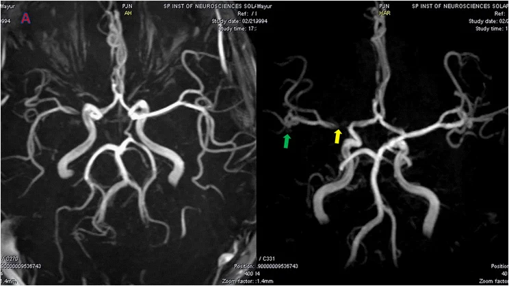
Long description
TOF MRI angiography demonstrating stenosis of the right middle cerebral artery.

Figure 2Aneurysma A cerebri media
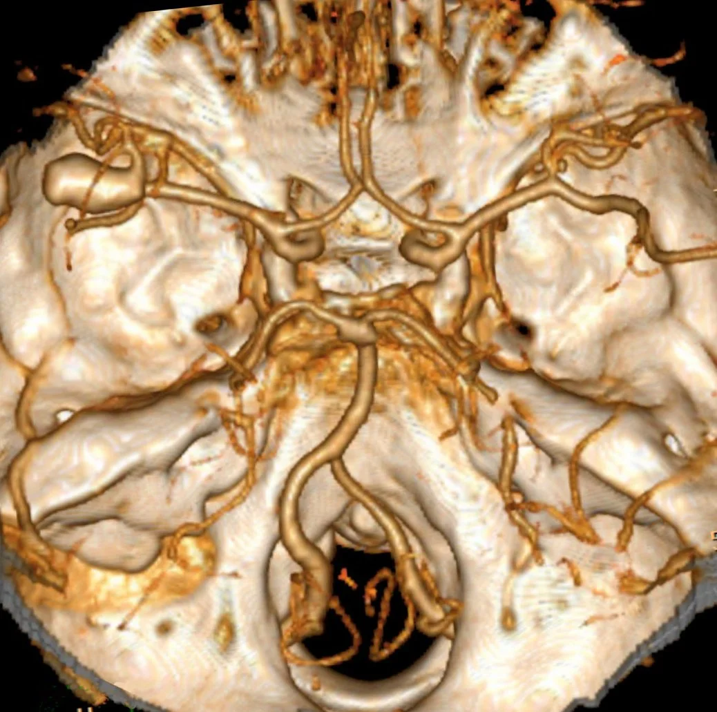
Long description
A left middle cerebral artery aneurysm shown in 3D volume rendering computed tomography of the brain.

Figure 3Aneurysem
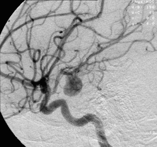
Long description
An angiogram of an aneurysm in a cerebral artery.

Figure 4SAB basal bei Aneurysma
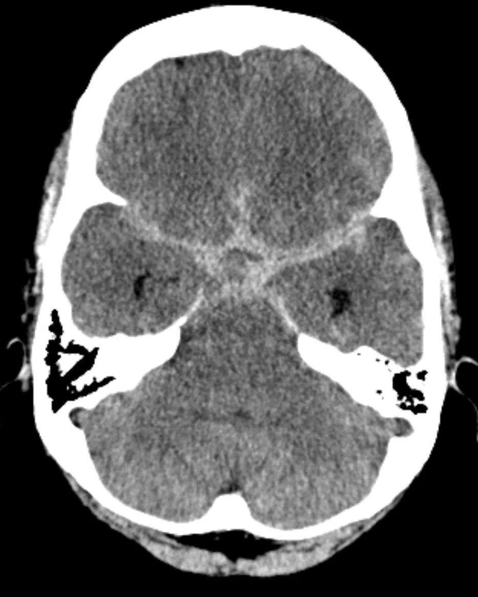
Long description
A CT image described as showing subarachnoid haemorrhage with hyperdense blood in the basal cisterns.

| Test | When and why | Key result or cut-off |
|---|---|---|
| Non-contrast CT head | First test in anyone with suspected SAH | Highest diagnostic accuracy within 6 hours of onset; modern CT is 100% specific if scanned in the first 6 hours; sensitivity falls to about 97% in the first 72 hours. [1] [3] |
| Lumbar puncture with spectrophotometry | When CT (done more than 6 hours after onset) is negative or equivocal | Wait at least 12 hours after onset (some sources note xanthochromia may appear from 6 to 12 hours); xanthochromia (elevated bilirubin) in the CSF confirms SAH; CSF xanthochromia can persist up to 2 weeks. [1] [3] |
| CT angiography of the head | Without delay in every confirmed SAH, to find the culprit aneurysm | Aneurysm plus a bleeding pattern that fits rupture confirms aSAH. [1] |
| Digital subtraction angiography (DSA) | When CT angiography is inconclusive and an aneurysm is still suspected | Gold standard for aneurysm detection; also allows endovascular treatment; complication rate about 1%. [3] |
| Magnetic resonance angiography (MRA) | Alternative when DSA is contraindicated (e.g. iodine allergy) | About 95% sensitive for aneurysms larger than 3 mm. [3] |
| Transcranial Doppler | Bedside monitoring tool only | Sensitivity over 90% but specificity about 71% and positive predictive value about 57%; not recommended to guide clinical management. [4] [1] |
Grading communicates severity, predicts outcome and structures multidisciplinary discussion. [3]
- WFNS grade (1 to 5): built on the Glasgow Coma Scale and the presence of a motor deficit. Grades 4 and 5 indicate a poor prognosis. [3]
- Hunt-Hess grade (I to V): the neurosurgical grade based on level of consciousness and focal signs, still widely quoted. [2]
- Modified Fisher score: a radiological score of the amount and location of blood on the first CT; higher grades predict a higher risk of vasospasm and DCI. [3]
- Other scores: SEBES, Hijdra and VASOGRADE are now also used. [3]
No single score should be used alone to decide transfer, treatment or suitability for surgery. [1]
The priorities are airway and oxygenation, blood pressure control, analgesia, seizure control and preparation for definitive treatment. [3]
- Airway: secure it if GCS is under 8 or falling. [3]
- Blood pressure: both European and US guidelines target control before securing. The ESO recommends treating if systolic blood pressure exceeds 180 mmHg, using analgesia, nimodipine and titratable antihypertensives, lowering slowly without overshoot. [3] The AHA/ASA 2023 guideline suggests a lower systolic target of under 160 mmHg with a titratable agent. [3] Mean arterial pressure above 95 mmHg may be detrimental. [3]
- Analgesia: effective pain relief, including opioids if needed, with the sedating and pupillary effects documented. [1]
- Antifibrinolytics: routine antifibrinolytic drugs before securing are not recommended; they reduced rebleeding but not poor outcome. [2]
- Drugs to avoid empirically: tirilazad, statins, magnesium sulphate and endothelin receptor antagonists are not recommended. [2]
- Early nimodipine for every patient (see below).
Rebleeding is most likely in the first 24 hours, so the aneurysm must be secured as soon as possible. [1] [3]
- The ESO consensus is to treat within 24 hours after ictus when the most experienced team is available. [2]
- The AHA/ASA 2023 guideline (as reported in the 2024 commentary by Al-Mufti and Mayer) recommends treating within 24 hours (class I, level B-NR), with complete obliteration of the rupture site whenever feasible. [4] [3]
- Delaying beyond 3 days is discouraged. [4]
Coiling versus clipping. Coiling (endovascular) and clipping (neurosurgical) are both options. After the ISAT trial, coiling is generally preferred where both are equally suitable, especially for good-grade anterior circulation ruptured aneurysms, because of better 1-year functional outcome; clipping remains preferred for some locations (for example middle cerebral artery aneurysms), for large parenchymal haematomas needing evacuation, for younger patients where durability matters, and when coiling is not feasible. [1] [3] [4] [2]
The choice must be made jointly by an interventional neuroradiologist and a neurosurgeon, weighing clinical condition, aneurysm anatomy and the blood pattern. [1] [4]
Nimodipine is the only drug shown to improve outcome in aSAH. Give it enterally to every patient as early as possible. [1] [3] [4]
- Dose: 60 mg by mouth every 4 hours, following the British aneurysm nimodipine trial schedule. [4]
- Intravenous nimodipine is reserved for specialist units when enteral administration is not possible. [1]
- Nimodipine does not reduce angiographic vasospasm; it reduces delayed cerebral ischaemia and improves outcome. [3]
- It causes systemic hypotension, so it is often reduced or stopped when hypotension complicates care. [4]
Keep euvolaemia and avoid hypotension. [1] [2] Neurocritical care bundles (ventilation, analgesia, delirium prevention, early mobilisation) improve outcomes in high-grade SAH. [4]
DCI most often appears 3 to 14 days after the bleed. [3] Management:
- Clinical detection: new focal deficit, decline in GCS of 2 or more, or unexplained fever. [3]
- Imaging: a non-contrast CT head is the first investigation for unexplained deterioration. [1]
- Fluids: ensure euvolaemia; [1]
- Vasopressors: if symptoms persist, an induced hypertension trial may be tried, but improvement is often temporary and the risk of complications (myocardial ischaemia, pulmonary oedema, worsening intracranial pressure) is real; hypervolaemia is not recommended. [1] [4]
- Endovascular treatment: angioplasty or intra-arterial vasodilators for refractory, critically reduced perfusion, as a rescue after multidisciplinary discussion; balloon angioplasty is last resort. [2]
- Transcranial Doppler: not recommended to guide management outside research. [1]
- Seizures: nonconvulsive seizures occur in up to 20% of patients (some sources report 10-30%); routine prophylactic anticonvulsants are not recommended because of cognitive harm. [3] [4]
Hydrocephalus develops in 20 to 30% of patients, often within 72 hours. [3] Drainage of CSF (via an external ventricular drain or, when safe, a lumbar drain) is considered when there is neurological deterioration from acute hydrocephalus. [1] Chronic hydrocephalus is treated with permanent CSF diversion. [1]
- Patients secured with coils should have follow-up imaging at least once, at or after 12 months. [2]
- Patients secured with clips usually need no long-term imaging if imaging within 6 months shows complete occlusion. [2]
- Partially occluded aneurysms, or those treated with intrasaccular or flow-diverting devices, need long-term follow-up imaging. [2]
- Patients at high risk of new aneurysms (young, strong family history, autosomal dominant polycystic kidney disease) should be scanned every 5 years. [2]
- Non-culprit (unruptured) aneurysms are managed after multidisciplinary review, considering size, location, lifetime rupture risk, treatment risk, comorbidity and the patient's wishes. [1]
- Rebleeding before securing (10 to 15%, half within 6 hours; about 40% by one month if untreated). [3] [2]
- Delayed cerebral ischaemia (about 30%, versus about 20% with symptoms in the ESO 2026 guideline). [3] [2]
- Vasospasm on imaging (about 70%). [3]
- Hydrocephalus (20 to 30%). [3]
- Seizures (about 6% early, versus about 4% cumulative convulsive in hospital; nonconvulsive seizures up to 20%, with some sources reporting 10-30%). [3] [4]
- Early brain injury and cerebral oedema (first 72 hours). [3]
- Terson syndrome (retinal haemorrhage). [3]
- Medical complications: fever, anaemia, hyperglycaemia, electrolyte disturbance, cardiac dysfunction. [3]
- Thrombosis/venous thromboembolism: manage according to risk; do not withhold antiplatelets or anticoagulants once the aneurysm is secured if they are otherwise indicated. [1]
- Long-term: cognitive and emotional impairment (memory, concentration, fatigue, depression); only about one-third of all patients regain independence in daily activities. [2]
Outcome depends on the severity of the initial bleed, complications in the first two weeks and the patient's age and comorbidity. Intact consciousness at presentation predicts a better outcome. [3] Survival has improved by about 17% over recent decades because of early diagnosis, early securing, nimodipine and intensive care. [3]
- Overall case fatality about 40%. [2]
- About one-third of all patients regain independence in daily activities. [2]
- Even survivors who recover physically often have cognitive deficits and do not return to their previous level of functioning. [2]
Most rebleeding, hydrocephalus and DCI occur in the first two weeks, which is why care is concentrated in a neurocritical care unit.
- Smoking cessation for everyone. [1]
- Blood pressure control after discharge, per standard guidelines. [1]
- Non-culprit aneurysms: multidisciplinary follow-up (see above). [1]
- Relatives: routine screening of all relatives is not supported; testing is usually limited to relatives with at least two first-degree relatives who have had an aSAH. [1]
- High-risk patients need 5-yearly scans for new aneurysms. [2]
- Older adults: outcome worsens with age; for patients over 70, the benefit of coiling versus clipping is uncertain. [4]
- Younger adults: surgical clipping may be favoured in patients under 40 for durability. [4]
- Pregnancy: aSAH in pregnancy is rare and requires combined obstetric, neurological and neurosurgical care; individual decisions about delivery mode and aneurysm securing follow the severity of the maternal condition.
- Children: aSAH is uncommon in childhood; familial aneurysm syndromes are more likely, so genetic and endocrine assessment is appropriate.
- A non-contrast CT head, available even in smaller hospitals, makes the diagnosis; learn to recognise thunderclap headache at the front door. [1]
- If CT is negative but suspicion remains, a lumbar puncture after 12 hours for xanthochromia confirms or refutes SAH. [1]
- Where neurovascular services are unavailable, secure transfer to a centre that treats aSAH is the priority; an untreated aneurysm can rebleed. [2]
- Nimodipine and blood pressure control are low-cost measures that can be started anywhere. [3] [4]
- Coiling and clipping require specialist infrastructure; referral pathways matter more than local heroics.
- SAH is a neurovascular emergency: severe sudden headache peaking within 1 to 5 minutes, often with neck stiffness and collapse. [1]
- CT within 6 hours of onset has the highest diagnostic accuracy; LP for xanthochromia (after at least 12 hours) is used when CT is negative later. [1] [3]
- Aneurysmal SAH demands finding and securing the culprit aneurysm, ideally within 24 hours. [2] [4]
- Coiling is generally preferred over clipping when both are suitable; multidisciplinary decision. [1] [4]
- Nimodipine 60 mg every 4 hours improves outcome despite not affecting vasospasm. [4]
- Blood pressure is controlled before securing; ESO recommends treating SBP over 180 mmHg, AHA/ASA 2023 suggests under 160 mmHg. [3]
- Complications that kill survivors are rebleeding, hydrocephalus and DCI; they occur mostly in the first two weeks. [2] [3]
- Hydrocephalus is treated with CSF drainage; DCI with euvolaemia and cautious, temporary induced hypertension. [1] [4]
- Follow-up imaging depends on treatment (coils at or after 12 months; clips usually need none if fully occluded within 6 months). [2]
- Screening relatives is usually restricted to families with at least two affected first-degree relatives. [1]
Answer each one in your head first, then open the answer.
A 45-year-old smoker is brought in with 'the worst headache of my life' of sudden, peak intensity, with vomiting and neck stiffness. Non-contrast CT head done 4 hours after onset is reported as normal. What should you do next?
Show answerHide answer
No further invasive testing is routinely required. A CT scan done within 6 hours of onset with highest diagnostic accuracy that is negative makes SAH unlikely; think of alternative diagnoses and seek specialist advice. [1] If the scan had been done more than 6 hours after onset, you would consider a lumbar puncture for xanthochromia performed at least 12 hours after onset.
Why is the aneurysm secured as early as possible after aneurysmal SAH?
Show answerHide answer
Rebleeding before securing occurs in about 10 to 15% of patients, with roughly half in the first 6 hours, and it carries very high morbidity and mortality. Securing the aneurysm is the only definitive prevention. [3]
A patient with good-grade anterior circulation aSAH has an aneurysm that could be coiled or clipped equally well. Which treatment is preferred and on what evidence?
Show answerHide answer
Primary coiling is preferred, because it gives superior 1-year functional outcome (class I, level A). Both remain reasonable for long-term outcome. [4]
Give the dose and duration of nimodipine in aSAH, and why it is given despite not affecting vasospasm.
A patient deteriorates on day 7 with a fall in GCS of 2 and no CT change. How do you define and manage this?
Show answerHide answer
A woman with aSAH has made a good recovery. Which of her relatives should be offered screening for aneurysms?
Show answerHide answer
Screening is usually limited to people with at least two first-degree relatives who have had an aneurysmal SAH. [1]
Acute headache evaluation and differential diagnosis · Intracranial aneurysm (unruptured) · Stroke: ischaemic stroke clinical features and treatment · Hydrocephalus · Cerebral oedema and raised intracranial pressure.
- National Institute for Health and Care Excellence (NICE). Subarachnoid haemorrhage caused by a ruptured aneurysm: diagnosis and management. NICE guideline NG228, published 23 November 2022. Accessed .
- Vergouwen MDI, Ramos-Pachon A, Terecoasa EO, Willett N, Cuadrado-Godia E, et al. European Stroke Organisation (ESO), European Association of Neurosurgical Societies (EANS) and European Society for Minimally Invasive Neurological Therapy (ESMINT) guideline on aneurysmal subarachnoid haemorrhage. European Stroke Journal 2026;11(5):aakag043. doi:10.1093/esj/aakag043. Accessed .
- Thilak S, Brown P, Whitehouse T, Gautam N, Lawrence E, Ahmed Z, Veenith T. Diagnosis and management of subarachnoid haemorrhage. Nature Communications 2024;15:1850. doi:10.1038/s41467-024-46015-2. Accessed .
- Al-Mufti F, Mayer S. Unpacking the 2023 American Heart Association Guidelines: The Ascendancy of Neuroendovascular and Neurocritical Care in Aneurysmal Subarachnoid Hemorrhage Management. Stroke: Vascular and Interventional Neurology 2024;4(4):e001264. doi:10.1161/SVIN.123.001264. Accessed .
Figure credits
- Figure 1: edit Shazia Mirza and Sankalp Gokhale See also source article for additional image creators., via Wikimedia Commons, CC BY 4.0, CC BY 4.0.
- Figure 2: Hellerhoff, via Wikimedia Commons, CC BY-SA 3.0, CC BY-SA 3.0.
- Figure 3: Lucien Monfils, via Wikimedia Commons, CC BY-SA 3.0, CC BY-SA 3.0.
- Figure 4: Hellerhoff, via Wikimedia Commons, CC BY-SA 3.0, CC BY-SA 3.0.
Educational content for medical students and doctors. It is not a substitute for clinical judgement or local guidelines.