Intracerebral haemorrhage
Non-traumatic bleeding into the brain parenchyma: causes by site, emergency imaging, blood pressure targets, reversal, surgery and prognosis.
- An intracerebral haemorrhage is a non-traumatic bleed into the brain parenchyma, with or without extension into the ventricles.
- Hypertensive small-vessel disease, cerebral amyloid angiopathy, vascular malformations and tumours are the main causes, and the site of the haematoma gives the first clue.
- The patient presents suddenly with focal deficit, headache and a fall in conscious level; non-contrast CT of the head makes the diagnosis within minutes.
- Early blood pressure lowering is offered for minor to moderate haematomas, anticoagulation is reversed urgently, and selected patients need neurosurgery.
- The ICH score estimates prognosis, but it must not become a self-fulfilling prophecy; most survivors need long-term blood pressure control and stroke-unit rehabilitation.
By the end of this page you should be able to:
- Define a spontaneous intracerebral haemorrhage and divide ICH into primary and secondary causes by anatomical site.
- Explain how small-vessel disease and cerebral amyloid angiopathy rupture, and why the haematoma expands in the first hours.
- Recognise the clinical picture and the bedside signs that separate a supratentorial from a posterior fossa or intraventricular bleed.
- Plan the investigations, interpret CT and CTA findings, and use coagulation results to choose a reversal strategy.
- Apply the current blood pressure targets, the ESO reversal recommendations and the surgical indications with their size thresholds.
- Explain the main prognostic scores, how complications are prevented, and secondary prevention after ICH.
DefinitionIntracerebral haemorrhage
A spontaneous, non-traumatic intraparenchymal bleed that may extend into the cerebral ventricles, and that is assumed to come from cerebral small-vessel disease when no macrovascular, neoplastic, infectious or haemodynamic cause is found. [1]
The distinction that matters most to management is primary versus secondary ICH. Primary ICH is the common kind: bleeding from a diseased small artery or arteriole in a hypertensive deep territory or in amyloid-laden cortical vessels. Secondary ICH is bleeding from a discrete structural lesion, such as an arteriovenous malformation, a cavernous malformation, a tumour or haemorrhagic transformation of an infarct. The clinical picture cannot separate the two, so imaging for an underlying cause is part of the routine work-up, especially in younger patients and in non-deep locations. [1]
Anatomical classification follows the haematoma site. A deep haematoma in the basal ganglia, thalamus, pons or cerebellum usually reflects hypertensive small-vessel disease. A lobar haematoma at the grey-white junction usually reflects cerebral amyloid angiopathy. A haematoma at an unusual site, at a young age, or with multiple simultaneous bleeds raises suspicion of a macrovascular lesion, a tumour or a coagulopathy. [1]
In 2019 the global incidence of spontaneous ICH was about 3.41 million cases, which is roughly 28.8% of all strokes, and the prevalence was about 20.6 million. ICH caused about 2.89 million deaths and 68.57 million disability-adjusted life years. Although age-standardised rates are falling, the absolute number of cases is rising with ageing populations. [1]
Geography matters. The relative importance of the five leading risk factors for DALYs lost to ICH, high systolic blood pressure, high body-mass index, ambient particulate matter pollution, high fasting glucose and smoking, varies between countries and regions. Up to 80% of patients have elevated blood pressure on admission. [1][2]
Spontaneous ICH today is best understood as a disease of the small vessels, with the site of the bleed predicting the vessel disease behind it. [1]
Hypertensive arteriopathy is the commonest cause in deep structures. Chronic pressure damages perforating arteries, producing lipohyalinosis and microaneurysms in vessels supplying the basal ganglia, thalamus, pons, cerebellum and deep white matter. The haematoma is therefore usually deep, single and occurs in a patient with long-standing hypertension. [1]
Cerebral amyloid angiopathy deposits amyloid-beta in the walls of cortical and leptomeningeal arterioles. The affected vessels become brittle and bleed into the lobar white matter, often as recurrent or multiple haematomas. Lobar ICH in an older patient, especially with multiple cerebral microbleeds or superficial siderosis on MRI, should make you think of this diagnosis. [1]
Vascular malformations include arteriovenous malformations, cavernous malformations, dural arteriovenous fistulae and aneurysms. They are the dominant cause of ICH in young adults and in children, and they can produce haematomas at any site, often with disproportionate subarachnoid or intraventricular blood. [1]
Intracranial tumours, metastases being more common than primary brain tumours, can present with haemorrhage into the mass or into the surrounding brain. A haematoma with unusual CT or MRI features, or one that does not fit the expected vascular distribution, needs a careful search for an underlying mass. [1]
Other relevant risk factors include older age, male sex in some populations, smoking, diabetes, dyslipidaemia of the low-cholesterol type reported in some cohorts, alcohol excess, anticoagulant and antiplatelet therapy, and very rarely vasculitis, moyamoya disease and reversible cerebral vasoconstriction syndrome. [1]
ICH is a two-stage disease. First the vessel leaks and creates the initial haematoma; then the haematoma expands and the clot itself damages surrounding tissue.
Small-vessel disease weakens the wall of perforating arterioles. When one ruptures, blood under arterial pressure dissects through the parenchyma and forms a haematoma. The haematoma destroys tissue directly by compression and disrupts axons and vessels in its path. Blood degradation products and plasma-derived factors trigger a local inflammatory response, adding secondary injury in the hours after ictus. [1]
Expansion is the reason for the early rush in management. Most haematoma enlargement occurs in the first 3 hours. It is associated with worse functional outcome and death. Early blood pressure control is aimed specifically at limiting this expansion. [1]
Raised intracranial pressure and herniation are the threats in large or posterior fossa bleeds. Intraventricular extension, which affects a substantial proportion of patients, can block cerebrospinal fluid flow and produce hydrocephalus. [1]
The typical onset is sudden, often during activity, but it may be indistinguishable from a major ischaemic stroke in the first minutes. The history and examination should answer three questions: where, how big, and why.
History. Sudden focal weakness or numbness, often maximal at onset. Headache is common and can be severe. Vomiting and a fall in conscious level are frequent, particularly with posterior fossa or intraventricular extension. A seizure may mark onset or follow minutes later. Cognitive symptoms and aphasia dominate in lobar bleeds.
Red flags for a large or expanding bleed include a sudden severe headache with vomiting, a decreasing Glasgow Coma Scale score, anisocoria, neck stiffness from intraventricular extension, and posturing or hypertension with bradycardia suggesting raised intracranial pressure. [1]
Examination should record the Glasgow Coma Scale, pupil reactions and the full set of cranial nerves, motor power, sensation and plantar responses, and then the circulation and respiratory pattern. Basal ganglia bleeds often give dense hemiparesis and hemisensory loss with gaze deviation. Thalamic bleeds can produce vertical gaze abnormalities and marked sensory loss with relatively spared power. Pontine bleeds classically cause coma with pinpoint pupils and long-tract signs. Cerebellar bleeds cause gait or limb ataxia, nystagmus, vomiting and an occipital headache, with a decreasing conscious level as the bleed expands. Intraventricular extension often presents with headache, meningism and a reduced conscious level. [1]
Always look for secondary causes at the bedside: tobacco stains and chronic hypertension, endocarditis or other infective clues, trauma marks, needle marks, anticoagulant tablets, pregnancy, or a history of radiation, tumour or known vascular lesion.
Figure 1CT Scan BGH
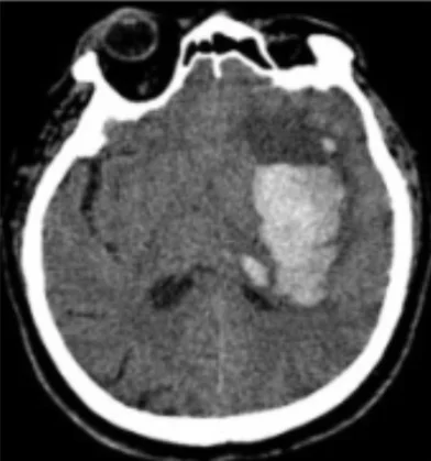
Long description
A CT scan illustrating a basal ganglia haemorrhage (BGH) on the left side of the brain, cropped from open-access source figure 2A in PMC2936928, whose caption describes a basal ganglia haemorrhage with perifocal oedema.

Every suspected ICH needs a non-contrast CT of the head as soon as possible. Acute blood appears hyperdense within minutes and the haematoma volume, intraventricular extension and mass effect are immediately visible. Repeat imaging is needed if the patient deteriorates, to look for expansion. [1]
| Test | What it is used for | Key cut-off or interpretation |
|---|---|---|
| Non-contrast CT head | Diagnosis; haematoma volume; IVH; herniation; hydrocephalus | Acute blood is hyperdense; reference first-line test [1] |
| CT angiography / CT venography | Spot sign; macrovascular cause; venous sinus thrombosis | Targeted screening before DSA in non-deep or young cases |
| MRI brain | Microbleeds, superficial siderosis, tumour, infarct, cavernoma, amyloid angiopathy | Best for cause; lobar microbleeds suggest amyloid angiopathy |
| Digital subtraction angiography | Definitive for AVM, aneurysm, fistula | Used when non-invasive imaging is suspicious or indeterminate |
| Full blood count, platelets | Anaemia, thrombocytopenia | Threshold for platelet cover by local policy |
| Coagulation (INR/PT/aPTT) | Warfarin and coagulopathy | INR 2.0 or higher needs PCC 30 to 50 IU/kg with IV vitamin K 10 mg; INR 1.3 to 1.9 needs 10 IU/kg [1] |
| Anti-Xa activity for FXa inhibitors | Quantify residual anticoagulant effect | Above 100 ng per mL supports use of andexanet alfa |
| Blood glucose, electrolytes, renal function | Hypo- or hyperglycaemia, hyponatraemia, drug clearance | Target a moderate glucose range; see below |
| ECG | Atrial fibrillation and other arrhythmias | Guides secondary prevention discussion |
| Blood pressure monitoring | Acute target and variability | Start treatment early; maintain below 140 mmHg in selected patients |
There is no separate diagnostic criterion for ICH beyond a correctly identified intraparenchymal bleed of non-traumatic origin on imaging, together with a search for its cause. The ESO/EANS 2025 guideline suggests using algorithms such as DIAGRAM to target investigation for a macrovascular cause rather than applying digital subtraction angiography to everyone. [1]
Two scores dominate prognosis.
- The ICH score combines age, haematoma volume, whether the bleed is infratentorial, the Glasgow Coma Scale and the presence of intraventricular haemorrhage. [1]
- The max-ICH score combines age, haematoma volume, intraventricular haemorrhage, the NIHSS at presentation and the use of oral anticoagulation. [1]
The original ICH score had an AUROC of 0.82 to 0.92 across six publications, and the max-ICH score an AUROC of 0.77 to 0.88 across three. [1]
These scores were developed and validated in an era before organised stroke units and modern care bundles, so they systematically overestimate death. The ESO/EANS 2025 guideline suggests limiting the ICH score to providing prognostic information, rather than using it as the primary or sole method for predicting outcome, because of the risk of a self-fulfilling prophecy. [1]
Several conditions look like or mimic ICH on first assessment. The table lists the commonest confounders.
| Condition | Feature that usually separates it from ICH |
|---|---|
| Acute ischaemic stroke | No blood on CT in the first hours; isodense or hypodense lesion rather than hyperdense bleed |
| Haemorrhagic transformation of an infarct | Prior infarct territory; petechial or gyral haemorrhage, often after reperfusion |
| Subarachnoid haemorrhage | Blood in basal cisterns and sulci; thunderclap headache; may coexist with lobar ICH |
| Subdural or extradural haematoma | Extra-axial crescent or lens-shaped collection, not intraparenchymal |
| Cerebral venous thrombosis | Haemorrhagic infarct in a venous territory; headache, seizures, papilloedema |
| Brain tumour with haemorrhage | Irregular mass-like lesion with disproportionate oedema or contrast enhancement |
| Hypertensive encephalopathy | Bilateral posterior white matter changes, often with visual symptoms; no focal bleed |
Suspected intracerebral haemorrhage: emergency approach
- Sudden focal deficit or fall in consciousnessTreat as stroke until proven otherwise
- Stabilise airway, breathing, circulation; check glucoseCorrect hypoglycaemia; do not chase blood pressure yet
- Urgent non-contrast CT headConfirms hyperdense blood; measure volume and look for IVH, hydrocephalus, mass effect
- Question: ICH confirmed?
Yes
- Question: Is the patient anticoagulated?
Yes
- Reverse urgently by drug: VKA with PCC plus IV vitamin K; FXa inhibitor with andexanet alfa if within 15 h and Anti-Xa above 100 ng/mL; dabigatran with idarucizumab 5 g IV total
No
- Continue to blood pressure and surgical assessmentAlso correct platelet count if markedly low
- Question: Minor to moderate haematoma (under 30 mL) with SBP 150 to 220 mmHg?
Yes
- Lower SBP toward under 140 mmHg within 6 hAvoid a drop of more than 70 mmHg and active reduction below 110 mmHg; start as early as possible, ideally within 2 h
No or large bleed
- Use cautionLarge haematoma, SBP above 220, planned evacuation or hydrocephalus need specialist judgement
- Question: Surgical indication?
Cerebellar ICH 15 mL or more, or brainstem compression or hydrocephalus
- Neurosurgical evaluation for evacuation plus or minus external ventricular drainage
Supratentorial, selected deep or lobar cases
- Consider minimally invasive removal in selected patients meeting ENRICH-type criteriaLobar or basal ganglia, 30 to 80 mL, GCS 5 to 14, surgery within 24 h
No
- Medical management on an organised stroke unitAvoid early treatment-limitation or universal DNR policies
- Prevent complicationsIntermittent pneumatic compression for DVT prophylaxis; moderate glucose control; normothermia within a care bundle; no routine prophylactic antiseizure medicines
No
- Reconsider diagnosisIschaemic stroke, venous thrombosis, SAH, tumour, SDH, encephalopathy
- Admit to organised stroke unit and start rehabilitationPlan secondary prevention before discharge
Admission to an organised stroke unit reduces death after ICH and is recommended when intensive care is not required. Early brain imaging, reversal and blood pressure therapy should happen in parallel rather than in sequence. [1]
A systolic pressure at or above 220 mmHg, a rapidly falling conscious level, signs of herniation, cerebellar signs with hydrocephalus, or a large intraventricular clot are all reasons to involve the neurointensive and neurosurgical teams immediately.
The ESO 2025 blood pressure update states that the net clinical benefit of intensive blood pressure lowering in acute ICH remains uncertain. Expert consensus supports early systolic blood pressure reduction to under 140 mmHg in small to moderate haematomas to limit haematoma expansion. [2]
The more detailed ESO/EANS 2025 recommendations are: [1]
- For adults with acute spontaneous ICH and systolic blood pressure of 150 to 220 mmHg, the overall balance of benefit and harm is uncertain, so patients should be offered enrolment in ongoing trials. [1]
- For minor or moderate ICH with haematoma volume under 30 mL, lower systolic pressure below 140 mmHg within 6 hours of onset, and ideally within 2 hours. Start as early as possible. [1]
- Avoid reducing systolic pressure by more than 70 mmHg from baseline and avoid actively pushing it below 110 mmHg.
- Be cautious in very high systolic pressure above 220 mmHg, in large haematomas above 30 mL, and when surgery is planned.
- Both during the acute phase and for up to 7 days in the subacute phase, keep systolic pressure below 140 mmHg once the target is reached. [1]
- Minimise variability; sustained control matters more than a single number.
The AHA/ASA 2022 guideline for spontaneous intracerebral haemorrhage is another major reference in this area, and the ESO 2025 update builds on the same trial base. Blood pressure management should always be tailored to the size of the bleed and the patient's baseline pressure rather than to a single fixed number.
Anticoagulant-associated ICH is an emergency of time and clotting factors. The ESO/EANS 2025 guideline and its predecessor on reversal recommend the following. [1]
Vitamin K antagonists. Give 4-factor prothrombin complex concentrate at 30 to 50 IU/kg when the INR is 2.0 or higher, or 10 IU/kg when the INR is 1.3 to 1.9, together with 10 mg intravenous vitamin K, to normalise and prevent rebound of the INR. Monitor the INR until sustained normalisation. [1]
Factor Xa inhibitors. For apixaban or rivaroxaban taken within 15 hours, or with proven anti-Xa activity above 100 ng/mL, and with symptom onset within 12 hours, consider andexanet alfa to reduce haematoma expansion. The ESO/EANS guideline notes continuing uncertainty about the balance of benefit and thromboembolic risk, and encourages enrolment in randomised trials. In hospitals without andexanet alfa, prothrombin complex concentrate may be considered, although its benefit is unproven and thromboembolic events are a concern. [1]
Dabigatran. Idarucizumab 5 g intravenously as two 2.5 g doses is recommended for ICH associated with this direct thrombin inhibitor. [1]
For ICH not related to antithrombotic drugs, the picture is uncertain. Recombinant activated factor VII remains experimental. Tranexamic acid reduces haematoma expansion at 24 hours but has not been shown to improve functional outcome or death, and its use is limited to situations where trial enrolment is not possible. [1]
For antiplatelet-associated ICH, platelet transfusion increases the likelihood of death or dependence at 90 days and is suggested against. Desmopressin has insufficient evidence and should be considered only in trials. [1]
Surgery aimed at haematoma removal reduces death after ICH overall, but the evidence is low certainty and much of it comes from older trials. [1]
- Standard craniotomy for supratentorial haemorrhage has not shown a clear benefit on death or functional outcome in major trials. [1]
- Minimally invasive surgery improved functional outcome and reduced death in the ENRICH trial for selected patients, predominantly lobar haemorrhage, with a haematoma volume of 30 to 80 mL, a Glasgow Coma Scale score of 5 to 14 and surgery within 24 hours of onset. It is the most promising surgical approach, but generalisability is limited. [1]
- Catheter placement plus thrombolysis, as in MISTIE III, did not significantly improve outcome; an end-of-treatment volume of 15 mL or less was, however, associated with better functional outcome. [1]
- Decompressive craniectomy without haematoma removal for severe deep supratentorial ICH showed a non-significant but clinically relevant reduction in death at the cost of more survivors with severe disability. [1]
- Cerebellar haemorrhage of 15 mL or more, or any cerebellar bleed with brainstem compression or hydrocephalus, is an indication for surgical evacuation, usually with external ventricular drainage when hydrocephalus is present. Survival was markedly higher with surgery in observational data. [1]
- Intraventricular haemorrhage is present in 30 to 40% of cases and hydrocephalus develops in about half of these. External ventricular drainage is a life-saving emergency procedure when hydrocephalus reduces consciousness. Adding intraventricular thrombolysis reduces death but at the cost of more survivors in a severely disabled state, and neuro-endoscopic removal reduces shunt dependence. [1]
Refer early when the patient deteriorates, when a secondary cause is suspected, when anticoagulation cannot be reversed locally, or when a cerebellar or posterior fossa bleed is present. Long-term follow-up should address blood pressure, rehabilitation, mood, epilepsy, cognition and the discussion of returning to work or driving.
Venous thromboembolism. Intermittent pneumatic compression is the evidence-based physical measure to reduce proximal DVT after stroke, including ICH, while graduated stockings alone are much less effective. Early prophylactic anticoagulation with low molecular weight heparin has not shown a clear benefit and comes with a possible bleeding risk; more evidence is needed. [1]
Fever. Active body temperature management as a single measure has not been shown to improve outcome, but managing fever is recommended as part of a care bundle. [1]
Glucose. Intensive control targeting 80 to 110 mg/dL is not recommended because of hypoglycaemia; a moderate range of 110 to 180 mg/dL is preferred. [1]
Intracranial pressure. Elevation is managed by standard measures: head elevation, avoiding hypoxia and hypercapnia, sedation, osmotherapy and, in selected cases, intracranial pressure monitoring or decompressive craniectomy. [1]
Seizures. Prophylactic antiseizure drugs are not recommended; treat clinically evident seizures. [1]
Infections. Antibiotic prophylaxis is not recommended outside bundles; treat proven infection. [1]
Care bundles for ICH combine several elements and are the direction of current practice: urgent reversal of anticoagulation within 90 minutes, invasive blood pressure control targeting a systolic pressure of 130 to 140 mmHg within 6 hours when it is above 150, and immediate neurosurgical referral in selected patients, together with fever and glucose management. [1]
ICH remains the deadliest major stroke subtype. Global 30-day case fatality is high and functional dependence is common among survivors. The prognosis depends on age, Glasgow Coma Scale at presentation, haematoma volume, intraventricular extension, infratentorial location and the cause. [1]
The ICH score remains the most extensively validated tool, but the ESO/EANS 2025 guideline warns against using it as the sole basis for withholding treatment. A low score should not be read as a guarantee of good recovery, and a high score should never be the only reason to limit care, especially in the first 24 hours. [1]
Case fatality is highest in the first days, and survivors commonly face cognitive, mood, fatigue and functional problems. Rehabilitation, mood screening and assessment of capacity, driving and work should be part of routine follow-up.
Secondary prevention centres on blood pressure. Intensive control after ICH reduced any stroke recurrence in pooled subgroup data, with a target at or below 130/80 mmHg recommended by general hypertension guidelines for patients with cerebrovascular disease. The ESO specifically notes that these post-acute targets come from general hypertension guidance rather than from an ICH-only trial. [1]
Other secondary prevention is genuinely uncertain: [1]
- Oral anticoagulation for atrial fibrillation after ICH reduces major cardiovascular events but increases recurrent ICH, and its net effect on death or dependence is uncertain. Decisions depend on whether the bleed was in a superficial lobar site suggesting amyloid angiopathy, the number of cerebral microbleeds, the level of blood pressure control and alcohol intake. Direct oral anticoagulants are preferred over vitamin K antagonists because of their lower intracranial haemorrhage risk. [1]
- Antiplatelet therapy was not associated with increased recurrent ICH in the RESTART trial, and it may be considered in patients with vascular comorbidity.
- Statins have mixed evidence: observational data associate treatment with lower mortality and recurrence, while the small ICH subgroup of SPARCL showed more recurrent bleeds. There is no ICH-only randomised evidence at present.
For the underlying cause, cerebral CT angiography, CT venography, MR angiography or MR venography are suggested to select patients for digital subtraction angiography. The ESO/EANS 2025 guideline recommends validated scores such as DIAGRAM for this pathway rather than universal angiography. [1]
Pregnancy. ICH in pregnancy and the puerperium is uncommon but serious, often from pre-eclampsia or eclampsia, reversible cerebral vasoconstriction syndrome, or a vascular lesion. Management should be multidisciplinary, involving obstetric, neurology, neurosurgery and anaesthetic teams. General ICH principles apply, but evidence for specific thresholds is limited.
Children. Structural lesions such as vascular malformations and tumours predominate over small-vessel disease. Consider inherited conditions and investigation for an underlying cause is mandatory. Rhabdomyoma aside, paediatric ICH is a pointer to a secondary cause.
Older adults. Cerebral amyloid angiopathy, falls and anticoagulation dominate. Drug interactions, frailty, polypharmacy and goals of care matter more, and the risk of a self-fulfilling prognosis is higher in this group.
- A non-contrast CT, even a single departmental scanner, is the single test that changes management. Learn to estimate haematoma volume using the ABC/2 method.
- If CT is unavailable, diagnose on clinical grounds and transfer; do not base blood pressure therapy on a guessed volume in a way that could cause harm.
- Prioritise stroke-unit principles: a dedicated area, vital sign monitoring, fever and glucose control, early mobilisation and swallowing assessment.
- Intermittent pneumatic compression or early mobilisation is a cheap and effective DVT measure.
- Keep simple reversal agents available: vitamin K and PCC where licensed; keep a protocol.
- Neurosurgery is often not available; early recognition of cerebellar and posterior fossa bleeds and prompt referral save lives.
- Discuss goals of care honestly; acknowledge the limits of prognostic scores.
- Intracerebral haemorrhage is a non-traumatic parenchymal bleed, often extending into the ventricles, usually from small-vessel disease.
- The haematoma site points to the cause: deep for hypertensive arteriopathy, lobar for cerebral amyloid angiopathy, any site in the young for a vascular malformation or tumour.
- Incidence is about 3.41 million cases a year worldwide, around 28.8% of all strokes, with about 2.89 million deaths. [1]
- Sudden focal deficit, severe headache and impaired consciousness are typical; non-contrast CT is the immediate diagnostic test.
- For minor to moderate haematomas under 30 mL, lower systolic pressure below 140 mmHg within 6 hours, avoiding a fall of more than 70 mmHg or active reduction below 110 mmHg. [1][2]
- Reverse VKA with 4-factor PCC plus IV vitamin K; idarucizumab for dabigatran; consider andexanet alfa for recent FXa inhibitor use. [1]
- Platelet transfusion worsens outcome in antiplatelet-associated ICH and is suggested against. [1]
- Minimally invasive surgery has the best recent evidence for selected lobar cases; standard craniotomy is not clearly beneficial. [1]
- Cerebellar bleeds of 15 mL or more with mass effect need surgical evacuation. [1]
- The ICH score overestimates death and must not be the sole basis for limiting care. [1]
- Secondary prevention centres on blood pressure control below 130/80 mmHg in the longer term; other measures remain uncertain. [1]
Answer each one in your head first, then open the answer.
A 62-year-old man with long-standing hypertension presents suddenly unable to move his right side and aphasic. Non-contrast CT shows a 12 mL hyperdense bleed in the left basal ganglia without intraventricular extension. SBP is 190 mmHg. What is the most likely cause and what is your blood pressure plan?
A 78-year-old patient is found with an INR of 3.4 after warfarin and a 22 mL lobar ICH. What reversal do you give?
Show answerHide answer
4-factor prothrombin complex concentrate 30 to 50 IU/kg with 10 mg intravenous vitamin K, then monitor the INR until sustained normalisation. [1]
A 55-year-old on rivaroxaban presents 4 hours after symptom onset with a 28 mL deep bleed. Anti-Xa activity is 180 ng/mL. What specific reversal should be considered?
Show answerHide answer
Andexanet alfa, because the last dose was within 15 hours and anti-Xa activity exceeds 100 ng/mL, provided onset was within 12 hours. Otherwise PCC may be considered, but its benefit is unproven. [1]
What haematoma volume threshold is used for early blood pressure lowering in the ESO 2025 small to moderate category?
Show answerHide answer
Under 30 mL. Above that threshold more caution is advised because the evidence for intensive lowering is limited. [1]
An ICH score of 4 is calculated for an 82-year-old patient. How should this score be used?
Show answerHide answer
As prognostic information only. It should not be the sole or primary method for predicting outcome, because of the risk of a self-fulfilling prophecy; scores from the pre-stroke-unit era systematically overestimate death. [1]
An 80-year-old has a 18 mL cerebellar haemorrhage with deteriorating consciousness and brainstem compression. What is the surgical threshold?
Show answerHide answer
Cerebellar ICH of 15 mL or more, or any size with neurological deterioration, brainstem compression or hydrocephalus, is an indication for surgical evacuation, usually with external ventricular drainage if hydrocephalus is present. [1]
Atrial fibrillation and anticoagulation. Hypertension. Subarachnoid haemorrhage. Stroke rehabilitation. Cerebral amyloid angiopathy. Anticoagulant drugs and reversal.
- Steiner T, Purrucker JC, Aguiar de Sousa D, et al. European Stroke Organisation (ESO) and European Association of Neurosurgical Societies (EANS) guideline on stroke due to spontaneous intracerebral haemorrhage. Eur Stroke J. 2025;10(4):1007–1086. doi:10.1177/23969873251340815. Accessed .
- Sandset EC, Palaiodimou L, Jahr SH, et al. 2025 update to European Stroke Organisation (ESO) guideline on blood pressure management in acute ischaemic stroke and intracerebral haemorrhage. Eur Stroke J. 2026;11(5):aakag004. doi:10.1093/esj/aakag004. Accessed .
Figure credits
Educational content for medical students and doctors. It is not a substitute for clinical judgement or local guidelines.