Intracranial haematomas and cavernous sinus thrombosis
Acute and chronic subdural and epidural haematomas after head injury, and septic or aseptic cavernous sinus thrombosis: imaging, surgery, antibiotics and.
- An intracranial haematoma is a collection of blood inside the skull. The three on this page are epidural haematoma (between skull and dura), subdural haematoma (between dura and arachnoid) and cavernous sinus thrombosis (clot in the cavernous sinus, usually septic).
- Epidural haematoma is classically arterial: a temporal skull fracture tears the middle meningeal artery, the clot is biconvex on CT and often confined by cranial sutures, and a lucid interval is seen in only about 14 to 21% of cases.
- Subdural haematoma is usually venous, from torn bridging veins; it is crescent-shaped on CT, crosses suture lines, and in older adults can follow a trivial fall. It is acute within 72 hours, subacute from 3 to 21 days, and chronic after 3 weeks. [2]
- Chronic subdural haematoma is a membrane-driven, inflammatory disease, not just old blood. It is commonest in frail older adults (about 17 per 100,000 per year overall, more than 350 per 100,000 in men aged 85 or over) and is projected to be the commonest cranial neurosurgical condition by 2030. [2]
- Cavernous sinus thrombosis usually spreads from an infection of the face or sinuses. It presents with fever, headache and an ophthalmoplegia that starts in one eye and spreads to the other; Staphylococcus aureus causes about two-thirds of cases. [3]
- For major head trauma, CT of the head is the first investigation, and the airway, breathing and circulation come before scanning. NICE (2023) advises a CT within 1 hour for adults with a Glasgow Coma Scale (GCS) score of 12 or less, and tranexamic acid 2 g intravenously within 2 hours when the GCS score is 12 or less and there is no active extracranial bleeding. [4]
- Emergency surgery is usually needed for an epidural haematoma larger than 30 mL, and for an acute subdural haematoma thicker than 10 mm or with a midline shift greater than 5 mm, regardless of GCS score. [2]
- Septic cavernous sinus thrombosis is treated with parenteral antibiotics (an anti-staphylococcal drug plus a third-generation cephalosporin and metronidazole) for 3 to 4 weeks, or at least 2 weeks beyond clinical resolution, together with anticoagulation, which retrospective reviews associate with lower mortality and morbidity. [3]
By the end of this page you should be able to:
- Distinguish epidural, subdural and cavernous sinus pathology by mechanism, anatomy and imaging shape.
- Explain the lucid interval and why it is not pathognomonic for an epidural haematoma.
- Recognise acute, subacute and chronic subdural haematomas, their risk factors and their CT appearances.
- State the operative and non-operative thresholds for epidural and acute subdural haematomas, and the role of burr holes, craniotomy, decompressive craniectomy and middle meningeal artery embolisation.
- Describe the clinical picture, microbiology, antibiotics and anticoagulation of septic cavernous sinus thrombosis.
- Apply a neurotrauma assessment and imaging sequence for head injury, including the emergency medicine, NICE, AAN and EAN/ESO guidance discussed here.
- Recognise how these conditions differ from each other and from mimics, and counsel on prognosis and prevention.
A head injury is any trauma to the head other than superficial injuries to the face. It may be closed or penetrating. The severity of an associated traumatic brain injury is usually graded by the initial post-resuscitation Glasgow Coma Scale (GCS) score: 13 to 15 mild, 9 to 12 moderate, and 8 or less severe, although some studies place patients with a score of 13 in the moderate category. [4][7]
Epidural haematoma is an extra-axial collection of blood in the potential space between the outer layer of the dura mater and the inner table of the skull. It is usually traumatic and most often arterial, classically from a branch of the middle meningeal artery torn by a temporal or parietal skull fracture. A venous epidural haematoma occurs when bleeding comes from a dural venous sinus or diploic vein, often after a skull fracture; these are commoner in children. Non-traumatic epidural haematomas are rare and may complicate frontal sinusitis, coagulopathy, vascular malformation or haemorrhagic tumour. [1]
Radiologically, an epidural haematoma may be classified by stage: type I is acute and occurs on the first day, sometimes with a swirl sign of uncoagulated blood; type II is subacute, days 2 to 4, often solid; type III is chronic, days 7 to 20, with mixed or lucent attenuation and contrast enhancement. It is usually supratentorial and most often occurs in the temporal region in adults. [1]
Subdural haematoma is a collection of blood in the plane between the dura mater and the arachnoid membrane. It most often comes from rupture of the thin-walled bridging veins that cross that space, under shearing forces from acceleration and deceleration. Acute subdural haematoma is usually an expanding extra-axial mass coupled with underlying parenchymal injury. Chronic subdural haematoma is now understood as a membrane-driven, inflammatory, vascular lesion: injury to the dural border cell layer leads to outer and inner membranes, with angiogenesis, hyperfibrinolysis, inflammation and repeated micro-haemorrhage into a new cavity. [2]
By convention an acute subdural haematoma is blood imaged within the first 72 hours after injury; subacute lesions occupy 3 to 21 days; chronic lesions are usually defined after 3 weeks. Mixed-density and acute-on-chronic collections mean that CT density reflects repeated bleeding rather than a simple single event. [2]
Cavernous sinus thrombosis is thrombosis within the cavernous sinus, the paired venous sinus beside the sella turcica. It is usually septic, arising from an underlying infection, but aseptic thrombosis occurs with trauma, surgery, pregnancy or a thrombophilia. [3]
DefinitionCavernous sinus thrombosis
Septic cavernous sinus thrombosis is usually a complication of an infection of the face, sinuses, orbit, pharynx, ear or teeth. The infection reaches the sinus through valveless facial and ophthalmic veins or by direct spread, and thrombosis then develops. Because the dural venous and cavernous system is valveless, infection can spread to the other cavernous sinus, to the meninges and brain, and via the jugular vein to the lungs.
Epidural haematoma occurs in about 2% of all head injuries and up to 15% of fatal head injuries. It is more frequent in adolescents and young adults, the mean age of affected patients ranges from 20 to 30 years, and it is rare after 50, because the dura adheres more firmly to the inner table with age. Males are more often affected. A 2022 meta-analysis estimated epidural haematoma in 8% of traumatic brain injuries worldwide and about 3.1 million people globally meet surgical indications for traumatic epidural haematoma each year. In the 65-centre CENTER-TBI study an epidural haematoma was present on the initial CT in 11% of all traumatic brain injuries and 16% of severe traumatic brain injuries. [1] [6]
Subdural haematoma remains one of the commonest focal lesions in severe traumatic brain injury and a major cause of early death and disability. Chronic subdural haematoma is predominantly a disease of older adults: one United States population study reported an incidence of 17.3 per 100,000 persons per year, reaching more than 350 per 100,000 in men aged 85 or over; if current demographic trends continue it is projected to be the commonest cranial neurosurgical condition by 2030. [2]
Traumatic brain injury itself is common. In the United States there were about 214,110 TBI-related hospitalisations in 2020 and 59,473 TBI-related deaths in 2021, and males were nearly twice as likely to be hospitalised and three times as likely to die as females. Among all patients with head trauma who seek medical attention, about 2% develop post-traumatic seizures, and about 12% of patients with severe traumatic brain injury develop them. The mortality of severe head injury in adults is 25 to 36% within the first 6 months, most deaths occurring within the first 2 weeks. [6][7]
Cavernous sinus thrombosis is rare: its annual incidence is estimated at 0.2 to 1.6 per 100,000 per year, partly because it accounts for only about 1 to 4% of cerebral venous and sinus thrombosis, which itself has an estimated incidence of 1.32 per 100,000 per year in Western Europe. It may be slightly more common in men in septic series (one review reported a 2 to 1 male to female ratio in 88 septic cases), despite the 3 to 1 female predominance of cerebral venous thrombosis overall. The incidence and mortality may be falling because of widespread antibiotics. [3][5] [6]
For epidural and subdural haematomas the essential risk factor is head trauma. Even minor trauma can cause a subdural haematoma in an older adult because cerebral atrophy stretches the bridging veins and enlarges the subdural space, so a ground-level fall may be enough. Other risk factors for traumatic intracranial bleeding include:
- alcohol use disorder
- seizures and falls
- anticoagulant or antiplatelet drugs (except aspirin monotherapy in the NICE head injury guideline)
- bleeding or clotting disorders, thrombocytopenia and liver disease
- cerebral atrophy and frailty
- birth trauma and, in infants, abusive head trauma ("shaken baby syndrome"), in which violent acceleration-deceleration ruptures bridging veins; retinal haemorrhage is a pathognomonic associated finding. [2][4]
For chronic subdural haematoma the initiating event may be so minor that it is not recalled. More important than the trauma are the cumulative risks: advanced age, cerebral atrophy, antithrombotic exposure and falls. Bilateral collections, hyperdense components, extensive membranes and poor brain re-expansion increase recurrence. [2]
Cavernous sinus thrombosis is usually septic. The facial infection classically lies in the nasolabial triangle (the area from the corners of the mouth to the bridge of the nose), and other sources are sinusitis (especially sphenoid and ethmoid sinusitis, possibly the commonest cause), otitis media, mastoiditis, periorbital or orbital cellulitis, pharyngitis, tonsillitis, tooth extraction or other dental work, and maxillofacial surgery. Aseptic causes are less common and include trauma, surgery, pregnancy and the puerperium, oral contraceptives or hormone replacement therapy, immunosuppression (diabetes mellitus, steroids, cancer or chemotherapy), severe dehydration, nephrotic syndrome and sickle cell disease. [3]
Thrombophilia increases the risk of both aseptic and, in some cases, septic cavernous sinus thrombosis. The list includes factor V Leiden mutation, prothrombin G20210A mutation, antithrombin III deficiency, protein C or protein S deficiency, elevated factor VIII levels, antiphospholipid antibody syndrome, hyperhomocysteinaemia and heparin-induced thrombocytopenia, as well as obesity. Infections can be bacterial (staphylococcus, streptococcus, pneumococcus, gram-negative and anaerobic organisms) or, less often, fungal (Aspergillus, Mucor, Coccidioides) or viral. [3]
The mechanism determines the clinical picture.
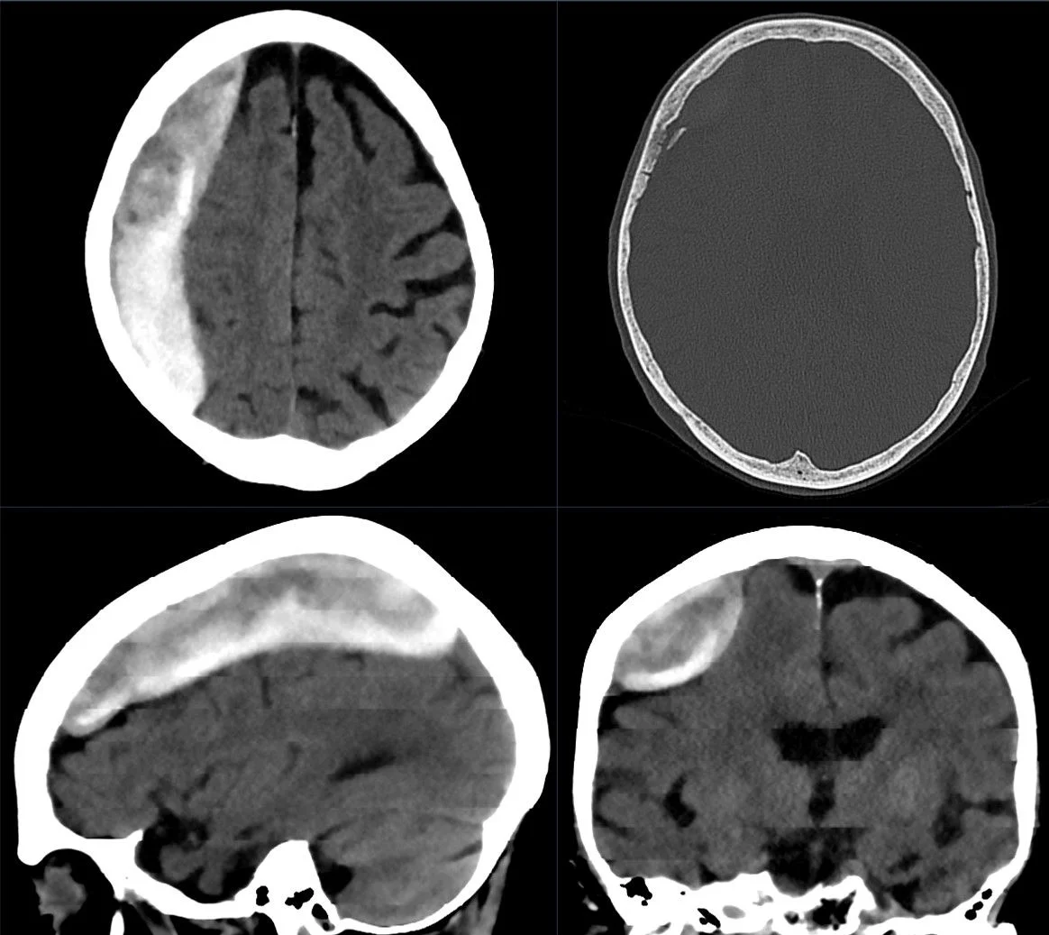
Traumatic epidural haematoma is classically an arterial bleed. Epidural haematomas occur most often in the temporal region in adults, and a fracture there classically tears a branch of the middle meningeal artery. Arterial blood then strips the dura from the inner table of the skull and forms a lentiform collection. Because the dura is firmly attached at cranial sutures, the haematoma is confined by sutures and has the characteristic biconvex shape, and active arterial bleeding explains the rapid expansion that can occur. Arteriovenous shunting within the haematoma has been proposed as one reason the classic lucid interval can occur, by transiently diverting blood flow and delaying the rise in intracranial pressure. Venous bleeding produces a slower-growing, later-detectable collection. [1]
Subdural haematoma follows the shearing forces that tear bridging veins between dura and arachnoid. A larger potential space, as in cerebral atrophy, lets cerebrospinal fluid accumulate and stretches these veins, so older and frailer patients bleed from minor trauma. Acutely, the clot expands as an extra-axial mass and is often accompanied by contusion, traumatic subarachnoid haemorrhage or diffuse axonal injury, which is why outcomes track overall injury severity, not clot volume alone. Chronic subdural haematoma enlarges instead by repeated micro-haemorrhage and exudation from the vascular outer membrane, driven by angiogenesis, hyperfibrinolysis and inflammation. [2]
Rising intracranial pressure and herniation are the dangerous steps in both. An expanding collection raises pressure, compresses or displaces brain tissue, effaces cisterns and, when compensation fails, causes uncal or transtentorial herniation with pupillary dilation, hyperreflexia, Babinski sign and depressed consciousness. Cushing reflex (hypertension, bradycardia and irregular respiration) is a late sign of brainstem compression. In adults a sustained intracranial pressure above 22 mmHg should be treated, cerebral perfusion pressure (mean arterial pressure minus intracranial pressure) should usually be kept between 60 and 70 mmHg, and systolic blood pressure should be kept at least 100 mmHg (ages 50 to 69) or at least 110 mmHg (younger or older). [1][7]
Cavernous sinus thrombosis follows from the anatomy. The cavernous sinuses lie on each side of the sella and contain the internal carotid artery, the sympathetic plexus and cranial nerve VI; the outer wall carries cranial nerves III, IV, V1 and V2. Because the system is valveless, an infection in the face can spread retrogradely and a thrombus can propagate through the intercavernous sinuses to the opposite side. Thrombosis and surrounding inflammation produce compressive cranial neuropathies and impaired venous drainage of the orbit (proptosis, chemosis, periorbital oedema and papilloedema). Carotid narrowing, vasculitis or haemorrhagic infarction can lead to stroke, and spread to the meninges or brain gives meningitis, empyema or abscess. [3]
The story matters more than almost any single finding.
- After head trauma, ask about the mechanism (falls, road-traffic collisions, assaults), loss of consciousness, the time of injury, alcohol or drug use, vomiting, seizures and any antithrombotic medication. An ill-fitting story in a child, or injuries inconsistent with the account, raises concern for abusive head trauma. [2][4]
- An epidural haematoma may cause an initial loss of consciousness, then a transient and complete recovery called a lucid interval, followed by rapid neurological deterioration. That sequence is seen in only about 14 to 21% of patients. Others are unconscious from the outset, or never lose consciousness at all, especially when the collection is small. The lucid interval is not pathognomonic for epidural haematoma: it can occur with any expanding intracranial mass. [1]
- Subdural bleeding may present acutely with headache, nausea, vomiting, somnolence, aphasia, hemiparesis, gaze deviation and a falling GCS. Seizures occur at presentation or during progression, especially with cortical contusions. [2]
- Chronic subdural haematoma presents over days to weeks. Gait decline is often more informative than headache in older adults. Patients may report repeated falls, slowed thinking, apathy, fluctuating confusion, urinary urgency, hand clumsiness, hemiparesis or facial droop. Bilateral collections are deceptive because lateralising signs are muted and the picture looks like global cognitive decline. Chronic subdural haematoma is regularly mistaken for ischaemic stroke, dementia, delirium, medication toxicity or normal-pressure hydrocephalus. [2]
- Cavernous sinus thrombosis begins with fever, headache, and periorbital pain and swelling, often with visual change. Symptoms may start abruptly or progress over days, usually in one eye first and then the other. Less common features include rigors, neck stiffness, facial numbness, confusion, seizures, stroke-like deficits or coma. [3]
- General neurotrauma survey. Secure the airway with cervical spine control, confirm breathing and oxygenation, and assess blood pressure and perfusion. A rapid neurological examination documents the GCS, pupil size and reactivity, and any asymmetry. Fixed or dilated pupils, extensor posturing or respiratory pattern change point to herniation and a time-critical need for decompression. [2][7]
- Focal pressure signs. In an enlarging epidural haematoma an ipsilateral dilated pupil from uncal herniation is often accompanied by contralateral hemiparesis, hyperreflexia and a positive Babinski sign; occasionally the weakness is ipsilateral (Kernohan phenomenon), a false localising sign. Posterior fossa haematomas are rare and patients may stay conscious until late, then abruptly lose consciousness, apnoea and die. [1]
- Chronic subdural examination blends cognitive and functional testing, with gait, baseline independence and frailty assessed alongside the GCS. [2]
- Cavernous sinus thrombosis almost always has eye findings (about 90% of patients): periorbital oedema (initially unilateral, usually bilateral), lid erythema, chemosis, ptosis, proptosis, restricted or painful eye movement, and less commonly papilloedema, retinal haemorrhages, decreased visual acuity (7 to 22%) and decreased corneal reflex. A "picket fence" fever pattern is described when the lateral wall of the sinus is involved. Ophthalmoplegia usually progresses rapidly from a sixth-nerve palsy (limited abduction, the most common early finding) to a complete external ophthalmoplegia (cranial nerves III, IV and VI), sometimes with internal ophthalmoplegia (a non-reactive pupil, either miotic from sympathetic fibre loss or dilated from parasympathetic loss), Horner syndrome, facial pain or numbness (V1 or V2) and decreased sensation over the cornea. [3]
- Numbers matter. In cavernous sinus thrombosis headache occurs in 50 to 90% of patients; decreased visual acuity in 7 to 22%; blindness in 8 to 15% of cases. [3]
The first question is which lesion the picture fits; the second is what else can mimic each. In trauma the CT shapes usually separate the two haematomas, but both can coexist.
| Condition | Shape on non-contrast CT | Crosses sutures / reflections | Typical source of bleeding or mechanism | Clinical course |
|---|---|---|---|---|
| Epidural haematoma | Biconvex or lens-shaped (lentiform), hyperdense | Does not cross sutures, may cross dural reflections (falx, tentorium) | Usually middle meningeal artery after temporal fracture; venous source possible | Rapid; lucid interval in 14 to 21%; can deteriorate within hours. [1] |
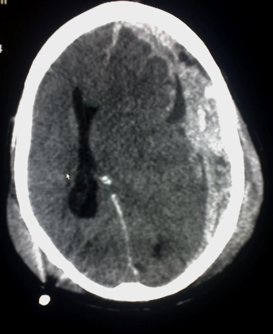
| Acute subdural haematoma | Crescentic, hyperdense | Crosses sutures, limited by falx and tentorium | Torn bridging veins; often combined with contusion | Gradual or rapid; mass effect and herniation; worse with low GCS and non-reactive pupils. [2] |
| Chronic subdural haematoma | Hypodense, mixed density or septated | Crosses sutures | Repeated micro-haemorrhage from outer membrane after minor trauma | Weeks of gait change, confusion or headache; mistaken for dementia or stroke. [2] |
| Subdural hygroma | CSF-density, uniform | Crosses sutures | CSF accumulation in subdural space | Low pressure-type headache; may evolve into chronic subdural haematoma. [2] |
| Cavernous sinus thrombosis | Filling defect or convex lateral wall of sinus on CTV/MRV; dilated ophthalmic veins | Bilateral sinus involvement possible | Septic spread or thrombophilia | Fever, headache and unilateral-to-bilateral ophthalmoplegia over days. [3] |
Mimics that should be kept in mind:
- For any extra-axial collection after apparently trivial trauma: ruptured aneurysm, cortical arteriovenous malformation, dural arteriovenous fistula, cerebral venous sinus thrombosis, tumour, coagulopathy. Consider vascular imaging when the history is atraumatic, discordant or radiographically atypical, when thunderclap headache precedes detection, or when subarachnoid blood accompanies the subdural clot. [2]
- For chronic subdural haematoma: normal-pressure hydrocephalus, dementia, delirium, medication toxicity, cerebrovascular disease, subdural empyema. [2]
- For cavernous sinus thrombosis: carotid cavernous fistula, meningioma and other tumours, metastasis, aspergillosis, Tolosa-Hunt syndrome, orbital apex syndrome, orbital cellulitis, sarcoidosis, syphilis and tuberculosis. MRI, blood cultures, lumbar puncture after imaging and sometimes angiography help to separate them. [3]
Figure 1Grosses epidurales Haematom nach Sturz 87W - CT - 001
Long description
Large epidural haematoma after a fall: a somewhat inhomogeneous, largely markedly hyperdense haematoma with a convex border to the brain parenchyma, with pronounced mass effect and initial subfalcine herniation; no skull fracture in this case.

Figure 2Trauma subdural
Long description
A CT scan described by its uploader as an example of subdural haemorrhage caused by trauma.

Non-contrast CT of the head is the first-line investigation for suspected traumatic intracranial injury: it is rapid, widely available and sensitive for significant extra-axial bleeding. MRI is reserved for cases where CT does not explain the neurological state, for vertex haematomas, isodense or bilateral collections, organised membranes or a suspected mimic, and for atypical histories. CT venography or MR venography is the right investigation when cavernous or cerebral venous sinus thrombosis is suspected. [2][3][4]
| Test or measure | What it tells you | Key value or cut-off |
|---|---|---|
| Non-contrast head CT | Primary investigation for acute extra-axial bleeding and mass effect | Biconvex (epidural), crescentic (subdural); acute blood hyperdense. [1] |
| Epidural haematoma volume | Size of collection | Non-operative if less than 30 mL, thickness less than 15 mm, midline shift less than 5 mm, GCS score greater than 8 and no focal deficit; operative if greater than 30 mL. [1] |
| Swirl sign on CT | Uncoagulated, actively bleeding blood within an epidural haematoma | Associated with larger volume and lower admission GCS; unfavourable 3-month outcome 29% versus 8% without the sign. [1] |
| Acute subdural thickness / midline shift | Mass effect of fresh subdural bleeding | Surgical if thickness greater than 10 mm or midline shift greater than 5 mm, regardless of GCS; in comatose patients (GCS less than 9) smaller collections need surgery if GCS falls by 2 or more points between injury and hospital assessment, if pupils are asymmetric or fixed and dilated, or if intracranial pressure is above 20 mmHg. [2] |
| Enhanced CTV / MRV | Detects cavernous and dural sinus thrombosis | Filling defect, widening or convexity of the lateral sinus wall, dilated ophthalmic veins; non-contrast CT may only suggest it. [3] |
| White blood cell count, C-reactive protein, ESR, D-dimer | Inflammation and thrombosis in cavernous sinus thrombosis | Often raised; blood cultures should be taken and are frequently positive. [3] |
| Glasgow Coma Scale | Severity of brain injury | 13 to 15 mild, 9 to 12 moderate, 8 or less severe. [7] |
| Coagulation studies, platelets, cross-match | Reversal and perioperative planning | INR, APTT, fibrinogen and drug-specific levels as indicated; reverse the relevant anticoagulant or antiplatelet drug. [2][6] |
United Kingdom (NICE)
NICE (NG232, 2023) sets out the CT pathway for head injury. For adults, a first CT is aimed for within 1 hour of any of: GCS score 12 or less on initial assessment; GCS less than 15 at 2 hours; suspected open or depressed skull fracture; signs of basal skull fracture; post-traumatic seizure; focal neurological deficit; more than one episode of vomiting. Where there has been loss of consciousness or amnesia, a CT is also needed within 8 hours (or within the hour if the patient presents more than 8 hours later) if age is 65 or over, if there is a bleeding or clotting disorder, after a dangerous mechanism of injury, or if there is more than 30 minutes of retrograde amnesia. Tranexamic acid 2 g intravenously is considered within 2 hours of injury when the GCS score is 12 or less and there is no active extracranial bleeding. A GCS score of 8 or less calls for early advanced airway management. [4]
United States
In the United States, CT of the head is the primary investigation for clinically important traumatic brain injury, and MRI is not the initial test. The Brain Trauma Foundation and Medscape emergency references continue to use the GCS (8 or less severe, 9 to 12 moderate, 13 to 15 mild) and treat intracranial pressure above 20 to 25 mmHg, hypotension and hypoxia as the principal secondary insults. [7]
Spinal imaging and vascular studies are needed in selected cases: a CT cervical spine when clinical criteria are met, MRI for ligamentous or spinal cord injury, and CT or MR angiography of the neck when vascular injury is suspected after a high-risk fracture or evidence of posterior circulation ischaemia. When a subdural haematoma is non-traumatic, discordant or atypical, CT or MR angiography or venography is suggested to exclude aneurysm, arteriovenous malformation, arterial rupture, dural fistula, tumour or cerebral venous sinus thrombosis. [2][4]
Lumbar puncture has no role in diagnosing the haematomas, and in cavernous sinus thrombosis it should be done only after imaging has excluded an intracranial mass, because a raised opening pressure and pleocytosis can be found even when cultures are negative. [3]
Repeat imaging is essential for any patient managed without immediate surgery: a monitored setting, serial neurological examinations, and a low threshold for repeat CT when consciousness, headache, pupils or focal deficits worsen. A follow-up CT about 6 hours after the first scan is typically recommended in conservatively managed epidural haematoma, and structured follow-up imaging is justified in chronic subdural haematoma with bilateral disease, hyperdense components, large postoperative cavities or limited access to urgent reassessment. [1][2]
These conditions have no single laboratory criterion. Diagnosis rests on compatible imaging.
- Imaging cut-offs for action are those given above and in the management tables. An epidural haematoma greater than 30 mL should be evacuated whatever the GCS. An acute subdural haematoma greater than 10 mm thick or with a midline shift greater than 5 mm is an indication for surgery regardless of conscious level. [1][2]
- Cavernous sinus thrombosis is diagnosed on neuroimaging (CTV or MRV) by a filling defect, widening and convexity of the lateral wall of the sinus (normally concave on coronal views), thrombosis of the superior ophthalmic vein and tributaries, and signs of venous congestion; non-contrast CT may only show orbital or sinus abnormalities. CT and MR imaging also exclude mimics such as abscess or tumour. [3]
- Transcranial Doppler or brain-tissue monitoring are not diagnostic on their own, and electroencephalography helps when non-convulsive status epilepticus is suspected or when AED decisions are needed. [7]
The principles are the same whatever the lesion: life-saving first, then prevention of secondary injury.
- Stabilise airway, breathing and circulation, with cervical spine control. Insert an advanced airway early in patients with a GCS score of 8 or less. [4][7]
- Give oxygen to keep saturation high; aim for a PaO2 above 13 kPa and a PaCO2 of 4.5 to 5.0 kPa unless raised intracranial pressure justifies brief hyperventilation. Avoid hypotension, because it increases mortality markedly, and avoid hypoxia. [4][7]
- Raise the head of the bed, treat pain, keep the bladder and fractures addressed, and correct glucose and temperature. Manage intracranial pressure with hyperosmolar therapy (mannitol or hypertonic saline) or, briefly, hyperventilation for impending herniation; cerebrospinal fluid drainage and decompression manage refractory pressure. [2] [7]
- Give tranexamic acid as in the NICE note above when the GCS score is 12 or less and there is no active extracranial bleeding. [4]
- Reverse anticoagulation and antiplatelet therapy when bleeding is present. For vitamin K antagonists this means intravenous vitamin K plus 4-factor prothrombin complex concentrate (or fresh frozen plasma); idarucizumab reverses dabigatran, andexanet alfa (where available) factor Xa inhibitors; protamine reverses unfractionated and low-molecular-weight heparin; platelet transfusion with desmopressin helps with aspirin or P2Y12 inhibitors. Treat thrombocytopenia, especially before surgery. [1][2]
- Involve the blood bank, anaesthetist and critical care team. [1]
- Discuss with a neurosurgeon any new, surgically significant abnormalities on imaging. Regardless of the scan, discussion is needed for a persisting GCS score of 8 or less, unexplained confusion lasting more than 4 hours, deterioration in the GCS score after admission, progressive focal deficits, a seizure without full recovery, a definite or suspected penetrating injury, or a cerebrospinal fluid leak. [4]
Non-operative management is appropriate only when all of the following hold: volume less than 30 mL, clot thickness less than 15 mm, midline shift less than 5 mm, GCS score greater than 8, and no focal neurological deficit. Such patients still need close observation with serial examinations and a follow-up CT, usually about 6 hours later. Some highly selected patients with volumes above 30 mL, high GCS (14 or more) and older age may still be observed in centres with close monitoring; conversely, even collections under 15 mL may need surgery when they lie in the temporal region, which carries a particular risk of deterioration. [1]
Surgical evacuation is recommended for an acute epidural haematoma larger than 30 mL, regardless of GCS score, and should be particularly early when the GCS score is below 9 with anisocoria. Outcomes worsen as the interval from first recorded neurological decline to decompression lengthens, especially beyond 2 hours. Standard treatment is craniotomy with clot evacuation and coagulation of the bleeding vessel; trephination (burr hole) is only a temporising measure where definitive neurosurgery is unavailable, because it rarely achieves adequate control or complete evacuation. Posterior fossa epidural haematoma management is individualised according to imaging and clinical state. [1]
Evacuation is recommended regardless of GCS score when either the haematoma thickness is greater than 10 mm or the midline shift is greater than 5 mm. In a comatose patient with a GCS score below 9 and a collection smaller than these thresholds, surgery is still indicated if the GCS score has fallen by 2 or more points between injury and assessment, if pupils are asymmetric or fixed and dilated, or if intracranial pressure is above 20 mmHg. Once indicated, surgery should proceed as soon as possible. [2]
Craniotomy is the standard evacuation technique because it allows rapid clot removal and haemostasis. If the brain is swollen and replacing the bone flap would recreate dangerous intracranial hypertension, decompressive craniectomy with duraplasty is appropriate; when swelling is modest, craniotomy with bone flap replacement avoids a later cranioplasty. Randomised evidence (RESCUE-ASDH) showed similar disability and quality of life at 12 months for primary decompressive craniectomy and craniotomy when both were feasible; craniotomy led to more later cranial surgery and craniectomy to more wound complications, so the choice is individualised. For patients in genuine clinical equipoise with modest lesions and a stable examination, conservative management is supported, although observation must include rescue surgery if the picture changes. [2]
The 2026 United Kingdom guidelines recommend surgery for patients with symptomatic chronic subdural haematoma, and it should also be considered in those with minimal or no symptoms if imaging shows a large-volume collection with mass effect (for example, a midline shift greater than 5 mm). [2]
- Burr-hole evacuation with closed-system drainage remains the reference operation for symptomatic chronic subdural haematoma because it combines high symptom relief with low invasiveness. One or two burr holes may be used; twist-drill craniostomy suits selected frail patients with liquefied, non-septated collections; craniotomy or mini-craniotomy is reserved for organised, solid, heavily septated, recurrent or calcified haematomas where simple drainage is unlikely to succeed. [2]
- Postoperative drainage reduces recurrence and improves survival compared with no drainage, and both subdural and subgaleal closed drainage are evidence-based. Intraoperative irrigation is supported by trial evidence showing higher reoperation rates when it is omitted, so washout plus drainage remains the most evidence-based standard approach. [2]
- Middle meningeal artery embolisation reduces treatment failure or reoperation in selected populations, such as high-recurrence-risk or recurrent chronic subdural haematoma and mildly symptomatic non-acute collections where rapid decompression is unnecessary. It should not replace surgery in significant mass effect, progressive deficits or impaired consciousness (it does not decompress), and UK guidance advises against its routine use. [2]
Adjunctive medical treatment is unsettled. Atorvastatin and tranexamic acid have shown promising signals but are not standard care. Corticosteroids should be avoided: dexamethasone worsened outcomes in Dex-CSDH and was not non-inferior to burr-hole drainage in DECSA. [2]
Reversal for acute or mixed subdural bleeding follows the same principles as for epidural haematoma: 4-factor prothrombin complex concentrate plus intravenous vitamin K for vitamin K antagonists; idarucizumab for dabigatran; andexanet alfa or PCC for factor Xa inhibitors; protamine for heparin; thrombocytopenia corrected, with individualised antiplatelet reversal. [2]
No randomised trials or expert guideline address this rare condition directly. Treatment rests on two primary modalities. [3]
Antimicrobial therapy: an anti-staphylococcal agent (vancomycin if methicillin resistance is present), a third-generation cephalosporin and metronidazole for anaerobic cover, plus antifungal treatment with amphotericin B where indicated. Parenteral therapy is usually given for 3 to 4 weeks, or at least 2 weeks beyond clinical resolution, and patients need close follow-up after antibiotics stop. [3]
Anticoagulation: most experts recommend unfractionated heparin or low-molecular-weight heparin for several weeks to several months in the absence of strong contraindications. The rationale is to stop thrombus propagation, prevent dissemination and possibly allow better antibiotic penetration. Retrospective reviews of septic cavernous sinus thrombosis suggest a fall in mortality from 40% to 14% and in neurological morbidity from 61% to 31% when anticoagulation is added to antibiotics, although this is not supported by prospective trials. Risk of systemic or intracranial bleeding or dissemination of septic emboli must be weighed. Cavernous sinus thrombosis is excluded from the usual cerebral venous thrombosis evidence base of the ESO guideline endorsed by the European Academy of Neurology, but the same general principles hold: parenteral anticoagulation in acute cerebral venous thrombosis, with a strong recommendation even in the presence of an intracerebral haemorrhage at baseline, and low-molecular-weight heparin suggested in preference to unfractionated heparin unless contraindicated (for example, severe renal insufficiency or a need for rapid reversal for neurosurgery). [3][5]
Surgical intervention: none directly on the cavernous sinus. Patients may need sphenoidectomy, ethmoidectomy, maxillary antrostomy, mastoidectomy, abscess drainage, craniotomy for empyema or brain abscess, orbital decompression, extensive debridement for mucormycosis or ventricular shunting. [3]
Corticosteroids are often given without proven benefit, but they are required if hypopituitarism is present, because infarction of the pituitary can occur. [3]
Supportive care includes management of raised intracranial pressure, seizures, stroke, venous infarction and hypopituitarism, plus ophthalmology and rehabilitation. Septic cavernous sinus thrombosis can spread to give meningitis, subdural empyema, brain abscess, stroke, cortical vein thrombosis, haemorrhagic infarction, septic emboli, pulmonary abscess and coma, so these complications must be anticipated. [3]
For moderate-to-severe traumatic brain injury, the AAN practice parameter (2003, reaffirmed 2024) concludes that prophylactic phenytoin is effective in reducing early post-traumatic seizures (relative risk 0.37, 95% confidence interval 0.18 to 0.74) but probably not late seizures (relative risk 1.05, 95% confidence interval 0.82 to 1.35). Phenytoin prophylaxis for early post-traumatic seizures in severe traumatic brain injury is supported by the AAN guideline. Current practice favours short courses of about 7 days or less, often using levetiracetam instead of phenytoin or fosphenytoin, which also has a more favourable adverse-effect profile; routine prolonged prophylaxis after uncomplicated chronic subdural drainage is not evidence-based. [2][6] [1]
Every patient who has had an intracranial haematoma needs planned follow-up:
- repeating imaging if symptoms persist, recur or worsen
- rehabilitation (physiotherapy for mobility and core strength, occupational therapy for independence, speech and swallowing, neuropsychological assessment where cognition is affected)
- repeat neurosurgical review, especially for patients who have been managed conservatively or who have a recurrence risk
- for chronic subdural haematoma, follow-up led by recurrence-risk phenotype, with surveillance imaging in bilateral disease, hyperdense components or large residual cavities
- for cavernous sinus thrombosis, follow-up of vision, cranial nerve deficits and endocrine function (to exclude panhypopituitarism), and of anticoagulation. [1][2][3]
- An abnormal CT after a head injury, or an unexplained embolus or focal deficit in a younger patient without an obvious source, should lead to urgent discussion with the relevant specialist team. [4]
- Anyone with a persisting GCS score of 8 or less, new anisocoria, worsening hemiparesis, recurrent vomiting or refractory severe headache after a known or suspected haematoma should be treated as progression until repeat imaging proves otherwise. [2]
- Neurosurgery should assess patients whose imaging shows surgically significant lesions or whose courses raise concern on the NICE criteria. [4]
- Treat cavernous sinus thrombosis as an emergency requiring simultaneous neurology, infectious disease, haematology and ophthalmology input. [3]
- Herniation and death from uncontrolled rise in intracranial pressure, most often with arterial epidural bleeding or a rapidly expanding acute subdural collection. [1][2]
- Recurrent or expansion haematoma, including acute-on-chronic subdural haematoma after a small re-bleed, and expansion of unstable arterial epidural clots. [1][2]
- Seizures from cortical irritation; prophylaxis is short-course only for severe traumatic brain injury. [6]
- Infection: meningitis, subdural empyema, brain abscess, sepsis and osteomyelitis after trauma or surgery. [3]
- Thrombotic, ocular and endocrine complications of cavernous sinus thrombosis: visual impairment or blindness, persistent cranial nerve deficits (about 50% with cavernous sinus thrombosis), stroke, panhypopituitarism, raised intracranial pressure. [3]
- Postoperative problems: atrial fibrillation after craniotomy, wound infection, recurrence of chronic subdural haematoma, and the long-term complications of decompressive craniectomy (wound problems, later bone replacement). [1][2]
- Systemic complications of critical care, such as pneumonia, delirium, acute kidney injury and venous thromboembolism. [1]
Prognosis follows severity, age and mechanism, and the speed of definitive care.
- Epidural haematoma generally has an excellent functional outcome when it is recognised and evacuated promptly; no deaths occurred in one prospective series when the pre-operative GCS score was 8 or higher. Delays worsen outcomes, especially when the interval from neurological deterioration to decompression exceeds 2 hours, and larger pre-operative volumes (above 50 mL) predict poorer recovery. In-hospital mortality for acute traumatic epidural haematoma is about 1.9%, with postoperative mortality around 6.8%. [1]
- Acute subdural haematoma is a major cause of death and disability in severe traumatic brain injury, with 25 to 36% six-month mortality in adults with severe head injury overall. Poor outcomes are predicted by advanced age, poor admission GCS, non-reactive pupils and associated injuries; about 80% of surgically treated elderly patients have a poor functional outcome. [2][7]
- Chronic subdural haematoma has a better but frailty-sensitive outlook: a good postoperative outcome was observed in 81% of cases in one Swiss multicentre cohort, yet baseline frailty is common and strongly influences later mortality and discharge destination. [2]
- Cavernous sinus thrombosis mortality is now roughly 8 to 13% with antibiotics and early diagnosis, but morbidity remains significant: fewer than 20% have some degree of visual impairment and about 50% persistent cranial nerve deficits, most often cranial nerves III and VI. [3]
- Recovery from traumatic brain injury overall is highly variable: even mild head injury can be followed by cognitive, emotional and somatic symptoms that usually settle over weeks to months, while moderate and severe injuries carry a substantial burden of long-term disability. [7]
- Primary prevention of head trauma is the most effective intervention: helmets for cyclists and motorcyclists, seat belts, fall prevention at home (removing trip hazards, grab bars, appropriate lighting), and safer sport and workplace practices. Public education about these measures is emphasised repeatedly in the source material. [1][7]
- Secondary prevention means recognising deterioration early: patients and families should be taught to return for urgent assessment with any new or worsening headache, focal neurological deficit or altered consciousness, and clinicians should not dismiss a subdural haematoma in an older adult as simple confusion. [1][2]
- Anticoagulation should be reviewed and monitored in patients at fall risk; no patient-level risk stratification can be given on this page, but any intracranial bleed in the presence of anticoagulants requires prompt reversal and imaging. [2][4]
- For cavernous sinus thrombosis, prevention consists of prompt treatment of facial, dental and sinus infections, good diabetic control and awareness of bilateral disease. Routine screening for occult malignancy or thrombophilia is not recommended after cerebral venous thrombosis, although thrombophilia testing may be considered in patients with high pre-test probability of severe thrombophilia (personal or family history, young age or no transient or permanent risk factor), because such screening has not been shown to reduce death, improve functional outcome or prevent recurrence. [3][5]
- Screening for abusive head trauma is a clinical responsibility rather than a population test: any infant or young child with an intracranial bleed and an inconsistent history or injuries should trigger child protection procedures and a full skeletal survey. [2][4]
Children. Subdural haematoma represents one of the intracranial injuries associated with abusive head trauma, with reported incidence of abusive head trauma up to 17 per 100,000 child-years. Overall, subdural haematoma in children younger than 2 years approaches 13 per 100,000 child-years, rising to 21 per 100,000 in those younger than 1 year. Infants are vulnerable because of weak neck muscles and large heads. Retinal haemorrhage frequently accompanies shaken baby syndrome. Birth trauma, coagulopathy and non-accidental injury must be considered. Epidural haematoma is rare in the elderly but occurs across the age range, including in children after skull fracture. NICE provides specific CT criteria for patients under 16, including suspicion of non-accidental injury, GCS score less than 14 (or less than 15 in babies under 1 year), or a bruise or laceration larger than 5 cm on the head in babies under 1 year. [2][4]
Young or paediatric cavernous sinus thrombosis is usually managed along the same principles, but evidence from trials and expert guidelines is limited, and specialist paediatric input is required. Routine paediatric indications such as lumbar puncture and airway management follow age-appropriate thresholds. [3][4]
Pregnancy and the puerperium. Pregnancy and the puerperium are transient risk factors for cavernous and other cerebral venous thrombosis, as are oral contraceptives and hormone replacement therapy. Patients with a previous cerebral venous thrombosis should avoid oestrogen-containing contraceptives, subsequent pregnancies appear to be safe, and prophylactic low-molecular-weight heparin should be considered throughout pregnancy and the puerperium. Intracranial haematomas in pregnancy are managed by the trauma and obstetric teams together, with the same neurological priorities. [5]
Older adults. Older adults are the group most likely to have chronic subdural haematoma after minor trauma, and outcomes are generally worse even when surgery is technically successful, because of frailty, anticoagulant use and reduced brain plasticity. Two patients with identical scans may merit different recommendations because the chance of returning to baseline differs markedly. Falls prevention, medication review and low thresholds for imaging in confusion or gait decline matter most here. [2][7]
Immunocompromised patients are at increased risk of both cavernous sinus thrombosis and its fungal causes (mucormycosis, aspergillosis), and of medical complications; imaging should be early and treatment prolonged. [3]
- CT is not always available. Where it is scarce, treat the clinical picture: after significant head injury a patient with a falling GCS, a unilateral dilated pupil or new focal deficits needs transfer to a centre with imaging and neurosurgery as fast as local transport allows. NICE emphasises direct transport to a trauma centre with the appropriate resources, and community referral should not be delayed. [4]
- Temporising burr holes may save a life in an acute epidural haematoma when definitive neurosurgery is unavailable, but they rarely achieve adequate haemorrhage control or complete evacuation; transfer should be arranged without waiting for complete resolution. [1]
- Careful clinical surveillance (GCS, pupils, vital signs) and repeat neurological assessment can be more valuable than a single low-quality scan. A monitored setting and serial examination are the minimum standard. [2]
- Cavernous sinus thrombosis can be treated without neurosurgery: source control, broad-spectrum antibiotics covering staphylococci, streptococci, gram-negative and anaerobic organisms, and anticoagulation unless contraindicated, with transfer arranged if complications develop. [3]
- Anticoagulation reversal requires planning in advance where PCC, vitamin K, protamine or andexanet may not be stocked. [1][2]
Suspected intracranial haematoma after head injury
- Primary survey and resuscitationAirway with cervical spine control, breathing, circulation, disability (GCS), exposure; avoid hypoxia and hypotension
- Suspect haematomaTrauma, altered consciousness, lucid interval, focal deficit, seizure, vomiting, bruising or depressed fracture
- Question: GCS score 8 or less, or falling?
Yes
- Advanced airway and urgent transferIntubate and ventilate, keep SBP at least 100-110 mmHg, correct anticoagulation, arrange neurosurgical discussion
No
- Neurological assessment and imaging decisionNICE/CT criteria; CT head within the time window
- CT of the head (and cervical spine if indicated)Confirm haematoma, volume, thickness, midline shift, skull fracture and concurrent injuries
- Question: What is the lesion?
Epidural >30 mL (or temporal risk)
- Craniotomy for evacuationEarlier if GCS <9 with anisocoria; <2 h from decline if possible
Acute subdural >10 mm or shift >5 mm
- Craniotomy; decompressive craniectomy if swollen brain
Small / stable collections
- Conservative management with close observationRepeat CT at about 6 hours for epidural; ready to convert to surgery
Chronic subdural, symptomatic
- Burr-hole evacuation with closed drainage; consider MMAE selectively
Cavernous sinus thrombosis suspected
- CTV or MRV plus blood culturesAntibiotics and anticoagulation without waiting for imaging result if strongly suspected
- Definitive care and preventionRehabilitation, recurrence surveillance, reversal of antithrombotics, fall and infection prevention
- Intracranial haematomas and cavernous sinus thrombosis are emergencies in which haematoma expansion and secondary insults (hypoxia, hypotension, raised intracranial pressure) determine outcome as much as the primary injury.
- Epidural haematoma is usually an arterial extra-axial collection with a biconvex, suture-confined appearance on CT; it causes only about a fifth of patients to have the classic lucid interval, and the lucid interval is not pathognomonic.
- Subdural haematoma is usually venous and crescent shaped, crossing sutures and bounded by dural reflections; acute collections behave as expanding masses, while chronic collections are inflammatory, membrane-driven cavities.
- Time windows matter: acute subdural is blood within 72 hours, subacute 3 to 21 days, chronic after 3 weeks; epidural volumes above 30 mL and acute subdural thickness above 10 mm or midline shift above 5 mm call for surgery.
- Non-operative thresholds (epidural under 30 mL, thickness under 15 mm, shift under 5 mm, GCS above 8, no focal deficit) identify patients who can be observed with repeat imaging.
- CT of the head is the primary investigation in acute trauma; MRI is reserved for unexplained deficits and mimics, and CTV or MRV diagnose cavernous and cerebral venous thrombosis.
- NICE (2023) sets CT time rules and a 2 g tranexamic acid bolus within 2 hours when the GCS score is 12 or less without active extracranial bleeding; a GCS score of 8 or less needs early advanced airway management.
- The AAN supports phenytoin for early post-traumatic seizures in severe traumatic brain injury, but not late seizures; modern practice favours levetiracetam for no more than 7 days.
- Chronic subdural haematoma is most common in frail older men after trivial trauma; burr-hole drainage with closed drainage is the reference operation, postoperative drainage reduces recurrence, and steroids should be avoided.
- Cavernous sinus thrombosis is usually septic and presents with fever, headache and a unilateral-to-bilateral ophthalmoplegia; Staphylococcus aureus causes about two-thirds. Treatment is antibiotics (anti-staphylococcal drug, third-generation cephalosporin, metronidazole; 3 to 4 weeks or 2 weeks beyond clinical resolution) plus anticoagulation (several weeks to several months).
- Prognosis is good when an arterial epidural haematoma is evacuated promptly and guarded in acute subdural and cavernous sinus thrombosis, where morbidity remains high despite adequate treatment.
- Prevention centres on head protection, fall prevention, infection control and early recognition, with prompt treatment of infections, good diabetic control and thrombophilia testing only for patients at high risk of severe thrombophilia.
Answer each one in your head first, then open the answer.
A 22-year-old cyclist is hit by a car and briefly knocked out. He is alert on arrival but returns an hour later with a headache and a dilated right pupil. What is the likely diagnosis, what shape will the bleed have on CT, and what must happen now?
Show answerHide answer
Epidural haematoma. The bleed is typically biconvex (lens-shaped) and confined by cranial sutures. It needs immediate neurosurgical evacuation, ideally within 2 hours of the decline in consciousness, because outcomes worsen with delay. A lucid interval is classic in only 14 to 21% of cases but should be suspected whenever recovery is followed by rapid deterioration.
State the CT thresholds that make an epidural haematoma and an acute subdural haematoma surgical diagnoses, independent of the GCS score.
Show answerHide answer
Epidural: volume greater than 30 mL. Acute subdural: thickness greater than 10 mm or midline shift greater than 5 mm. In a comatose acute subdural patient with smaller collections, surgery is still indicated if the GCS score has fallen by 2 or more points between injury and assessment, if pupils are asymmetric or fixed and dilated, or if intracranial pressure is above 20 mmHg.
A 78-year-old man on warfarin falls from standing height and over 3 weeks develops fluctuating confusion, gait decline and a mild left hemiparesis. CT shows a low-attenuation crescentic collection. Why is this called chronic, and what is the reference operation?
Show answerHide answer
The collection is chronic because it is older than 3 weeks (acute is under 72 hours, subacute is 3 to 21 days). The chronic form is a membrane-driven, inflammatory lesion rather than simple old blood, so clot volume does not explain it fully. The reference operation is burr-hole evacuation with closed-system drainage; steroids should be avoided.
Which clinical sequence best suggests septic cavernous sinus thrombosis, and how does it usually spread?
Show answerHide answer
Fever with headache and painful periorbital swelling and proptosis, starting in one eye and progressing to the other, accompanied by a cranial nerve palsy (most often limited eye abduction from cranial nerve VI). The infection usually originates in the nasolabial triangle or sinuses and spreads through valveless facial and ophthalmic veins into the cavernous sinus, then across the intercavernous sinuses.
A patient has septic cavernous sinus thrombosis after ethmoid sinusitis. What empiric antibiotics and what anticoagulant strategy are appropriate, and for how long?
Show answerHide answer
Empiric parenteral antibiotics: an anti-staphylococcal agent (vancomycin if methicillin-resistant Staphylococcus aureus is possible), a third-generation cephalosporin and metronidazole, plus amphotericin B if fungal disease is suspected. Duration is 3 to 4 weeks, or at least 2 weeks beyond clinical resolution. Most experts add unfractionated or low-molecular-weight heparin for several weeks to several months, which retrospective reviews associate with lower mortality and neurological morbidity.
Which organism causes about two-thirds of cavernous sinus thrombosis cases, and which additional groups should be remembered?
Show answerHide answer
Staphylococcus aureus (including methicillin-resistant strains). Also remember Streptococcus species (about 20%), Streptococcus pneumoniae (about 5%), gram-negative organisms such as Proteus, Haemophilus and Pseudomonas, anaerobes such as Bacteroides, Fusobacterium and Actinomyces, and, in immunocompromised patients, Aspergillus, Mucor and Coccidioides.
How do the imaging shapes of epidural and acute subdural haematoma differ, and why does that follow from their anatomy?
Show answerHide answer
Epidural bleeding strips the dura from the inner table and is limited by its firm attachment at cranial sutures, so it is biconvex. Subdural blood accumulates beneath the dura where it is not suture-limited, so it layers along the brain surface as a crescent and crosses sutures (though it is limited by the falx and tentorium).
Traumatic brain injury; Emergency management of head injury; Cerebral venous thrombosis; Intracerebral haemorrhage; Stroke; Child maltreatment and abusive head trauma; Anticoagulation; Coagulation and thrombosis; Neurocritical care.
- Khairat A, Margetis K, Waseem M. Epidural Hematoma (StatPearls, last update 7 July 2025). StatPearls [Internet]. Treasure Island (FL): StatPearls Publishing; 2025 Jan-. Accessed .
- Pierre L, Julien C, Margetis K, Kondamudi NP. Subdural Hematoma (StatPearls, last update 24 May 2026). StatPearls [Internet]. Treasure Island (FL): StatPearls Publishing; 2026 Jan-. Accessed .
- Plewa MC, Hall WA. Cavernous Sinus Thrombosis (StatPearls, last update 16 June 2025). StatPearls [Internet]. Treasure Island (FL): StatPearls Publishing; 2025 Jan-. Accessed .
- National Institute for Health and Care Excellence. Head injury: assessment and early management (NG232). NICE guideline, published 18 May 2023. Accessed .
- Ferro JM, Bousser MG, Canhao P, Coutinho JM, Crassard I, Dentali F, di Minno M, Maino A, Martinelli I, Masuhr F, Aguiar de Sousa D, Stam J. European Stroke Organization guideline for the diagnosis and treatment of cerebral venous thrombosis, endorsed by the European Academy of Neurology. Eur Stroke J. 2017;2(3):195-221. doi:10.1177/2396987317719364. Accessed .
- Chang BS, Lowenstein DH, Quality Standards Subcommittee of the American Academy of Neurology. Practice parameter: antiepileptic drug prophylaxis in severe traumatic brain injury. Neurology. 2003;60(1):10-6. doi:10.1212/01.WNL.0000031432.05543.14. Accessed .
- Olson DA, Berman SA. Head Injury (Medscape reference, updated 24 December 2024). Medscape, neurology reference. Accessed .
Figure credits
Educational content for medical students and doctors. It is not a substitute for clinical judgement or local guidelines.