Neurocutaneous disorders
Neurofibromatosis 1 and 2, tuberous sclerosis complex, Sturge-Weber syndrome and von Hippel-Lindau disease: genetics, features, surveillance and treatment.
- A neurocutaneous disorder is a group of multisystem hereditary conditions in which benign or malignant tumours and malformations develop in the skin and the nervous system together; the classic examples are neurofibromatosis 1 and 2, tuberous sclerosis complex, Sturge-Weber syndrome and von Hippel-Lindau disease.
- Each is a germline tumour-suppressor or mosaic-activating disorder, inherited in an autosomal dominant pattern or arising de novo, with a distinctive skin marker that usually appears before the brain, eye or kidney complications.
- The skin sign is the examination clue: café-au-lait macules and neurofibromas in neurofibromatosis 1; vestibular schwannomas and posterior subcapsular cataracts in neurofibromatosis 2; hypopigmented macules and facial angiofibromas in tuberous sclerosis complex; a facial port-wine birthmark in Sturge-Weber syndrome; and retinal haemangioblastomas in von Hippel-Lindau disease.
- There is no cure for the underlying tendency, so management is surveillance and early treatment: magnetic resonance imaging (MRI) and clinical examination on set schedules, surgical or medical reduction of dangerous tumours, and, for tuberous sclerosis complex, medicines that block the mammalian target of rapamycin (mTOR) pathway.
- Early diagnosis matters because each condition has a treatable complication that is dangerous if missed: malignant peripheral nerve sheath tumour in neurofibromatosis 1, bilateral acoustic loss and brainstem compression in neurofibromatosis 2, renal angiomyolipoma bleeding and subependymal giant cell astrocytoma in tuberous sclerosis complex, glaucoma and epilepsy in Sturge-Weber syndrome, and renal cell carcinoma and central nervous system haemangioblastoma haemorrhage in von Hippel-Lindau disease.
By the end of this page you should be able to:
- Recognise and classify the five classic neurocutaneous disorders from their skin, neurological and systemic features.
- Explain the genetic basis of each condition and its inheritance pattern.
- Plan the structured surveillance (imaging and screening) that each condition needs, with the correct intervals.
- Select current first-line management, including doses where guidelines state them, for the key complications of each disorder.
- Distinguish each disorder from its close mimics using a table of separating features.
- Explain how the conditions differ in prognosis and in the special situations of pregnancy and childhood.
DefinitionNeurocutaneous disorders (phakomatoses)
A group of hereditary or mosaic conditions in which a predisposition to tumours and malformations involves both the skin and the central and peripheral nervous systems. The five classic disorders are neurofibromatosis type 1 (NF1), neurofibromatosis type 2 (NF2, now termed NF2-related schwannomatosis), tuberous sclerosis complex (TSC), Sturge-Weber syndrome (SWS) and von Hippel-Lindau disease (VHL). They differ in genetics, inheritance, organs involved and tumour risk.
NF1 is caused by pathogenic variants in the NF1 tumour-suppressor gene on chromosome 17q and is autosomal dominant, with high penetrance and variable expression. [4]
NF2-related schwannomatosis (NF2-SWN) is caused by pathogenic variants in the NF2 gene on chromosome 22q12.2 and is also autosomal dominant. It was formerly called neurofibromatosis type 2, but the 2022 international consensus renamed it to reflect that its tumours are schwannomas, meningiomas and ependymomas, not neurofibromas. [3]
TSC is caused by pathogenic variants in TSC1 or TSC2, which normally restrain the mTOR growth-signalling pathway; loss of either gene causes hamartoma formation in brain, heart, skin, lungs and kidneys. [5] [6]
SWS is not inherited in the usual sense: it results from a somatic, post-zygotic, gain-of-function variant in GNAQ or GNA11 in cells derived from the developing prosencephalon, producing a mosaic disorder. [2]
VHL disease is caused by germline variants in the VHL tumour-suppressor gene on chromosome 3p. It is autosomal dominant, with a penetrance greater than 90% by age 65. [1]
A useful clinical grouping is by the cell or tissue of origin: neural-crest derived tumours in NF1 and NF2-SWN, mTOR-driven hamartomas in TSC, vascular malformations from GNAQ/GNA11 signalling in SWS, and hypoxia-inducible-factor driven tumours in VHL. The NIH consensus statement of 1987 formally separated NF1 from NF2, and the 2022 consensus group (I-NF-DC) updated NF2 nomenclature and diagnostic criteria. [3]
NF1 occurs in about 1 in 2,500 to 3,000 births, with an estimated global prevalence of about 1 in 3,000 to 7,000 people. [4]
NF2-SWN has an estimated birth prevalence of about 1 in 25,000 to 30,000 worldwide, with higher de novo rates in populations with good ascertainment; about 73% of cases are de novo and roughly 60% of de novo cases are mosaic. [3]
TSC occurs in about 1 in 6,000 to 10,000 live births. Penetrance is effectively complete, but expression is very variable; TSC2 variants are more common than TSC1 variants and tend to produce more severe disease. [5] [6]
SWS occurs in about 1 in 20,000 to 50,000 live births, affects males and females equally, and has no racial predilection. [2]
VHL disease affects about 1 in 36,000 live births; about 20% of cases arise from de novo mutations. [1]
Risk factors are mostly genetic: a confirmed pathogenic variant in the relevant gene, or a mosaic variant in affected tissue for SWS. For SWS, the main clinical risk factor for brain and eye involvement is an extensive facial port-wine birthmark involving the forehead or upper eyelid. [2] For VHL, type 1 disease (truncating or some missense variants) carries a very low risk of phaeochromocytoma, while type 2 (missense) carries a higher phaeochromocytoma risk. [1] For NF1, NF1 microdeletion, high internal tumour burden and prior radiotherapy identify a subgroup at higher risk of malignant peripheral nerve sheath tumour. [4]
In NF1, loss of neurofibromin, a Ras-GTPase activating protein, removes normal restraint on Ras signalling, leading to proliferation of neural-crest derived cells and to café-au-lait macules, Lisch nodules, neurofibromas and tumours of the optic pathway. [4]
In NF2-SWN, loss of merlin (schwannomin), the NF2 gene product, disrupts contact-dependent growth inhibition in Schwann cells, producing schwannomas, particularly of cranial nerve VIII. [3]
In TSC, loss of hamartin or tuberin disinhibits the mTORC1 kinase, driving uncontrolled cell growth and protein synthesis and producing hamartomas in the brain, skin, heart, kidneys, lungs and retina. [5]
In SWS, the somatic GNAQ/GNA11 variant leaves Gαq/Gα11 signalling switched on, activating phospholipase C and the downstream MAPK and PI3K pathways, producing malformed, dilated capillary-venous vessels in the skin, leptomeninges and choroid. [2]
In VHL, loss of pVHL prevents normal ubiquitin-mediated degradation of hypoxia-inducible factor, so vascular growth factors such as vascular endothelial growth factor (VEGF) and erythropoietin are overproduced, driving haemangioblastomas, renal clear cell carcinoma, pheochromocytoma and other tumours. [1]
Figure 1Facial angiofibromas of tuberous sclerosis
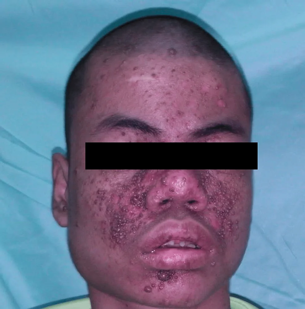
Long description
Figure titled 'Facial angiofibromas of tuberous sclerosis' from an open-access Cureus case report by Mustaqim et al. (2023).

Figure 2Whitemacules
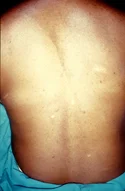
Long description
Clinical photograph of hypomelanotic macules in tuberous sclerosis: dull-white, lance-oval, ash-leaf-shaped patches on the skin.

Figure 3Early neurofibromatosis
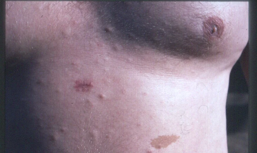
Long description
A clinical photograph of a patient with multiple small cutaneous neurofibromas and a cafe-au-lait spot, one of which has been biopsied.

NF1 presents with skin and eye findings: six or more café-au-lait macules, axillary or inguinal freckling, two or more neurofibromas or one plexiform neurofibroma, an optic pathway glioma, two or more Lisch nodules (iris hamartomas), or a distinctive bone lesion such as sphenoid dysplasia. [4] In adults, cutaneous neurofibromas become the most obvious feature. A minority develop plexiform neurofibromas, which can distort the head and neck or limbs, and some malignant peripheral nerve sheath tumours. Families often notice a bony deformity, scoliosis or learning problems rather than the tumours themselves.
NF2-SWN usually presents in adolescence or early adult life with tinnitus, hearing loss and balance disturbance from bilateral vestibular schwannomas; only rarely with facial palsy or trigeminal sensory loss. About 60–80% of patients have posterior subcapsular or cortical cataracts on specialist ophthalmic examination. Cutaneous findings are subtle: about 70% have cutaneous schwannomas, but only 10% have 10 or more. Other features include meningiomas, spinal ependymomas and, in children, muscle-wasting mononeuropathy. Two clinical patterns are described: the milder Feiling-Gardner type, with late onset and few tumours, and the more severe Wishart type, with many tumours from childhood. [3]
TSC produces seizures (often in infancy), developmental delay, skin lesions and tumours of several organs. Mucocutaneous findings include hypomelanotic macules, facial angiofibromas, ungual fibromas (Koenen tumours), a shagreen patch (connective tissue naevus) and a fibrous cephalic plaque. Cardiac rhabdomyomas are often the first clue in infancy. Renal angiomyolipomas, retinal astrocytic hamartomas, cortical tubers and subependymal nodules complete the picture. [6]
SWS presents with a facial port-wine birthmark, leptomeningeal angiomatosis and seizures (often starting before the first year of age, frequently as infantile spasms); about 70–80% of patients develop neurological complications. Encephalic involvement is usually unilateral and posterior. Glaucoma develops in 30–70% of patients and 60% of these develop it in infancy. Choroidal haemangiomas occur in 40–50%. Migraine-like headaches, stroke-like episodes and developmental delay are common. Vascular malformations extend to the oral mucosa and often produce gingival bleeding. [2]
VHL disease presents most often with cerebellar haemangioblastoma or retinal haemangioblastoma, followed by clear cell renal cell carcinoma, pheochromocytoma/paraganglioma, pancreatic neuroendocrine tumours, endolymphatic sac tumours and cysts of the kidney and pancreas. [1] Early manifestations usually appear in the second decade and cerebellar lesions are the commonest presenting symptom. [1]
How to examine: always inspect the whole skin in daylight with a Wood's lamp for hypopigmented macules in TSC, examine the irides for Lisch nodules in NF1, check the facial port-wine stain distribution in SWS (forehead and upper eyelid, high risk area), and perform fundoscopy for retinal haemangioblastomas in VHL. Blood pressure must be checked at every NF1 visit and VHL patients require regular blood pressure measurement for phaeochromocytoma.
The table compares the five disorders on the features that matter at the bedside. The features overlap with isolated café-au-lait macules, Legius syndrome, non-NF2 schwannomatosis, tuberous sclerosis mimics and sporadic vascular malformations, all of which must be excluded clinically or genetically.
| Feature | NF1 | NF2-SWN | TSC | SWS | VHL |
|---|---|---|---|---|---|
| Inheritance | Autosomal dominant | Autosomal dominant | Autosomal dominant (mostly de novo) | Mosaic, somatic | Autosomal dominant |
| Gene | NF1 | NF2 | TSC1 or TSC2 | GNAQ or GNA11 (somatic) | VHL |
| Pathology | Neurofibromas, Lisch nodules, optic glioma | Schwannomas (CN VIII), meningiomas, ependymomas | Hamartomas (brain, skin, heart, kidney, eye) | Capillary-venous malformation (skin, brain, choroid) | Haemangioblastomas, clear cell renal carcinoma, phaeochromocytoma |
| Key skin marker | Café-au-lait macules, neurofibromas, freckling | Subtle skin schwannomas | Hypopigmented macules, angiofibromas, shagreen patch | Facial port-wine birthmark | None typical; rare skin angiomas |
| Nervous system marker | Optic pathway glioma, spinal neurofibromas | Bilateral vestibular schwannomas, cataracts, meningiomas | Cortical tubers, subependymal nodules, SEGA, epilepsy | Leptomeningeal angiomatosis, seizures, stroke-like episodes | Retinal/cerebellar/spinal haemangioblastomas |
| Major eye finding | Lisch nodules; optic glioma | Posterior subcapsular cataracts, retinal hamartoma | Retinal astrocytic hamartoma | Glaucoma, choroidal haemangioma | Retinal haemangioblastoma |
| Other organs | Bones, vessels, learning problems | Spinal roots, peripheral nerves | Heart (rhabdomyoma), kidneys (AML), lungs (LAM), teeth | Mouth, orbit, endocrine axes | Kidneys, pancreas, adrenals, epididymis |
| Main mimic | Legius syndrome (SPRED1) | Non-NF2 schwannomatosis (SMARCB1, LZTR1) | Focal cortical dysplasia; isolated seizures | Port-wine stain without brain/eye involvement | Sporadic clear cell renal carcinoma |
Fine discriminating points: NF1 is distinguished from Legius syndrome by the presence of neurofibromas, optic glioma, Lisch nodules or a characteristic bone lesion, and by SPRED1 genetic testing when needed. [4] NF2-SWN is distinguished from non-NF2 schwannomatosis by the presence of vestibular schwannoma and other NF2-related tumours, cataracts or retinal hamartomas, and by NF2 mutation testing. [3] TSC skin lesions (hypopigmented macules, facial angiofibromas) are not seen in isolated epilepsy, and mosaic GNAQ changes produce the dermatomal distribution typical of a port-wine birthmark in SWS. [2]
Diagnostic criteria (all paraphrased) are below.
NF1 (revised NIH criteria, as accepted in the 2026 Asia-Pacific consensus): a definite diagnosis needs 2 or more of: six or more café-au-lait macules at least 5 mm across in prepubertal children (15 mm in adults), two or more neurofibromas of any type or one plexiform neurofibroma, axillary or inguinal freckling, an optic pathway glioma, two or more Lisch nodules or two or more choroidal abnormalities, a distinctive osseous lesion, or a heterozygous pathogenic NF1 variant with a variant allele fraction of 50%. [4]
NF2-SWN (2022 international consensus, Manchester-based): bilateral vestibular schwannomas; or an identical NF2 pathogenic variant in 2 anatomically distinct NF2-related tumours; or 2 major or 1 major plus 2 minor criteria. Major criteria are a unilateral vestibular schwannoma, a first-degree relative (other than a sibling) with NF2-SWN, 2 or more meningiomas, or a pathogenic NF2 variant in blood or tumour. Minor criteria (counted with repeats allowed as specified) include ependymoma, meningioma, schwannoma, juvenile cataract under age 40, retinal hamartoma and epiretinal membrane under age 40. [3]
TSC (2021 international consensus): diagnosis uses 11 major and 6 minor clinical features plus a genetic criterion. Major features include 3 or more hypomelanotic macules at least 5 mm in diameter, 3 or more facial angiofibromas or a fibrous cephalic plaque, 2 or more ungual fibromas, a shagreen patch, multiple retinal hamartomas, cortical dysplasias (including tubers and cerebral white matter radial migration lines), subependymal nodules, subependymal giant cell astrocytoma, cardiac rhabdomyoma, renal angiomyolipomas (2 or more) and/or lymphangioleiomyomatosis. Minor features include multiple renal cysts, confetti skin lesions, 3 or more dental enamel pits, 2 or more intraoral fibromas, retinal achromic patch, non-renal hamartoma, renal cysts (multiple) and sclerotic bone lesions. A definite diagnosis is made with 2 major features, or 1 major plus 2 or more minor features, or an identified pathogenic TSC1 or TSC2 variant. [6]
SWS has no formal genetic test in routine use; diagnosis is clinical and imaging-based on a facial port-wine birthmark in the high-risk forehead distribution, plus neurological or ocular involvement. [2]
VHL is diagnosed in a person with a family history and 1 associated tumour, or, without a family history, 2 VHL-associated tumours; molecular testing confirms it. [1]
Staging and severity are described separately: many NF1 centres use the Common Terminology Criteria for Adverse Events or a clinical Complex/PN status, and TSC has no single staging score; instead disease activity is tracked by organ, using tuber and tumour imaging, seizure frequency and renal function. [5]
| Test or measure | When used | Cut-off or expected finding |
|---|---|---|
| NF1 NF1 sequencing and deletion analysis | Confirm atypical or mosaic NF1; family planning | Pathogenic NF1 variant; MLPA/SNP array detects large deletions when sequencing is negative [4] |
| NF1 whole-body or targeted MRI | Tumour burden in complex NF1 | One-time baseline WBMRI at child-to-adult transition; annual if high burden, microdeletion, prior radiotherapy or distinct nodular lesions [4] |
| NF2 MRI of internal auditory canals and brain | Confirm bilateral vestibular schwannomas, meningiomas, ependymomas | Bilateral CP angle masses (vestibular schwannomas) are the hallmark [3] |
| NF2 NF2 mutation testing | Confirm NF2-SWN, especially de novo or mosaic cases | Sensitivity ~95% in subsequent generations; as low as 37% in sporadic cases; test blood plus at least 2 tumours [3] |
| TSC cranial MRI | Cortical tubers, subependymal nodules, SEGA | Repeat every 1 to 3 years from childhood according to consensus; earlier if new symptoms [5] |
| TSC renal MRI | Angiomyolipoma and cysts | Repeat every 1 to 3 years [5] |
| TSC echocardiography | Cardiac rhabdomyoma in infants | Not routinely repeated in adults; at diagnosis in infants, since most rhabdomyomas regress in early childhood [5] |
| SWS brain MRI (with contrast) | Leptomeningeal angiomatosis, cortical change | Performed at diagnosis; choroid plexus thickness measured as a marker for early cerebral disease [2] |
| SWS ophthalmology | Glaucoma and choroidal haemangioma | Every 3 months in the first year, then yearly [2] |
| VHL plasma/urinary metanephrines | Phaeochromocytoma screening | 24-hour urine or plasma, annually from age 5 [1] |
| VHL MRI of brain, spine and abdomen | Haemangioblastomas, renal cell carcinoma, pancreatic tumours | Brain and spinal cord: every 2 years from age 11; abdomen with and without contrast at least every other year from age 15 [8] |
| VHL eye examination | Retinal haemangioblastoma | Every 6 to 12 months from birth to age 30, then at least yearly (indirect ophthalmoscopy) [8] [1] |
NF1: there is no single international guideline with a version number that all regions accept. Management follows the Legius 2021 revised diagnostic criteria, the ERN GENTURIS tumour-surveillance recommendations and, in this text, the 2026 Asia-Pacific NF1 consensus, which introduced a modified Delphi, 75% consensus threshold for 141 statements. [4] Key points:
- Symptomatic, inoperable plexiform neurofibromas (or plexiform neurofibromas still symptomatic after partial surgery) are treated with mitogen-activated protein kinase (MEK) inhibitors (selumetinib, trametinib as available), with echocardiographic and ophthalmic monitoring at baseline and on treatment. [4]
- Cutaneous neurofibromas are treated by surgical excision, laser, electrodessication or radiofrequency ablation; pharmacological options such as topical MEK inhibitors are emerging. [4]
- Malignant peripheral nerve sheath tumours require complete surgical resection with negative margins, which is the only curative strategy. [4]
- Optic pathway gliomas are managed by a neuro-oncology team, with surveillance imaging and treatment decisions made in a specialist paediatric neuro-oncology service.
- Surveillance: children aged 0–5 years should have a general examination every 6 to 12 months and every year from 6 to 10 years, with annual eye and vision assessment to age 8 and annual blood pressure; adults with non-complex NF1 every 2 to 3 years, with safety-netting for new symptoms; complex disease requires at least annual review and imaging. [4] Adults with asymptomatic NF1 need one-time whole-body MRI to document internal tumour burden, then imaging only if indicated. [4]
NF2-SWN: updated diagnostic criteria and nomenclature published in 2022 (Genetics in Medicine, international consensus group led by Plotkin). Treatment is multidisciplinary and there is no single approved systemic therapy; management follows 2012 consensus recommendations for NF2 treatments and accelerating clinical trials. [3]
- Active surveillance for vestibular schwannomas with serial MRI is common first-line when hearing and neurological findings are stable. [3]
- Surgery (retrosigmoid, translabyrinthine, middle fossa) is reserved for tumours causing brainstem compression, progressive neurological deficit or failed medical therapy; the aim is maximal safe resection, and inner-ear surgery causes hearing loss. [3]
- Bevacizumab (an antibody against vascular endothelial growth factor) has shown particular efficacy in NF2-SWN and is often used in the UK through national health agencies, although it is unlicensed in this indication. [3]
- Stereotactic radiosurgery or fractionated radiotherapy are options, particularly for growing tumours after failed medical therapy, and the modality and dose are selected by the neuro-oncology and neurosurgical team. [3]
- Meningiomas and ependymomas are managed as in other patients, with surgery or radiotherapy by site; anticonvulsants and rehabilitation for functional deficits.
Radiation therapy is an option for NF2-SWN tumours that progress despite surveillance and medical therapy, with modality and dose chosen by the neuro-oncology and neurosurgical team.
TSC: the International TSC Consensus (Northrup et al., Updated International Tuberous Sclerosis Complex Diagnostic Criteria and Surveillance and Management Recommendations, 2021) sets the approach; NICE technology appraisal TA873 (March 2023) addresses cannabidiol in this population. [6] [7]
- Everolimus is the first-line medicine for asymptomatic renal angiomyolipomas larger than 3 cm in diameter, at 10 mg daily in adults; lower doses such as 5 mg daily and sequential dosing (10 mg daily for 4 months, then 5 mg daily) have shown similar benefit with fewer adverse effects. [5]
- In the EXIST-2 trial, everolimus produced at least a 50% reduction in angiomyolipoma volume in 42% of patients (33 of 79), compared with 0% (0 of 39) with placebo. [5]
- Everolimus reduces the size of subependymal giant cell astrocytomas and cutaneous angiofibromas and has an effect on seizures. [5] [6]
- Cannabidiol is recommended (NICE TA873, March 2023) as an add-on treatment for seizures caused by TSC in people aged 2 years and over, only if seizures are not controlled by 2 or more antiseizure medicines and seizure frequency is checked every 6 months, stopping cannabidiol if frequency has not fallen by at least 30% compared with the 6 months before starting treatment. [7]
- Epilepsy surgery (tuberectomy) is considered for drug-resistant epilepsy, and everolimus is not used routinely for this; vigabatrin is used for infantile spasms.
- Selective arterial embolisation or partial nephrectomy is second-line for angiomyolipoma larger than 3 cm; arterial embolisation with corticoid cover is used for acute haemorrhage. [5]
- Renal cystic disease, cystic renal cell carcinoma and rare renal complications are managed in a nephrology/oncology service. [5]
- Surveillance: brain MRI (or cranial CT if MRI is unavailable) to assess tubers and SEGA; repeat at 1 to 3 years; renal MRI every 1 to 3 years; echocardiogram in the first 2 years of life for rhabdomyoma; ophthalmology and dermatology reviews; blood pressure and renal function monitoring. [5]
SWS: management is supportive and complication-led, following the current review of approaches. [2] Local neurology and ophthalmology pathways organise the multidisciplinary surveillance.
- Skin: early pulsed-dye laser for the facial port-wine birthmark improves cosmetic outcome; nodular or thickened areas may need surgical excision later. Soft tissue or bony hypertrophy occurs in about two-thirds of patients by age 50. [2]
- Brain: anticonvulsants for epilepsy; low-dose aspirin is used in many centres to delay seizure onset and stroke-like events, although evidence is still limited. Refractory focal epilepsy may be treated by focal resection of the affected lobe, hemispherectomy in severe infantile cases, or vagus nerve stimulation.
- Eye: glaucoma requires regular pressure lowering and often surgery; diffuse choroidal haemangioma may need photodynamic therapy, laser or radiotherapy. Screening every 3 months in the first year, then yearly. [2]
- Stroke prevention: head injury avoidance, migraine management, and neurological review; there is no evidence-based primary prevention of stroke-like events. [2]
- A multidisciplinary team (neurology, ophthalmology, dermatology, neuroradiology, neuropsychology) reviews each child.
VHL: management follows the surveillance protocol summarised in the 2017 review; tumours are managed surgically or with ablation as they become symptomatic or reach the size thresholds below. [1]
- Haemangioblastomas of the cerebellum, spine and retina are treated by microsurgical resection when symptomatic or enlarging; radiotherapy is used for inoperable lesions.
- Renal cell carcinoma: nephron-sparing surgery or ablation once growth or size thresholds are met on serial imaging, with careful preservation of renal function; active surveillance for smaller lesions. [1]
- Clear cell renal cysts and pancreatic neuroendocrine tumours are followed with imaging; enucleation or resection when size or growth demands.
- Phaeochromocytoma/paraganglioma: biochemical screening and surgical removal after adequate alpha blockade.
- Surveillance, following the 2023 international VHL consensus and the 2017 review: retinal examination every 6 to 12 months from birth to age 30, then at least yearly; abdominal imaging: annual ultrasound from age 8 per the 2017 review and MRI at least every other year from age 15 per the 2023 consensus; brain and full-spine MRI every 2 years from age 11; metanephrines yearly from age 5; and, in pregnancy, regular retinal review, phaeochromocytoma testing in early, mid and late pregnancy, and non-contrast MRI at 4 months. [1] [8]
- Endolymphatic sac tumours are evaluated with MRI of the internal auditory canals if indicated, and treated surgically when hearing is threatened. [1]
NF1: optic pathway glioma (usually childhood), plexiform neurofibroma with risk of malignant peripheral nerve sheath tumour, scoliosis and tibial pseudarthrosis, learning difficulties and attention deficit. [4]
NF2-SWN: progressive bilateral deafness, facial palsy, trigeminal sensory loss, raised intracranial pressure from large tumours, blindness from cataracts or optic nerve sheath meningioma, and spinal cord compromise from ependymoma or meningioma. [3]
TSC: refractory epilepsy, subependymal giant cell astrocytoma, renal angiomyolipoma bleeding, progressive renal failure, cardiac rhabdomyoma-related arrhythmia in infants, neuropsychiatric disorders (TAND: tuberous sclerosis complex-associated neuropsychiatric disorders) and lymphangioleiomyomatosis in women. [5] [6]
SWS: intractable epilepsy, hemiparesis, stroke-like episodes, intellectual disability, glaucoma and visual loss, and the psychological burden of a facial birthmark. [2]
VHL: haemorrhage from haemangioblastoma or renal tumour, renal failure or metastatic renal cell carcinoma, and blindness; these remain the main causes of premature death. [1]
NF1 shortens life expectancy by 8 to 20 years and carries a 2.7 to 4 times higher risk of cancer than the general population, largely through malignant peripheral nerve sheath tumour. [4]
NF2-SWN had an average actuarial survival of about 60 years before 1990; survival has improved with specialist centres and new therapies, but remains reduced, and genotype predicts severity: truncating variants in exons 2 to 13 have the worst outcome. [3]
TSC survival has improved markedly with mTOR inhibitors and imaging surveillance; the main determinants of quality of life are epilepsy control and renal function. [5]
SWS survival is good; morbidity rather than mortality dominates, although bilateral intracranial involvement and early, intractable seizures predict poorer cognitive outcomes. [2]
VHL survival historically averaged 40 to 52 years, but has lengthened with surveillance programmes; renal cell carcinoma and central nervous system tumours remain the main causes of VHL-related death. [1]
There is no primary prevention for any of these germline or mosaic conditions. Practical prevention consists of:
- Genetic counselling and preimplantation or prenatal testing for at-risk families with NF1, NF2-SWN, TSC or VHL. [1] [3] [4] [5]
- Regular surveillance as tabulated, beginning at diagnosis or birth for congenital conditions.
- Lifestyle measures: sun protection after laser for port-wine birthmark; avoidance of head trauma in SWS; not smoking, to reduce lymphangioleiomyomatosis risk in women with TSC.
- Screening relatives: first-degree relatives of a person with an NF1 or NF2-SWN pathogenic variant, or with VHL, should be offered mutation testing; relatives of a person with TSC should be examined clinically and by brain and renal imaging, and genetic testing if a variant is known. [1] [4]
Pregnancy. Women with NF1 can usually carry a pregnancy but may experience growth of neurofibromas; surveillance should be maintained. Women with TSC and lymphangioleiomyomatosis or large renal angiomyolipomas need pulmonary and renal assessment before conception. Women with VHL need retinal assessment and phaeochromocytoma screening in early, mid and late pregnancy, plus non-contrast MRI at 4 months if indicated, because of lesion growth in pregnancy. [1] SWS pregnancy is managed by a neurology and obstetric team; seizure control and stroke-prevention plans are individualised. [2]
Children. TSC is most often recognised in infants through cardiac rhabdomyoma or seizures, and NF1 through multiple café-au-lait macules; NF2-SWN is rare before adolescence. Children with NF1 should have annual ophthalmology to age 8, developmental surveillance and blood pressure at every visit. [4] Children with TSC need brain and renal MRI from diagnosis. [5] Infants with SWS need early seizure control and frequent eye checks. [2]
Older adults. Late or missed diagnosis is common in adults with mild TSC or NF2-SWN; cutaneous markers can be the clue. Long-term surveillance remains relevant, particularly renal imaging in TSC and VHL, because new tumours can present in later life. [1] [6]
Approach to a suspected neurocutaneous disorder
- Skin and clinical cluesCALMs, neurofibromas; Lisch nodules → NF1. Hypopigmented macules, angiofibromas → TSC. Port-wine birthmark (forehead/eyelid) → SWS. Bilateral hearing loss, cataracts → NF2-SWN. Retinal haemangioblastoma, clear cell renal mass → VHL
- Confirm the diagnosisClinical criteria first; genetic testing where atypical, mosaic or for family planning
- Question: Which system is threatened now?
Brain/eye (NF1, NF2-SWN, TSC, SWS)
- MRI brain/spine and ophthalmology
- Manage seizures, glaucoma, Raised ICP; refer neuro-oncology/surgery
Kidney/vascular (TSC, VHL)
- Renal imaging; metanephrines if VHL
- mTOR inhibitor for TSC AML >3 cm; nephron-sparing surgery at ~3 cm for VHL
- Start lifelong surveillanceUse the organ-specific intervals in the tests table above
- The five classic neurocutaneous disorders share a predisposition to tumours or malformations of skin and nervous system, but they have different genes and natural histories; identify each from its skin marker and organ pattern.
- NF1 (NF1 gene, chromosome 17) is the most common, with café-au-lait macules, neurofibromas, Lisch nodules and optic glioma; plexiform neurofibromas are treated with MEK inhibitors when symptomatic and inoperable, and malignant peripheral nerve sheath tumours by complete surgical resection. [4]
- NF2-SWN (NF2 gene, chromosome 22) is defined by bilateral vestibular schwannomas; bevacizumab and radiotherapy are used off-licence or off-label, and surgery is used for compressive tumours.
- TSC (TSC1/TSC2) releases mTOR from restraint; everolimus 10 mg daily is first-line treatment for renal angiomyolipoma larger than 3 cm, and surveillance imaging is repeated every 1 to 3 years. [5]
- SWS is a mosaic GNAQ/GNA11 disorder producing a facial port-wine birthmark, glaucoma and seizures; laser, anticonvulsants, glaucoma therapy and aspirin (in many centres) form the main treatment.
- VHL (VHL gene, chromosome 3) drives HIF-dependent tumours; surveillance starts in infancy with retinal checks every 6 to 12 months, expanding to brain and spine imaging from age 11 and abdominal imaging from age 15. [8]
- Diagnosis is clinical first, with genetic confirmation where atypical or familial; each condition has published consensus criteria that should be cited rather than memorised.
- Prevention is surveillance, not cure: counselling, organ-specific imaging schedules and early treatment of complications lengthen life and protect function.
Answer each one in your head first, then open the answer.
A 4-year-old child has six café-au-lait macules, axillary freckling and an optic pathway glioma. What is the likely diagnosis and the gene involved?
Show answerHide answer
Neurofibromatosis type 1, from pathogenic variants in the NF1 gene on chromosome 17q. The diagnostic threshold is two or more of the NIH features; here three are present. Genetic testing may be reserved for atypical or mosaic presentations and for family planning.
A 16-year-old has bilateral sensorineural hearing loss and posterior subcapsular cataracts. What syndrome and tumour type do you expect, and what test would confirm it?
Show answerHide answer
Neurofibromatosis type 2-related schwannomatosis (NF2-SWN), caused by NF2 variants, with bilateral vestibular schwannomas. MRI of the internal auditory canals confirms the tumours; NF2 mutation testing of blood plus at least two tumours is most sensitive, since sensitivity in sporadic cases can be as low as 37%.
An adult with TSC has an asymptomatic renal angiomyolipoma measuring 4 cm. What is the recommended first-line treatment and its usual adult dose?
Show answerHide answer
mTOR inhibition, usually everolimus 10 mg daily, with renal MRI repeated every 1 to 3 years. Selective embolisation or partial nephrectomy are second-line. Lower doses such as 5 mg daily and sequential regimens have shown similar benefit with fewer adverse effects.
An infant has a facial port-wine birthmark in the forehead and upper eyelid distribution and new-onset seizures. What condition should you suspect, what is the first eye check, and how often should it be repeated?
Show answerHide answer
Sturge-Weber syndrome. Urgent brain MRI with contrast is needed to assess leptomeningeal angiomatosis. Ophthalmological examination for glaucoma should be done promptly, then every 3 months in the first year and yearly afterwards.
A young adult presents with a cerebellar haemangioblastoma and clear cell renal carcinoma. What is the unifying diagnosis and at what age should routine VHL surveillance have begun for the eyes?
Show answerHide answer
Von Hippel-Lindau disease. Routine indirect ophthalmoscopy for retinal haemangioblastoma should begin at birth, every 6 to 12 months to age 30 (then at least yearly), with brain and whole-spine MRI added from age 11 and abdominal imaging from age 15.
A patient has 2 major TSC features. How many major features (or what combination) of TSC is needed for a definite diagnosis under the 2021 consensus, and which 3 findings make the commonest clinically obvious triad?
Show answerHide answer
A definite diagnosis requires either 2 major features, or 1 major plus 2 or more minor features, or an identified pathogenic TSC1 or TSC2 variant. Three of the most recognisable major features are 3 or more hypomelanotic macules of at least 5 mm, 3 or more facial angiofibromas or a fibrous cephalic plaque, and renal angiomyolipomas.
Neurofibromatosis type 1, neurofibromatosis type 2-related schwannomatosis, tuberous sclerosis complex, Sturge-Weber syndrome and von Hippel-Lindau disease are often grouped as phakomatoses. They overlap clinically with epilepsy, stroke, renal malignancy, neurocutaneous tumours and visual loss, so cross-reference those topics where they exist.
- Varshney N, Kebede AA, Owusu-Dapaah H, Lather J, Kaushik M, Bhullar JS. A Review of Von Hippel-Lindau Syndrome. Journal of Kidney Cancer and VHL 2017;4(3):20–29. Accessed .
- Sánchez-Espino LF, Ivars M, Antoñanzas J, Baselga E. Sturge-Weber Syndrome: A Review of Pathophysiology, Genetics, Clinical Features, and Current Management Approaches. The Application of Clinical Genetics 2023;16:63–81. Accessed .
- Evans DG, Blakeley JO, Plotkin SR. History and clinical epidemiology of NF2-related schwannomatosis. Familial Cancer 2025;24(4):79. Accessed .
- Berman Y, Lee BH, Liu APY, Nishida Y, Tan ES, Wang Z, APAC NF1 Expert Panel. Consensus recommendations for neurofibromatosis type 1 management in Asia Pacific. iScience 2026;29(10):117002. Accessed .
- Monich AG, Bissler JJ, Barreto FC. Tuberous Sclerosis Complex and the kidneys: what nephrologists need to know. Jornal Brasileiro de Nefrologia 2024;46(3):e20240013. Accessed .
- Kundak S, Yalabik H. Delayed Diagnosis of Tuberous Sclerosis Complex: A Case Report. Cureus 2026;18(7):e112466. Accessed .
- NICE. Cannabidiol for treating seizures caused by tuberous sclerosis complex (TA873). Technology appraisal guidance, published 1 March 2023. Accessed .
- Daniels AB, Tirosh A, Huntoon K, Mehta GU, Spiess PE, Friedman DL, Waguespack SG, et al, International VHL Surveillance Guidelines Consortium. Guidelines for surveillance of patients with von Hippel-Lindau disease: consensus statement of the International VHL Surveillance Guidelines Consortium and VHL Alliance. Cancer 2023;129(19):2927-2940. Accessed .
Figure credits
Educational content for medical students and doctors. It is not a substitute for clinical judgement or local guidelines.