Visual field defects and glaucoma
How the visual pathway explains field defects, and how glaucoma is recognised, measured and treated with drops, laser and surgery.
- A visual field defect is not a single disease: it is a pattern of vision loss that tells you where along the visual pathway the problem lies.
- Glaucoma is a progressive optic neuropathy in which retinal ganglion cells die, the optic disc cups, and the visual field is lost (usually starting peripherally), most often in association with raised intraocular pressure (IOP).
- Raised IOP is the only modifiable risk factor, so every treatment — drops, laser, operation — aims at lowering it.
- In primary open-angle glaucoma (POAG) the eye's drainage meshwork looks open but drains poorly; in angle-closure glaucoma the drainage angle is physically blocked, which can become an emergency.
- Goldmann applanation tonometry is the reference for measuring IOP; perimetry (formal visual field testing) shows the functional loss and gonioscopy shows whether the angle is open or closed.
By the end of this page you should be able to:
- Name the defect that each major site along the visual pathway produces (monocular, bitemporal, homonymous, quadrantanopic) and localise it.
- Distinguish primary open-angle glaucoma, ocular hypertension, angle-closure disease and acute angle-closure crisis, and explain how raised IOP damages the nerve.
- Plan case-finding and diagnostic testing for glaucoma, with the key thresholds for referral and reassessment.
- Choose first-line, ongoing and rescue treatment for chronic open-angle glaucoma and for acute angle-closure, using the current NICE, AAO and EGS recommendations.
- Describe laser and surgical options, their indications and the main complications.
- Recognise the gaps between guidelines and the management of the fellow eye, children, pregnancy and older adults.
DefinitionVisual field
The visual field is everything you can see without moving the eye or head. A visual field defect is a localised loss within it. Because the retina, optic nerve, chiasm, radiations and cortex each carry information from a particular part of the visual world in a predictable order, the shape of the defect usually reveals the site of the lesion.
DefinitionGlaucoma
Glaucoma is a progressive optic neuropathy in which retinal ganglion cells undergo apoptosis, the optic disc develops pathologic cupping and thinning of the neuroretinal rim, and the retinal nerve fibre layer thins, leading to characteristic visual field loss. Raised IOP is the only modifiable risk factor, but a substantial minority of patients have normal IOP (normal-tension glaucoma).[4]
Glaucoma is classified first by whether the iridocorneal angle is open or closed, and second by whether it is primary or secondary:
- Open-angle glaucoma (OAG): the angle looks open on gonioscopy, but aqueous outflow is reduced through the trabecular meshwork.
- Primary open-angle glaucoma (POAG) is the commonest form, about three-quarters of all glaucoma.[4]
- Secondary forms include pigmentary, exfoliative (pseudoexfoliative), uveitic, traumatic, steroid-induced and others.
- Normal-tension glaucoma: open angle with typical glaucomatous damage despite IOP consistently within the normal range; lowering IOP is still beneficial.[2][4]
- Angle-closure glaucoma (ACG): the angle is closed (occluded by at least 270 degrees in the acute/chronic angle-closure glaucomas) by the iris or another mechanism, so aqueous cannot reach the trabecular meshwork at all.[4]
- Acute angle-closure crisis (AACC): a sudden, symptomatic occlusion with very high IOP — an ocular emergency.
- Chronic angle-closure glaucoma.
- Secondary angle-closure forms include neovascular, phacomorphic, uveitic, endothelial and tumour-related.
Figure 1Hemianopsia en
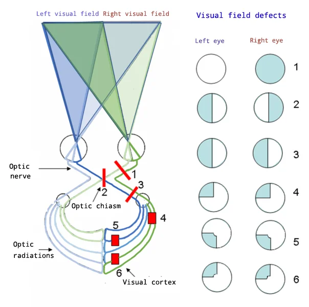
Long description
Visual field defects in visual pathway lesions, shown as paired left/right visual-field circles for lesions at successive points along the visual pathway.

The route from eye to brain is the reason a clinician can read a visual field like a map:
Retina (ganglion cells) → optic nerve → optic chiasm (nasal fibres crossing) → optic tract → lateral geniculate nucleus (LGN) → optic radiations (Meyer's loop in the temporal lobe and parietal fibres) → primary visual cortex in the occipital lobe.
Two anatomical facts decode most defects:
- The nasal retina sees the temporal visual field and its fibres cross at the chiasm; the temporal retina sees the nasal visual field and its fibres do not cross.
- Each structure beyond the chiasm represents the contralateral visual hemifield.
| Pattern of defect | Most likely lesion site |
|---|---|
| Loss in one eye only (monocular) | Ipsilateral optic nerve, retina or globe |
| Bitemporal hemianopia (loss of both temporal halves) | Optic chiasm (classically a compressive pituitary or suprasellar mass) |
| Homonymous hemianopia (same side lost in both eyes) | Contralateral optic tract (or, more posteriorly, occipital cortex with macular sparing) |
| Homonymous sectoranopia | Lateral geniculate nucleus |
| Upper homonymous quadrantanopia ("pie in the sky") | Contralateral temporal optic radiation (Meyer's loop) |
| Lower homonymous quadrantanopia ("pie on the floor") | Contralateral parietal optic radiation |
| Homonymous hemianopia with macular sparing | Contralateral occipital lobe (vascular supply often spares the pole) |
A defect confined to one eye localises anterior to the chiasm: optic neuropathy (for example, glaucoma, ischaemic optic neuropathy, optic neuritis, compression), retinal disease or disorders of the media and globe. Glaucoma typically produces arcuate scotomas and nasal steps that respect the horizontal midline, because it damages the retinal nerve fibres as they arc around the macula. A central scotoma from a papillomacular bundle lesion is early and, once established, usually irreversible.[6]
Loss of the temporal visual field in both eyes means the crossing nasal fibres are compressed at the optic chiasm. The commonest causes are pituitary macroadenoma, craniopharyngioma and parasellar masses; the onset is usually gradual and may be detected only on formal perimetry long before the patient notices it. Standard automated perimetry can show temporal depression and a vertical step very early in chiasmal disease, even with normal Goldmann fields.[6]
Defects that are congruent between the two eyes lie behind the chiasm: optic tract (complete homonymous hemianopia, often with a relative afferent pupillary defect), LGN (sectoranopia), optic radiations (quadrantanopia; temporal lobe involvement gives an upper defect, parietal involvement a lower one) or occipital cortex (hemianopia with macular sparing). About 38% of homonymous hemianopias improve spontaneously over the first weeks to months after stroke.[6]
Clinical pearlLocalising clue
If the defect is the same side in both eyes and affects a whole quadrant, ask whether it is upper or lower: upper points to the temporal lobe (Meyer's loop), lower to the parietal lobe. Macular sparing strongly suggests an occipital lesion.
Glaucoma is the leading cause of irreversible blindness worldwide, behind cataract as a cause of vision loss overall.[4]
- It affects about 3.5% of people aged 40–80 years and nearly 112 million people are expected to be affected by 2040.[4]
- POAG is the commonest form of glaucoma.[4]
- Angle-closure glaucoma is less common than open-angle glaucoma but causes roughly half of all glaucoma-induced blindness.[4]
- Primary angle-closure glaucoma has a global prevalence of about 0.6%.[4]
- Normal-tension glaucoma accounts for about one third of patients with glaucoma.[4]
- Up to 50% of glaucomatous eyes have IOP within the normal range, which is why measuring IOP alone never screens for glaucoma.[4]
- IOP in the normal eye is conventionally quoted as about 11 to 21 mmHg, but the distribution varies and there is no single cut-off between normal and abnormal.[4]
POAG is more common with older age, Black or Latino/Hispanic ethnicity, a family history, diabetes mellitus, lower blood pressure and ocular perfusion pressure, myopia, thinner cornea and lower corneal hysteresis, optic disc haemorrhage, a larger cup-to-disc ratio and higher pattern standard deviation on threshold perimetry.[2]
Glaucoma is usually primary (no identifiable ocular cause). Risk factors fall into two groups:
- Non-modifiable: older age, family history, Black or Latino/Hispanic ethnicity (POAG), Asian or Inuit ancestry, female sex and hypermetropia (angle-closure disease), shallow anterior chamber, shorter axial length, family history of angle closure.[2][3]
- Modifiable and local: raised IOP, thinner central cornea, steroid use (topical or systemic), and the anatomical risk of pupil block or plateau iris in angle-closure disease.[2][3]
There is no known primary prevention for POAG beyond controlling IOP where it is raised and avoiding unnecessary steroid exposure. For angle-closure disease, anatomical predisposition can be identified before a crisis occurs.
Aqueous humour is produced by the ciliary body, passes from the posterior chamber round the lens, via the pupil to the front of the eye, and drains the eye mainly through the trabecular meshwork into Schlemm's canal and the episcleral veins.[4] IOP rises when production and outflow are out of balance.
The lamina cribrosa of the sclera is mechanically weak. Raised IOP (and, in normal-tension glaucoma, reduced perfusion or individual susceptibility) stresses the axons of the retinal ganglion cells at this point, leading to blocked axoplasmic flow, ganglion cell apoptosis, excavation of the optic cup and progressive thinning of the neuroretinal rim — classically more marked superiorly and inferiorly. Two theories describe this: a biomechanical one (narrow scleral pores compress axons) and a vascular one (reduced perfusion causes ischaemic damage).[4] In open-angle disease the primary resistance is in the juxtacanalicular tissue of the trabecular meshwork; in angle-closure the iris physically blocks the angle (pupillary block or plateau iris) and the IOP can rise to very high levels within hours.[4]
Individuals differ greatly in how much IOP their optic nerve can withstand: this is why the same IOP can be harmless in one person and blinding in another, and why treatment targets are titrated to disease behaviour rather than to a universal number.[2]
Figure 2Acute angle closure glaucoma
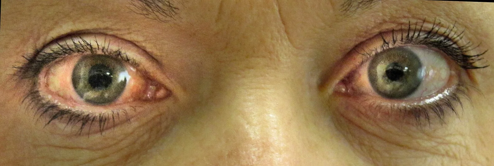
Long description
Acute angle-closure glaucoma of the right eye (intraocular pressure 42) with a mid-sized pupil that is non-reactive to light and conjunctival redness.

Figure 3Glaukompapille2
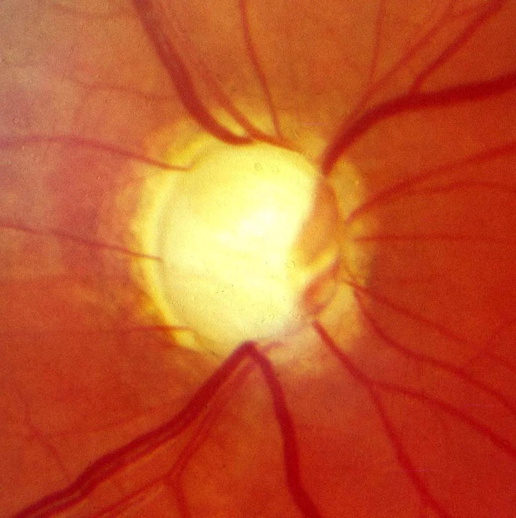
Long description
Fundus photograph described as the optic nerve in advanced glaucoma disease.

Figure 4Fundus photograph of a healthy right eye (OD) from a myopic Asian male patient. Age 23
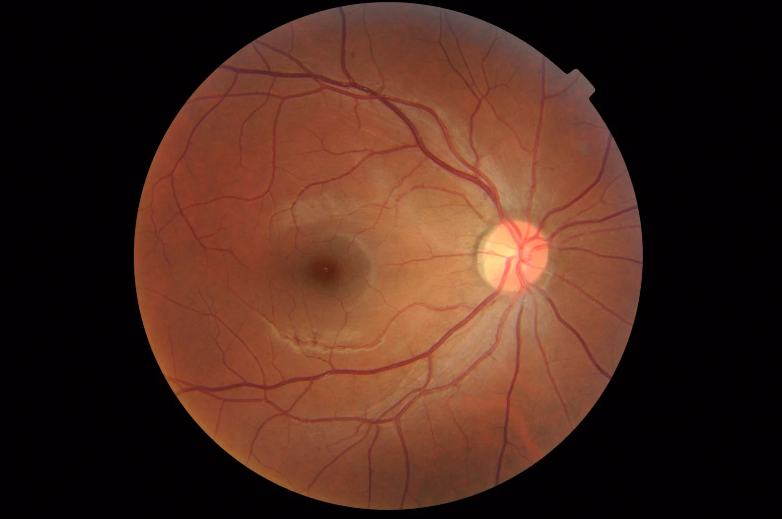
Long description
Fundus photograph of a healthy right eye from a myopic 23-year-old Asian male patient in which the optic disc contains a small cup whose margins are indistinct.

Early POAG is asymptomatic. Patients often notice nothing until advanced, because central acuity is preserved while peripheral field is lost. Features include:
- Gradual, usually bilateral but asymmetric peripheral visual field loss with nasal steps and arcuate scotomas.
- Optic disc cupping with thinning of the neuroretinal rim, often vertical.
- RNFL thinning on imaging (OCT).
- IOP that may be raised or normal.
- Occasionally an optic disc haemorrhage, a marker of activity.
The diagnosis is made on examination, not on symptoms.
AACC is unmistakable once you have seen it:
- Severe eye pain with headache, often with nausea and vomiting (which can misdiagnose as migraine or an acute abdomen).
- Blurred vision with coloured haloes.
- Markedly raised IOP, a mid-dilated, fixed pupil, a hazy cornea (oedema) and conjunctival/episcleral injection.
- A shallow anterior chamber.[3]
Chronic angle-closure may be silent, like POAG, or present with intermittent blurred vision and haloes.
Every glaucoma assessment needs:
- Visual acuity and refraction.
- IOP by Goldmann applanation tonometry (the reference standard).
- Slit-lamp examination of the anterior segment, including van Herick estimation of peripheral anterior chamber depth.
- Gonioscopy in a dark room: the gold-standard test for open versus closed angle (Shaffer grades) and the only way to classify the disease.
- Stereoscopic slit-lamp biomicroscopy of the optic disc: cup-to-disc ratio, rim, haemorrhages, asymmetry.
- Central visual field by standard automated perimetry (SAP), usually a central 24-degree threshold test, repeated to establish severity.
- OCT of the RNFL and ganglion cell layer for structure.
- Central corneal thickness (CCT), which modulates IOP interpretation.
- Dilated examination of the fundus and retinal vessels.
NICE recommends that an optic nerve head image be obtained at diagnosis as a baseline for future comparison.[1]
| Condition | Intraocular pressure | Angle on gonioscopy | Optic disc / nerve fibre layer | Visual field pattern |
|---|---|---|---|---|
| Ocular hypertension (OHT) | Raised (24 mmHg or more) | Open | Normal | Normal |
| Primary open-angle glaucoma (POAG) | Raised or normal | Open | Cupping, rim thinning, RNFL thinning | Glaucomatous scotomas/nasal steps |
| Normal-tension glaucoma | Normal (always) | Open | Glaucomatous cupping | Glaucomatous defects |
| Suspected COAG | Variable | Open | Suspicious disc or borderline field | Borderline or normal |
| Primary angle-closure (PAC/PACG) | Raised | Closed (180 degrees or more ITC) | Normal or glaucomatous | Normal or glaucomatous |
| Acute angle-closure crisis | Very high | Closed | May be normal initially | May be normal initially |
| Secondary glaucomas (pigmentary, exfoliative, steroid-induced, uveitic, traumatic, neovascular) | Raised | Open or closed | Glaucomatous (if chronic) | Depends on duration |
| Non-glaucomatous optic neuropathy (ischaemic, compressive, inflammatory) | Normal | Open | Pallor, altitudinal or central loss | Usually does not match glaucoma pattern; may be altitudinal or central |
Eye pain with a red, hard eye and haloes is not glaucoma until proven otherwise: it may be acute angle-closure crisis, but also uveitis, keratitis, scleritis, endophthalmitis or (rarely) a tumour. Retinal vascular occlusion, ischaemic optic neuropathy and optic disc drusen also enter the differential of an optic disc that looks cupped.
| Test | What it answers | Cut-off or key value |
|---|---|---|
| Goldmann applanation tonometry | IOP for referral, treatment and follow-up | Commonly cited normal range 11–21 mmHg; 24 mmHg or more using Goldmann tonometry triggers referral and treatment decisions; case-finding uses repeated measurements rather than a single value[1][4] |
| Rebound tonometry (iCare) | Screening/monitoring IOP | Agrees well with Goldmann; reliability falls at high IOP and with thick corneas. Do not base a referral decision on non-contact tonometry alone[1][4] |
| Standard automated perimetry | Functional loss; severity | Used for case-finding (full threshold or supra-threshold) and diagnosis (central thresholding). Repeats needed to confirm. Severity: see below[1] |
| Gonioscopy | Open vs closed angle | PACD defined by 180 degrees or more iridotrabecular contact; dark-room dynamic gonioscopy is required to diagnose and verify improvement[3] |
| Slit-lamp cup/disc assessment and OCT | Structural damage | Cupping, neuroretinal rim thinning, RNFL/ganglion cell thinning; imaging complements perimetry |
| Central corneal thickness | Refines IOP and risk | Measured at diagnosis; thinner corneas carry higher risk (no single treatment threshold) |
| Ultrasound biomicroscopy / AS-OCT | Mechanism of angle closure | UBM most reliably identifies plateau iris |
Visual field severity (ICD-10 grading, 2015): mild glaucoma corresponds to a general absence of visual field defects; moderate to defects in one hemifield but outside 5 degrees from fixation; severe to abnormalities in both hemifields with loss within 5 degrees of fixation.[4]
Approach to diagnosis (NICE NG81): before referral, case-finding offers SAP, stereoscopic optic nerve examination (plus OCT or optic nerve image if available), Goldmann-type applanation tonometry and gonioscopy (or van Herick/OCT if gonioscopy is unavailable). A decision to refer should not rest on IOP alone, and where IOP is below 24 mmHg the patient continues regular primary eye care. Referral (to a suitably trained professional) is appropriate with optic nerve head damage, a visual field defect consistent with glaucoma, or IOP of 24 mmHg or more on Goldmann tonometry; repeat measurements are considered first unless urgent referral is indicated.[1]
At diagnosis all patients are offered SAP (central thresholding, repeated as needed), stereoscopic slit-lamp biomicroscopy with dilated examination, Goldmann applanation tonometry, gonioscopy, CCT and an optic nerve image for baseline. For someone with ocular hypertension, the risk of future visual impairment is weighed from IOP level, CCT, family history and life expectancy.[1]
- The sole modifiable target is IOP. The AAO preferred practice pattern suggests a reasonable initial goal of 20% to 30% reduction below baseline, adjusted thereafter according to disease course and severity, including in normal-tension glaucoma.[4][2]
- Aim for the fewest effective medications and minimal adverse effects; adherence and drop-instillation technique dominate outcomes.[4]
- NICE NG81 newly recommends first-line 360-degree selective laser trabeculoplasty (SLT) for OHT with IOP of 24 mmHg or more and for non-advanced COAG (excluding pigment dispersion syndrome), with a generic prostaglandin analogue (PGA) offered if SLT is declined, unsuitable, awaited or insufficient.[1]
- The EGS 6th Edition (2025) provides an evidence-based, Europe-wide framework that supports the same stepwise approach of diagnosis, target IOP and staged treatment, recognising that guidance should be personalised to the individual patient.[7]
- Surgery is generally reserved for advanced disease at presentation, failure of drugs and laser, or progression despite two drug classes.
This is a medical then laser/surgical emergency:
- Lower IOP urgently with a combination of topical agents (beta-blocker, alpha-agonist, carbonic anhydrase inhibitor) plus a systemic carbonic anhydrase inhibitor or hyperosmotic agent, with caution for renal, cardiac and respiratory disease.
- Control pain, nausea and vomiting; hospitalise if needed.
- Perform laser peripheral iridotomy (or surgical iridectomy/paracentesis/iridoplasty if the cornea is too oedematous for laser) to break pupillary block and reopen the angle. Once the IOP falls, iridotomy is the definitive step.[3][4]
- Examine and treat the fellow eye prophylactically with LPI, because the risk of a similar crisis is high.[3]
- Once the crisis settles, consider lens extraction for primary angle-closure disease with persistently high IOP or significant PAS. LPI alone may not suffice, particularly in plateau iris.[3][4]
Long-term follow-up is needed to watch for progressive peripheral anterior synechiae and glaucomatous optic neuropathy.[3]
Drug classes and how they work (mechanisms and practical points from current reviews and guidelines):
| Class | Examples | Typical use | Principal issues |
|---|---|---|---|
| Prostaglandin analogues (PGAs) | Latanoprost, bimatoprost, travoprost, tafluprost, unoprostone, latanoprostene bunod | First line for OHT/COAG; once daily, uveoscleral outflow; well tolerated | Eyelash growth, iris pigmentation, uveitis; long-term concerns about non-trabecular targeting [4] |
| Beta-blockers | Timolol (non-selective), betaxolol (β1-selective) | One or more times daily; reduce aqueous production. Cardioselective agents safer in asthma/COPD | Contraindicated in asthma, COPD, bradycardia, heart failure, some diabetes; systemic absorption possible despite topical use |
| Carbonic anhydrase inhibitors (CAIs) | Dorzolamide 2%, brinzolamide 1% (topical); acetazolamide, methazolamide (systemic) | 2–3 times daily (topical); systemic agents highly effective but poorly tolerated | Topical: sulfa allergy; systemic: renal calculi, electrolyte disturbance; about half of patients discontinue systemic CAI within a month [4] |
| Alpha-agonists | Brimonidine, apraclonidine | 2–3 times daily; decrease production and increase outflow; often second-line in combination | Allergic conjunctivitis, fatigue, hypotension; avoid with MAOI therapy [4] |
| Rho-kinase inhibitors | Netarsudil 0.02% | Once daily; increases conventional outflow | Conjunctival haemorrhage, keratitis, corneal verticillata [4] |
| Miotics (cholinergics) | Pilocarpine, carbachol | Frequent dosing; increase conventional outflow | Miosis, accommodation spasm, retinal detachment risk |
| Hyperosmotic agents | Glycerol, mannitol, isosorbide | Systemic, for acute IOP crises, not chronic therapy | Cardiac/renal contraindications, nausea |
Ongoing care: encourage patients to stay on the same regimen unless IOP, disc or field progresses, or the drug is not tolerated. If IOP remains high despite a PGA, reassess adherence and drop technique, then add a drug from another class, offer SLT, or offer surgery. Where two drug classes have failed, or surgery is refused or unsuitable, options are trabeculectomy (with mitomycin C), SLT or cyclodiode laser treatment.[1]
Preservative-free drops are reserved for genuine preservative allergy or clinically significant symptomatic ocular surface disease. Latanoprost–netarsudil is recommended by NICE as an option for reducing IOP in adults with primary open-angle glaucoma when a PGA alone, then a fixed-dose combination, has failed or is unsuitable.[1]
Laser is an in-office, low-risk step and is now, where NICE guidance applies, the recommended first-line intervention for eligible patients.
- Selective laser trabeculoplasty (SLT): 360-degree SLT lowers IOP by targeting melanin-containing cells in the trabecular meshwork, sparing tissue and allowing repeat treatment; it has largely superseded argon laser trabeculoplasty because of its safety and reproducibility.[1][4]
- Laser peripheral iridotomy: creates an opening in the peripheral iris; the standard treatment for pupillary-block angle closure and prophylaxis in the fellow eye after a crisis.[3][4]
- Laser peripheral iridoplasty: low-power burns shrink the peripheral iris to reopen the angle, when iridotomy is insufficient (for example, plateau iris). Combined iridotomy and iridoplasty is effective in acute primary ACG refractory to drugs.[3][4]
- Cyclodestructive procedures (transscleral or endoscopic laser to the ciliary body): reserved for glaucoma refractory to other therapy and for secondary glaucomas.
SLT can be repeated if its effect wanes over time. NICE recommends considering a second 360-degree application in OHT and in COAG.[1]
Indicated for advanced disease at presentation, failure of medication and laser, or progression on two drug classes:
- Trabeculectomy (creation of a scleral flap and excision of trabecular meshwork) with antimetabolite augmentation (mitomycin C) — historically the gold standard, with excellent IOP control including when very low IOPs are needed; performed with or without cataract extraction.[1][4]
- Tube/shunt (glaucoma drainage device) implantation, valved or non-valved; now favoured by many surgeons for refractory disease, with good IOP and medication reduction over 5 years.
- Minimally invasive glaucoma surgery (MIGS) (trabecular bypass stents, canaloplasty, goniotomy/viscodilation systems, subconjunctival micro-shunts): conjunctiva-sparing, suitable for mild-to-moderate disease, often combined with cataract surgery; lower IOP reduction than trabeculectomy but better safety.
- Cyclodiode as a later option after unsuccessful surgery.
In primary angle-closure with markedly raised IOP, and in primary angle closure with glaucoma, lens extraction is an effective treatment.[3]
Reassess control of IOP at every visit; re-evaluate the risk of conversion or progression to set the next interval, and ask about adherence, drop technique and side effects.[1] Tests at each assessment are Goldmann applanation tonometry and anterior segment slit-lamp examination, with van Herick assessment; visual fields and optic nerve head imaging are repeated when clinically indicated, with the same field strategy each time.[1]
NICE time-to-next-assessment tables support clinical judgement within an interval:[1]
- Treated OHT (baseline IOP 24 mmHg or more, normal optic nerve/field): 1–4 months if not controlled or conversion uncertain; 6–12 months if uncertain conversion with control; 18–24 months if no conversion with control. [1]
- Suspected COAG: 1–4 months if not controlled/uncertain; 6–12 months if uncertain with control; 12–18 months if none with control. [1]
- COAG: 1–4 months if no progression without control; 1–2 months if progression without control; 6–12 months if no progression (high risk) with control; 12–18 months if no progression (low risk) with control; 2–6 months if progression with control. [1]
- Avoidable blindness from late diagnosis and poor control.
- In acute crisis, prolonged high IOP can cause irreversible optic nerve damage, corneal decompensation and synechial angle closure.
- Medication adverse effects: topical (conjunctival hyperaemia, corneal oedema, allergy, uveitis) and systemic (beta-blocker bronchospasm/heart block, CAI stones, alpha-agonist allergy).
- Laser complications: IOP spikes, anterior uveitis, atrophic iris scarring, cataract progression.
- Surgical complications: hypotony (shallow anterior chamber, choroidal effusion), bleb leaks and infection, bleb encapsulation, persistent corneal oedema, fibrosis, endophthalmitis.
- Poor adherence is in itself a cause of progression: fewer than half of glaucoma patients use drops as prescribed after a year.[4]
Most people treated appropriately for COAG keep good vision for life. Glaucoma is, however, progressive and irreversible: once nerve fibres are lost they do not regenerate, and lost sight cannot be recovered. Severity at presentation and life expectancy drive the risk of bilateral blindness, so aggressive early targets are warranted for young patients and for advanced disease.[1] A small but real risk of progression persists even with maximal treatment.
- There is no population-wide screening programme for glaucoma recommended in these guidelines. Detection depends on case-finding during routine sight tests and on evaluating at-risk individuals.[1]
- Case-finding rests on all three of: SAP, stereoscopic optic nerve assessment with OCT or optic nerve image when available, and Goldmann-type applanation tonometry, plus gonioscopy (or van Herick/OCT) for anterior chamber configuration.[1]
- Anyone with an affected first-degree relative, raised or asymmetric IOP, suspicious disc or field, steroid exposure, or suggestive symptoms should be examined fully; people with IOP below 24 mmHg and a low risk are advised to continue regular primary eye care.[1][2]
- Eye protection, adherence education and (in angle closure) prophylactic iridotomy in the fellow eye are the practical preventive actions.
- Pregnancy. Topical agents can be absorbed systemically; management of pregnant and breastfeeding women with glaucoma is individualised under both ophthalmology and obstetric input, with the smallest effective regimen and punctal occlusion. No single regimen from the guidelines cited here is universally suitable — consider side-effect profiles (especially beta-blockers and CAIs) and check the product information.
- Children. Childhood glaucoma is usually secondary (for example congenital, uveitic, steroid-induced, or related to ocular or systemic disease) rather than primary open-angle. In younger children, PACD and POAG concepts are extrapolated with specialist paediatric ophthalmology input; congenital cataract and syndromes associated with ocular hypertension (such as Sturge–Weber) warrant early pressure assessment.
- Older adults. Uptake and correct instillation of drops are major determinants of outcome. Cognitive and physical impairments (tremor, arthritis, reduced vision, polypharmacy) should shape the drug choice, dose frequency, compliance aids and follow-up interval. NICE specifically asks professionals to account for these.[1]
- Immunocompromised/other. Secondary glaucomas (steroid, uveitic, neovascular) need evaluation for their underlying cause; treatment targets the cause as well as the IOP.
Case-finding does not require expensive imaging: a slit lamp, a Goldmann tonometer, a field machine (even tangent screen or Humphrey where available) and a gonioscopy lens are the backbone. Where tonometry is unavailable, refer on the basis of a suspicious disc or field, or when clinical circumstances demand. Where SLT, surgery or MIGS are unavailable, suppressive medical therapy still reduces progression; surgery (trabeculectomy) should be arranged early where advanced disease is present. Preservative-free drops are a luxury, not a necessity, in most settings; if generic drugs are the only option, use them and review tolerability regularly.
Glaucoma is a pre-chiasmal optic neuropathy; it therefore never produces a bitemporal or homonymous hemianopia. Its field defects start as arcuate scotomas and nasal steps respecting the horizontal meridian, then coalesce into concentric tunnel vision in advanced disease. A bitemporal defect in a patient with a glaucoma risk profile should prompt imaging of the chiasm (for a pituitary mass) before any treatment is started. Perimetry is the bridge between the anatomy of the visual pathway and the clinical monitoring of glaucoma: the same SAP test is used to detect defects, to classify severity and to judge progression.[5][6]
Approach to a person with suspected glaucoma
- Case-finding in primary eye careSAP (full threshold or supra-threshold), stereoscopic optic nerve exam +/- OCT/optic nerve image, Goldmann applanation tonometry, gonioscopy (or van Herick/OCT)
- Question: IOP 24 mmHg or more, or visual field defect, or optic nerve head damage?
No
- Low-risk ocular hypertension or normalAdvise regular primary eye care; repeat measures and review if clinical circumstances change
Yes
- Refer to hospital eye serviceProvide all examination results with the referral
- Question: Diagnosis after full assessment (SAP, dilated disc exam, Goldmann IOP, gonioscopy, CCT, optic nerve image)
OHT or suspected COAG
- Risk assessmentIOP level, CCT, family history, life expectancy
- Treat only if at risk of visual impairment within lifetime360-degree SLT if IOP 24 mmHg or more (or generic PGA if unsuitable/declined)
COAG (non-advanced)
- First-line: 360-degree SLT or generic PGATarget 20–30% IOP reduction; reassess per NICE tables
Advanced COAG
- Offer surgery with mitomycin C; interim PGA if on waiting listAlternatives if surgery unsuitable: drugs, SLT, cyclodiode
Angle-closure disease (PAC/PACG or AACC)
- AACC: treat as an emergencyTopical/systemic IOP-lowering, then LPI or iridectomy; prophylactic LPI to fellow eye
- PAC/PACG: LPI +/- iridoplasty, lens extraction if neededLong-term monitoring for PAS and glaucomatous progression
- Reassess at every visitIOP, anterior segment, visual fields/OCT when indicated; adjust target if progression occurs
- The shape of a visual field defect maps onto a site: monocular (optic nerve/globe), bitemporal (chiasm), homonymous (post-chiasmal), quadrantanopic (temporal or parietal radiations); macular sparing suggests occipital disease.
- Bitemporal hemianopia is the hallmark of a compressive chiasmal lesion (commonly pituitary macroadenoma).
- Glaucoma is a progressive, irreversible optic neuropathy; raised IOP is its only modifiable risk factor, but many patients have normal IOP.
- POAG is the commonest form of glaucoma; angle-closure is less common worldwide but causes about half of glaucoma blindness.
- IOP is commonly quoted as 11–21 mmHg; up to half of glaucomatous eyes have "normal" IOP; a single raised IOP is never a diagnosis.
- Case-finding and diagnosis require SAP, a stereoscopic optic disc assessment (with OCT/optic nerve imaging), Goldmann applanation tonometry and gonioscopy; refer at IOP 24 mmHg or more, or with nerve or field damage.
- Initial treatment target for POAG is a 20–30% reduction below baseline; NICE now recommends 360-degree SLT first for eligible OHT and non-advanced COAG, with generic PGA as the alternative.
- Angle-closure crisis is an emergency: lower IOP medically, then LPI; always treat the fellow eye prophylactically.
- Drug choice balances mechanism, frequency and contraindications; adherence and drop technique determine outcome.
- Follow-up intervals are titrated to risk and control: short when uncontrolled or uncertain, long once stable.
Answer each one in your head first, then open the answer.
A 62-year-old man notices bumping into objects on his left side with both eyes. Perimetry shows a congruent loss of the left hemifield in both eyes with macular sparing. Where is the lesion?
Show answerHide answer
The lesion is in the right (contralateral) primary visual cortex of the occipital lobe. Homonymous hemianopia localises to the post-chiasmal pathway, and macular sparing strongly suggests an occipital lesion rather than the optic tract.
A 45-year-old woman complains of severe eye pain, headache, nausea and coloured haloes, with a fixed mid-dilated pupil and cloudy cornea. What is the diagnosis and what must be done for the fellow eye?
Show answerHide answer
Acute angle-closure crisis. Treat as an emergency by lowering the IOP medically then performing laser peripheral iridotomy; once the crisis settles, examine and treat the fellow eye prophylactically with iridotomy, because the risk of a similar crisis is high.
A screening examination finds an IOP of 26 mmHg on non-contact tonometry, a normal optic disc and normal fields. What is the appropriate next step?
Show answerHide answer
Do not base a referral decision on non-contact tonometry alone. Repeat the IOP with Goldmann-type applanation tonometry (and consider repeat visual field measurement) to confirm, and refer only if the IOP is 24 mmHg or more on Goldmann tonometry, or there is disc or field damage, or clinical circumstances indicate urgent referral. Advise about regular primary eye care if IOP remains below 24 mmHg.
Why does glaucoma cause arcuate scotomas and nasal steps rather than a hemianopia?
Show answerHide answer
Glaucoma damages retinal ganglion cell axons as they arch around the macula (the arcuate nerve fibres), producing arcuate scotomas and nasal steps that respect the horizontal meridian. Because the lesion is in the eye, pre-chiasmal, it never produces a bitemporal or homonymous hemianopia.
According to NICE NG81, what is first-line treatment for someone newly diagnosed with non-advanced chronic open-angle glaucoma, and what if SLT is unsuitable?
Show answerHide answer
First line is 360-degree selective laser trabeculoplasty; if SLT is declined, unsuitable (for example in pigment dispersion syndrome), awaited, or insufficient, offer a generic prostaglandin analogue. In all cases the target is to reduce the person's clinical risk of progression to sight loss, typically starting with a 20–30% IOP reduction.
A patient with treated ocular hypertension has stable pressures and no conversion to COAG detected on the most recent assessment. Within what interval should they be reassessed?
Show answerHide answer
Reassess between 18 and 24 months. The shorter intervals in the NICE tables apply to uncontrolled pressure, uncertain conversion or suspected disease.
Plain-text cross-references (no links): clinical neuroanatomy and spinal cord syndromes; ischaemic stroke and TIA (homonymous field defects); hypertensive, diabetic and other systemic disorders; optic neuritis and multiple sclerosis; neuro-ophthalmology and disorders of the pituitary; paediatric vision disorders; eye emergencies (acute glaucoma); pregnancy pharmacology. Glaucoma suspects, normal-tension glaucoma and secondary glaucomas are discussed within this page rather than on separate pages.
- National Institute for Health and Care Excellence (NICE). Glaucoma: diagnosis and management. NICE guideline NG81; published 1 November 2017, last updated 26 January 2022, last reviewed 26 March 2025. Accessed .
- American Academy of Ophthalmology Preferred Practice Pattern Glaucoma Committee. Primary Open-Angle Glaucoma Preferred Practice Pattern (2025). American Academy of Ophthalmology; February 2026 web edition. Accessed .
- American Academy of Ophthalmology Preferred Practice Pattern Glaucoma Committee. Primary Angle-Closure Disease Preferred Practice Pattern (2025). American Academy of Ophthalmology; February 2026 web edition. Accessed .
- Wagner IV, Stewart MW, Dorairaj SK. Updates on the Diagnosis and Management of Glaucoma. Mayo Clinic Proceedings: Innovations, Quality & Outcomes 2022;6(6):618-635. doi:10.1016/j.mayocpiqo.2022.09.007. Accessed .
- Patel SC, Smith SM, Kessler AT, Bhatt AA. Imaging of the Primary Visual Pathway based on Visual Deficits. Journal of Clinical Imaging Science 2021;11:19. doi:10.25259/JCIS_12_2021. Accessed .
- Kedar S, Ghate D, Corbett JJ. Visual fields in neuro-ophthalmology. Indian Journal of Ophthalmology 2011;59:103-109. Accessed .
- European Glaucoma Society (Pazos M, Traverso CE, Viswanathan A, editors). Terminology and Guidelines for Glaucoma, 6th Edition. European Glaucoma Society; published 8 September 2025 in British Journal of Ophthalmology 2025;109(Suppl 1):1-212. Accessed .
Figure credits
- Figure 1: As eo, via Wikimedia Commons, CC BY-SA 4.0, CC BY-SA 4.0.
- Figure 2: James Heilman, MD, via Wikimedia Commons, CC BY-SA 3.0, CC BY-SA 3.0. resized.
- Figure 3: Snoop at German Wikipedia (Original text: Snoop), via Wikimedia Commons, CC BY-SA 3.0, CC BY-SA 3.0.
- Figure 4: OptometrusPrime, via Wikimedia Commons, CC BY-SA 2.0, CC BY-SA 2.0.
Educational content for medical students and doctors. It is not a substitute for clinical judgement or local guidelines.