Ectopic pregnancy
Ectopic pregnancy: risk factors, presentation, hCG and ultrasound diagnosis, and expectant, medical and surgical management with Rh care.
- An ectopic pregnancy is any pregnancy that implants outside the endometrial cavity of the uterus; it cannot become a live birth and, if it ruptures, can kill.
- It is uncommon but not rare: in the UK RCOG estimates about 11 ectopic pregnancies per 1,000 pregnancies, with similar or higher rates where infection and assisted conception are common. [1]
- The classic picture is missed periods with pain and bleeding, but many women have few or no symptoms until rupture; a ruptured tube presents as an acute abdomen with shock. [1][2]
- Diagnosis rests on transvaginal ultrasound plus serial serum beta-human chorionic gonadotrophin (hCG): an empty uterus with an adnexal mass strongly suggests an ectopic pregnancy; the serum hCG discriminatory zone is now set conservatively high (up to 3,500 IU/L) so that a viable intrauterine pregnancy is not misdiagnosed. [5]
- Stable, small, low-hCG tubal ectopics can be managed expectantly or with a single injection of methotrexate; unstable, large, symptomatic or high-hCG cases need surgery. RhD-negative women at or beyond 12 weeks should receive anti-D immunoglobulin according to local protocol after surgery. [2][5][6]
By the end of this page you should be able to:
- Define ectopic pregnancy and list the main implantation sites, identifying tubal pregnancy as the commonest.
- Explain how prior tubal damage, infection, smoking and assisted conception raise risk, and why most cases have no known risk factor.
- Interpret hCG kinetics and transvaginal ultrasound findings to diagnose or exclude ectopic pregnancy, including the modern discriminatory zone.
- Apply current expectant, medical and surgical algorithms to a stable or unstable woman, including methotrexate doses and follow-up.
- Recognise non-tubal and heterotopic ectopics and how their management differs.
- Counsel on complications, recurrence, future fertility and Rhesus immunoglobulin.
DefinitionEctopic pregnancy
The commonest site is the fallopian tube (tubal ectopic pregnancy), accounting for the large majority of cases. [3] RCOG lists tubal, cervical, caesarean scar, interstitial, cornual, ovarian, abdominal and heterotopic ectopics as covered topics. [1]
- Ampullary ectopics are the most common tubal site and the type that is most often managed medically or expectantly because they are distal and distensible.
- Isthmic ectopics lie near the uterus, are narrow, and tend to rupture earlier.
- Interstitial (cornual) ectopics implant in the intramural portion of the tube that passes through the uterine wall; they are rare but rupture later and bleed heavily because of the blood supply of the corner of the uterus. [1]
- Fimbrial ectopics may present later and can sometimes be expelled into the peritoneal cavity.
- Cervical pregnancy implants in the cervical canal.
- Caesarean scar pregnancy implants in a previous caesarean scar defect.
- Ovarian pregnancy implants on or in the ovary and can mimic ruptured corpus luteum.
- Abdominal pregnancy implants on peritoneal surfaces; some continue for months but require surgery.
- Heterotopic pregnancy is a co-existing intrauterine and ectopic pregnancy. It is rare in spontaneous conception (roughly 1 in 30,000) but occurs more often after assisted reproduction; an intrauterine pregnancy on ultrasound does not exclude an ectopic. [5]
- Incidence. In the UK, RCOG estimates approximately 11 ectopic pregnancies per 1,000 pregnancies, about 11,000 each year. [1]
- Maternal mortality. Six maternal deaths from ectopic pregnancy were reported in the UK between 2006 and 2008; the case fatality rate has fallen, which RCOG attributes to earlier diagnosis and treatment. [1]
- Demography. Tubal pregnancy is a disease of women of reproductive age; most present in the first trimester, typically 6 to 10 weeks' gestation. [1]
- No identifiable risk factor. The majority of women with an ectopic pregnancy have no identifiable risk factor. [1]
Fertilisation occurs in the ampulla of the fallopian tube. The conceptus is normally propelled toward the uterus by tubal cilia and muscular contractions. Anything that delays or blocks that passage gives the embryo time to implant in the tubal mucosa. [1][5]
- Previous pelvic inflammatory disease (PID), most often Chlamydia trachomatis, produces tubal inflammation, epithelial loss and scarring that disturbs transport and ciliary action. [1]
- Previous tubal surgery (salpingostomy, salpingotomy, reversal of sterilisation, adhesiolysis).
- Previous ectopic pregnancy itself: the risk of a further ectopic is higher after a first one, and higher again with tubal damage or more than one previous ectopic. [1]
- Endometriosis and prior abdominal or pelvic surgery that produces adhesions.
- Assisted reproduction. In-vitro fertilisation (IVF) and intracytoplasmic sperm injection (ICSI) raise the risk; tubal factor infertility and multiple embryo transfer add to it. [1] Heterotopic pregnancy is more common after assisted reproduction.
- Intrauterine device (IUD/coil) and progestogen-only pill. Conception while using a progesterone-only method or IUD is more likely to be ectopic if it occurs, even though the overall pregnancy rate is low. [1]
- Smoking. A modifiable risk factor that impairs tubal ciliary function. [1]
- Age over 35 and a history of infertility.
- Previous damage to the contralateral tube, previous pelvic infection, previous surgery and previous ectopic pregnancy are the "fertility-reducing factors" that modify surgical choices. [1]
Exam trapNo risk factors does not exclude it
Because more than half of women with an ectopic pregnancy have no known risk factor, a normal history must not lower vigilance. A woman of reproductive age with abdominal pain, vaginal bleeding, shoulder-tip pain, diarrhoea, or collapse in early pregnancy should be treated as having an ectopic until it is excluded. [1][2]
Implantation of a blastocyst in tubal epithelium differs from implantation in the endometrium. The trophoblast invades the thin tubal wall, which lacks a decidua cap; bleeding occurs into the wall and peritoneum, producing the characteristic clinical picture.
- Haemorrhage and rupture. As the embryo grows, the tube stretches and the wall becomes vascular and friable. Rupture releases blood into the peritoneum (haemoperitoneum). A woman can lose several litres into the abdomen before the presentation is obvious; right-sided pain mimics appendicitis, and referred shoulder-tip pain is due to diaphragmatic irritation by blood. [1][2]
- hCG production. Ectopic trophoblast produces hCG, but often less and less predictably than an intrauterine pregnancy. Serial hCG therefore shows a subnormal rise. [5]
- Tubal pregnancy of unknown location. Some ectopics are below ultrasound resolution; these patients need serial hCG and repeat scanning until the diagnosis becomes clear.
- Progesterone. A single serum progesterone level is not useful in predicting ectopic pregnancy. [1]
Symptoms usually appear 6 to 10 weeks after the last menstrual period, but any woman in early pregnancy can present atypically. RCOG lists the following. [1]
- Abdominal pain, often one-sided, colicky or sharp; it can be gradual or sudden. Sudden severe pain suggests rupture.
- Vaginal bleeding, usually different from a normal period: lighter, darker, or "spotting", sometimes after a missed period.
- Missed periods or pregnancy symptoms (nausea, breast tenderness).
- Shoulder-tip pain, from blood irritating the diaphragm; an ominous sign of bleeding. [1]
- Gastrointestinal symptoms such as diarrhoea or pain on defaecation, from blood in the pelvis or irritation of the bowel.
- Syncope or collapse, the presentation of rupture with haemorrhagic shock.
- Minimal or absent symptoms, in early or asymptomatic cases; many women have only mild or atypical symptoms, so a missed diagnosis is common in early pregnancy. [2]
A UK patient-facing summary also notes that some women are entirely asymptomatic until rupture. [1]
- Vital signs. Tachycardia, hypotension, pallor are red flags for rupture and shock. A woman can be haemodynamically unstable despite modest apparent bleeding. [2]
- Abdominal examination. Tenderness, often unilateral in the iliac fossa; guarding and rebound if irritated peritoneum; distension if significant haemoperitoneum. [2]
- Pelvic examination. Cervical excitation (pain on moving the cervix), adnexal tenderness, and rarely a palpable adnexal mass. Uterine size is consistent with dates or smaller. An empty-uterus bleeding pattern with a positive pregnancy test is dangerous.
- Signs of shock (rapid weak pulse, low blood pressure, cold clammy skin, tachypnoea) require immediate resuscitation and surgery. [1]
Abdominal pain, amenorrhoea and vaginal bleeding in early pregnancy. This triad is often taught, but it is neither sensitive nor specific; reliance on it causes missed diagnoses. [1]
Many conditions mimic ectopic pregnancy in early pregnancy. The table lists the common ones and the features that separate them. [1][2][5]
| Condition | Typical presentation | How it is separated |
|---|---|---|
| Early viable intrauterine pregnancy | Pain or bleeding in a wanted pregnancy | Gestational sac with yolk sac/embryo seen in the uterus on transvaginal ultrasound; hCG rises normally. |
| Threatened or inevitable miscarriage | Bleeding in early pregnancy | Products of conception in the uterus, or empty uterus with falling hCG; no adnexal mass "ring of fire". |
| Incomplete or complete miscarriage | Heavy bleeding, cramping, tissue passed | Histology confirms chorionic villi; declining hCG. |
| Pelvic inflammatory disease | Lower abdominal pain, discharge, fever | Negative pregnancy test; raised inflammatory markers; no adnexal gestational ring. |
| Ruptured corpus luteum haemorrhage | Sudden unilateral pain in a cycle | Often mid-cycle; may have free fluid; pregnancy test negative. |
| Ovarian torsion | Sudden severe unilateral pain, nausea | Pregnancy test usually negative; Doppler shows absent flow; ovary enlarged. |
| Appendicitis | Right iliac fossa pain, fever, anorexia | Negative pregnancy test (unless coincident); surgical opinion; ultrasound/CT. |
| Urinary tract infection or pyelonephritis | Dysuria, fever, loin pain | Negative pregnancy test; urinalysis positive; no adnexal mass. |
| Gastroenteritis | Vomiting, diarrhoea, colicky pain | Negative test; self-limited. |
| Molar pregnancy | Heavy bleeding, hyperemesis, large uterus | Very high hCG; "snowstorm" on ultrasound. |
- A urinary pregnancy test should be performed whenever a woman of childbearing age comes in with abdominal pain, vaginal bleeding, dizziness or collapse. [2]
- A serum hCG is more sensitive and quantifies the level for follow-up; a negative serum hCG effectively rules out an ongoing pregnancy. [5]
- Do not delay resuscitation for a urinary or serum hCG result in haemodynamic instability. [2]
In a viable intrauterine pregnancy with an initial hCG below 1,500 IU/L, there is a 99% chance the level rises by at least 49% over 48 hours. As the initial level rises, the expected rise slows (at least 40% from 1,500 to 3,000 IU/L; 33% above 3,000 IU/L). A rise slower than this, or any fall, suggests early pregnancy loss or ectopic pregnancy. [5] [2]
- A fall of at least 21% over 48 hours suggests a likely failed intrauterine pregnancy. [5]
- A smaller fall raises concern for ectopic pregnancy. [5]
A single hCG measurement cannot distinguish a viable from a non-viable intrauterine pregnancy or an ectopic; serial values 48 hours apart are required. [5]
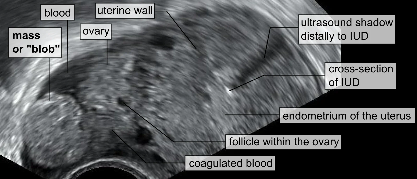
Figure 1Blob sign of ectopic pregnancy
Long description
Transvaginal ultrasonography of an ectopic pregnancy showing the blob sign, a spherical adnexal mass (the ectopic pregnancy) adjacent to the right ovary, with an intrauterine contraceptive device visible in the uterine cavity and no gestational sac in the uterus.

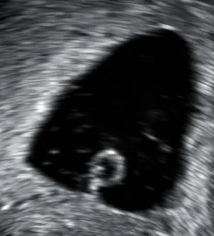
Figure 2Anembryonic gestation
Long description
Transvaginal ultrasonography showing a gestational sac about 28 mm in diameter (approximately 7 weeks 5 days) that contains a yolk sac but no embryo.

TVUS is the first-line imaging test and, when performed by an experienced operator, the gold standard for location. [1][2]
- An intrauterine pregnancy (gestational sac with yolk sac or embryo) on TVUS essentially excludes ectopic pregnancy, except in the exceedingly rare heterotopic case. [5]
- A tubal ectopic is diagnosed positively when an adnexal mass is visualised moving separately from the ovary, often with a tubal ring of peripheral vascularity ("ring of fire"). [1]
- Definitive signs: a yolk sac or embryo in the adnexa (with or without cardiac activity).
- An adnexal mass is identified in most ectopics, but 15 to 35% show no extrauterine mass on TVUS; these need close follow-up. [5]
- Free fluid or haemoperitoneum (non-clotting blood on culdocentesis) supports rupture.
The discriminatory zone is the hCG level above which an intrauterine gestational sac should be visible on TVUS. For two decades the figure used was 1,500 to 2,000 IU/L, and pregnancies above it with an empty uterus were treated presumptively as ectopic, sometimes with methotrexate. [5]
Modern guidance is more cautious. Both ACOG and AFP advise that if a discriminatory level is used at all in a woman who hopes to continue a pregnancy, it should be conservatively high, as high as 3,500 IU/L, so that a viable pregnancy is not misdiagnosed and exposed to methotrexate. [3][5] Most viable singletons are nonetheless seen by 1,500 IU/L. [5]
Practical points:
- Below the discriminatory zone with no mass and normal adnexa, the woman may simply have a very early pregnancy below US resolution; repeat hCG and rescan. [5]
- Above the discriminatory zone with an empty uterus, ectopic (or non-viable intrauterine) pregnancy is likely, but a single value must not trigger treatment; serial hCG and repeat imaging come first. [3][5]
- Transabdominal ultrasound is less sensitive, so TVUS is preferred whenever it is available. [5]
A serum progesterone level does not predict ectopic pregnancy. [1]
In a pregnancy of unknown location, endometrial sampling can demonstrate chorionic villi, proving an intrauterine pregnancy (miscarriage) and avoiding unnecessary methotrexate. It is performed as part of diagnostic pathways in early pregnancy assessment units. [3]
If the diagnosis remains unclear, laparoscopy under general anaesthetic is both diagnostic and therapeutic: the surgeon visualises the pelvis and, if an ectopic is found, treats it in the same operation. [1]
Aspiration of non-clotting blood from the posterior fornix under local anaesthesia indicates haemoperitoneum and supports the diagnosis of ruptured ectopic pregnancy. [1]
Approach to suspected ectopic pregnancy
- Woman of reproductive age; abdominal pain, bleeding, shoulder pain, collapse in early pregnancyUrine then serum hCG; if negative, ectopic unlikely
- Positive hCGTransvaginal ultrasound; assess stability, gestation, hCG level and mass size
- Question: Haemodynamically stable?
No (shock, peritonism, rupture)
- Immediate resuscitationIV access, cross-match, activate massive transfusion if needed
- Emergency laparotomy or laparoscopy by senior teamSalpingectomy usually; treat shock in parallel
Yes
- Question: TVUS findings
Intrauterine pregnancy (viable)
- Manage as miscarriage/IUP; heterotopic if assisted conception
Adnexal mass separate from ovary
- Tubal ectopic confirmed; serum hCG guides path
- Question: hCG and size
hCG ≤ 1,000 (up to 1,500) IU/L, mass < 35 mm, no heartbeat, stable, pain free
- Offer expectant management with hCG days 2, 4, 7, then weekly until < 20 IU/L
hCG < 1,500 IU/L, mass < 35 mm, no heartbeat, no significant pain
- Offer single-dose methotrexate 50 mg/m2 IM; follow hCG (details below)
hCG 1,500-5,000 IU/L, or no/failed medical management
- Offer choice methotrexate vs surgery; surgery if hCG ≥ 5,000, mass ≥ 35 mm, heartbeat visible, significant pain
No mass, empty uterus (PUL)
- Serial hCG at 48 h; repeat TVUS; uterine aspiration/Iaparoscopy if location still unknown
This is an emergency. Priority is resuscitation and control of bleeding. [2]
- Two large-bore IV lines (16-18G), crystalloid (e.g. Hartmann's/Ringer's lactate), warmed blood, activate massive transfusion.
- Keep nil by mouth; urgent senior/obstetric review; theatre team.
- Laparotomy (or laparoscopy in experienced hands) without waiting for all test results; treat shock in parallel.
- After stabilising haemodynamics, salpingectomy (removal of the affected tube) is usually performed because it is quicker and more reliable than repair in unstable women. [2]
- Azithromycin or erythromycin may be given post-operatively in low-resource settings for infection prophylaxis (as per local policy). [1]
Suitable for carefully selected, compliant, clinically stable women: [2]
- Clinically stable and pain free and
- Tubal ectopic under 35 mm with no visible heartbeat on TVUS and [2]
- Serum hCG of 1,000 IU/L or less (offer); or 1,000 to 1,500 IU/L (consider).
Monitoring: repeat hCG on days 2, 4 and 7. If each drops by at least 15%, repeat weekly until below 20 IU/L. If the level fails to fall by 15%, stays the same or rises, review the clinical condition and seek senior advice. [2]
By RCOG, expectant management is an option for clinically stable women with an ultrasound diagnosis of ectopic pregnancy and a decreasing hCG initially below 1,500 IU/L. [1] [2]
Advise women that, on limited evidence, there appears to be no difference between expectant and medical management in resolution rate, tubal rupture risk, need for further treatment, or mental health scores; the time to resolution and future fertility are likely to be similar. [2]
Stop expectant management if pain develops, hCG does not fall, or rupture is suspected. [2]
Systemic methotrexate can be offered to women who: [2]
- have no significant pain and
- have an unruptured tubal ectopic with an adnexal mass smaller than 35 mm with no visible heartbeat and [2]
- have a serum hCG below 1,500 IU/L and
- have no intrauterine pregnancy on ultrasound and
- can return for follow-up.
Methotrexate should be given on a first visit only when the diagnosis is definite and a viable intrauterine pregnancy excluded. [2] NICE advises giving a choice of methotrexate or surgery for hCG of at least 1,500 but below 5,000 IU/L when the other criteria are met, and offering surgery first when hCG is 5,000 IU/L or more, the mass is 35 mm or more, there is a fetal heartbeat, pain is significant, or follow-up cannot be guaranteed. [2]
Intramuscular methotrexate is the only medical treatment for ectopic pregnancy. [5] The three published protocols are: [3][5]
| Regimen | Dose | Monitoring and re-dosing |
|---|---|---|
| Single-dose | 50 mg/m2 IM on day 1 | hCG on days 4 and 7. If fall > 15%, weekly hCG to negative. If fall ≤ 15%, repeat 50 mg/m2; if still no fall after 2 doses, operate. |
| Two-dose | 50 mg/m2 IM on day 1 and day 4 | hCG on days 4 and 7. If fall > 15%, weekly to negative. If fall ≤ 15%, give further 50 mg/m2 on day 7 with hCG day 11; if ≤ 15% between days 7 and 11, dose again; operate if no fall after 4 doses. [5] |
| Multi-dose (with leucovorin rescue) | Methotrexate 1 mg/kg IM on days 1, 3, 5, 7 with folinic acid 0.1 mg/kg on days 2, 4, 6, 8 | Give up to 4 doses until hCG falls by 15%; operate if no fall after 4 doses. [5] |
A single dose is the simplest and carries the lowest risk of side-effects; the two-dose regimen is more effective in patients with higher initial hCG. [5] Multidose protocols are more toxic and are not preferred. [5] AFP suggests single-dose for hCG below 3,600 IU/L and considering two-dose above that, especially above 5,000 IU/L, while acknowledging no consistent consensus on exact cut-offs. [5] Medical management success overall is roughly 70 to 95%. [3] (Local outcomes vary by regimen and patient selection.)
- hCG often rises slightly in the first few days, then falls; a failure to fall by at least 15% from day 4 to day 7 carries a high risk of failure. [3]
- After methotrexate, take two hCG measurements in the first week (days 4 and 7) and then weekly until negative. [2]
- If hCG plateaus or rises, reassess for further treatment or surgery. [2]
- Pain 2 to 3 days after methotrexate, presumably from its cytotoxic effect on trophoblast, is common. In a stable woman without a significant fall in haemoglobin or haemoperitoneum, it can be managed expectantly with monitoring. [3]
- Patients must avoid alcohol, folic acid (not folinic acid rescue in the multi-dose protocol), NSAIDs and sexual intercourse while hCG is positive; and must avoid conception for at least 3 months, because of the teratogenic and cytotoxic effects of the drug. [2][3] [1]
ACOG lists absolute contraindications: intrauterine pregnancy, immunodeficiency, moderate to severe anaemia, leukopenia or thrombocytopenia, sensitivity to methotrexate, active pulmonary disease, active peptic ulcer disease, clinically important hepatic or renal dysfunction, breastfeeding, ruptured ectopic pregnancy, haemodynamic instability, and inability to follow up. [3] Relative contraindications include tubal mass 35 mm or larger, embryonic cardiac activity, a high initial hCG, and patient refusal of blood transfusion; many clinicians add hepatic or renal disease, and alcohol use. [3][5] A full blood count, urea and electrolytes, and liver function tests are advisable before administration. [2][5]
- Indications: ruptured ectopic; haemodynamic instability; significant pain; mass 35 mm or more; visible fetal heartbeat; hCG at least 5,000 IU/L; inability to guarantee follow-up; failed medical/expectant management; or patient preference. [2]
- Approach: laparoscopic, whenever possible, by a surgeon competent in laparoscopic techniques; laparotomy for the unstable woman or complex surgery. [1][2]
- Conservative vs radical: the surgeon either removes the gestation and preserves the tube (salpingotomy) or removes the tube (salpingectomy). NICE recommends offering salpingectomy unless other risk factors for infertility exist; salpingotomy is considered when there is contralateral tubal damage. [2] RCOG advises salpingectomy where a healthy contralateral tube is present, and salpingotomy where fertility-reducing factors exist, because tube-preserving surgery leaves a higher risk of persistent trophoblast. [1]
- Follow-up after salpingotomy: serum hCG at day 7 then weekly until negative; persistent trophoblast occurs in up to 1 in 5 women and may need methotrexate or salpingectomy. [2]
- After salpingectomy: a urine pregnancy test at 3 weeks; attend if positive. [2]
- Ovarian sparing: preserve the ovary where safely possible.
- Cervical, interstitial, ovarian, abdominal and caesarean scar pregnancies. [1]
- Evidence is mostly case series; management is individualised by a senior obstetrician/gynaecologist. [2]
- General rules:
- Interstitial ectopics can be managed non-surgically (methotrexate, local or systemic) when stable and asymptomatic, but rupture is dangerous because of heavy bleeding; expectant management is used with caution. [1][2]
- Cervical pregnancy may be treated medically or by evacuation with haemostatic measures (cervical suture, balloon tamponade); total hysterectomy is a last resort for uncontrollable bleeding. [1]
- Abdominal pregnancy: at laparotomy, leave the placenta in place if removal risks severe bleeding. [1]
- Caesarean scar pregnancy: options include methotrexate, surgical resection, uterine artery embolisation and hysteroscopy.
- WHO context: in abortion care, known or suspected ectopic pregnancy is a contraindication to medical (mifepristone/misoprostol) and surgical abortion, because neither terminates an ectopic; missing the diagnosis is life-threatening. [4]
Co-existing intrauterine and ectopic pregnancy. Surgical treatment of the ectopic is usual because methotrexate would endanger the intrauterine pregnancy; methotrexate is not given when a continuing intrauterine pregnancy is desired. [5]
- Rupture with intra-abdominal haemorrhage and hypovolaemic shock, the immediate life threat.
- Persistent trophoblast after salpingotomy or methotrexate.
- Tubal rupture requiring salpingectomy and its effect on future fertility.
- Psychological sequelae: grief, anxiety and PTSD symptoms after pregnancy loss; provide psychological support and follow-up. [2]
- Anti-D sensitisation in Rhesus-negative women.
- Infection after surgery (particularly after low-resource procedures).
- Mortality has fallen with earlier diagnosis, but deaths still occur. [1]
- Recurrence. A previous ectopic makes a further ectopic more likely where there is tubal damage, although most women still have a normal pregnancy next time.
- Fertility. Salpingectomy on one side leaves the other tube; salpingotomy preserves the tube but carries a higher risk of recurrent ectopic and persistent trophoblast. Women should be counselled about risks to future pregnancy and offered a clear follow-up plan. [1][2] Available evidence suggests methotrexate does not adversely affect subsequent fertility or ovarian reserve. [3]
- Future pregnancies. Women should self-refer to an early pregnancy assessment service in future pregnancies if they have any concerns; NICE recommends written information, emergency contacts and an early scan. [2]
- BCSH recommends a minimum of 250 IU anti-D immunoglobulin after a sensitising event in pregnancies under 12 weeks, including ectopic pregnancy, within 72 hours (and up to 10 days); a minimum of 250 IU from 12 to 20 weeks, and 500 IU after 20 weeks with a feto-maternal haemorrhage test. [6]
- RCOG (Green-top No. 22) recommends anti-D for all non-sensitised RhD-negative women with an ectopic pregnancy, regardless of whether it is managed medically, expectantly or surgically, because of the potential for sensitisation; a 2011 case series found significant fetal cells in 25% of ruptured tubal ectopics. [1][6]
- NICE NG126 (updated 2026) is more conservative: anti-D at least 250 IU for rhesus-negative women only if the ectopic or miscarriage is surgically managed at 12 weeks 0 days to 12 weeks 6 days; not for medical or expectant management at under 12 weeks; do not use a Kleihauer test at this stage. [2]
- WHO recommends against routine anti-D before 11 weeks 6 days gestation for abortion and miscarriage, aligning with the NICE change. [2][4]
- Local protocol prevails. The D negative woman's clinicians should follow their national or unit guideline, give anti-D within 72 hours of surgery or bleeding, and document administration. (BCSH notes the minimum is 250 IU; real vial doses are often 500 or 1,500 IU.) [6]
- There is no true primary prevention, but treating pelvic infection early (especially chlamydia) and smoking cessation reduce tubal damage. [1]
- Early pregnancy assessment services and a low threshold for ultrasound in women with pain or bleeding in early pregnancy are the main tools for reducing ruptured presentations. [2]
- Women with prior ectopic pregnancy or tubal pathology should have an early scan in any subsequent pregnancy. [2]
- The protective effect of chlamydia screening on ectopic incidence is supported conceptually but not quantified here.
- Adolescents. Ectopic pregnancy is rare but possible; pregnancy testing is essential in any adolescent with lower abdominal pain, because teenage pregnancies more often present late. [2]
- Women using assisted reproduction. Higher index of suspicion for heterotopic and non-tubal ectopics; early scanning.
- Trans men and non-binary people. NICE guidance applies to all women, trans men and non-binary people with early pregnancy complications; use inclusive language and offer the same care pathways. [2]
- Older adults/peri-menopausal women. Ectopic pregnancy must still be considered with a positive test; post-menopausal bleeding requires a different workup, but the pregnancy test stays non-negotiable.
- Women with severe anaemia, coagulopathy or immunosuppression. Surgery may be preferred over methotrexate (contraindications). [3]
- The diagnosis is made clinically and with the tests available: urine hCG, ultrasound where possible, and culdocentesis if ultrasound is unavailable.
- A ruptured ectopic is a surgical emergency; many districts lack laparoscopy. Urgent laparotomy (salpingectomy) and blood transfusion (screened for HIV-1, HIV-2, hepatitis B, hepatitis C, syphilis and malaria where endemic) are the standard of care; transfer to a comprehensive emergency obstetric centre if surgery is not possible on site. [1]
- If only medical (methotrexate) management is available, ensure strict suitability criteria (no pain, small mass, low hCG, no heartbeat) and a reliable follow-up system.
- Anti-D immunoglobulin is given per national protocol to non-sensitised RhD-negative women.
- Send all pregnancy tissue for histopathology where available, to exclude molar pregnancy and confirm the diagnosis.
- An ectopic pregnancy is a pregnancy outside the endometrial cavity; the tube is the commonest site (tubal, usually ampullary).
- Incidence is roughly 11 per 1,000 pregnancies in the UK. [1]
- Most women have no identifiable risk factor, but tubal damage (PID, surgery, prior ectopic), smoking, IVF and in-utero-conception with an IUD or progestogen-only pill raise risk. [1]
- Presentation ranges from asymptomatic to rupture with shock; the classic triad of pain, amenorrhoea and bleeding is non-specific. Shoulder-tip pain signals bleeding. [1]
- Transvaginal ultrasound is the key test; intrauterine pregnancy excludes ectopic (except heterotopic); an adnexal mass moving separately from the ovary is strongly suggestive. [1][5] Do not rely on a single hCG value.
- The hCG discriminatory zone is now set conservatively high (up to 3,500 IU/L) to avoid misdiagnosing a viable pregnancy. [3][5]
- Management is by stability, size, heartbeat and hCG: expectant (≤ 1,000-1,500 IU/L, < 35 mm, no heartbeat), methotrexate (< 1,500 IU/L, < 35 mm, no heartbeat) or surgery (rupture, unstable, pain, ≥ 5,000 IU/L, ≥ 35 mm, heartbeat, failed follow-up). [2]
- Methotrexate is 50 mg/m2 IM single dose, with hCG on days 4 and 7; re-dose or operate if the fall is 15% or less. [3]
- Surgery is laparoscopic where possible; salpingectomy unless fertility is at risk, salpingotomy with weekly hCG follow-up if the tube is preserved. [1][2]
- Non-tubal and heterotopic ectopics need individualised senior care. [1][2]
- RhD-negative women receive anti-D immunoglobulin per local protocol (minimum 250 IU below 12-20 weeks) within 72 hours; WHO and NICE no longer recommend routine anti-D below about 12 weeks. [2][4][6]
- Recurrence after one ectopic is more likely with tubal damage; most women later have a normal pregnancy; psychological support matters. [1][2]
Answer each one in your head first, then open the answer.
A 26-year-old woman, 7 weeks amenorrhoeic, presents with unilateral pelvic pain and light vaginal bleeding. Her blood pressure is 118/76 mmHg. A urine pregnancy test is positive. Transvaginal ultrasound shows an empty uterus, an adnexal mass 28 mm separate from the ovary with no visible heartbeat. Serum hCG is 900 IU/L. What is the most appropriate management?
Show answerHide answer
This is a stable tubal ectopic pregnancy under 35 mm with no heartbeat and hCG under 1,000 IU/L. Offer expectant management with hCG on days 2, 4 and 7 (fall of at least 15% each) then weekly until below 20 IU/L, or offer single-dose methotrexate (50 mg/m2 IM) if she prefers active treatment. Surgery is not first-line. Counsel about return precautions and give written emergency information.
Why is the hCG discriminatory zone now commonly quoted as high as 3,500 IU/L rather than 1,500 to 2,000 IU/L?
Show answerHide answer
Using a low threshold led to misdiagnosing viable intrauterine pregnancies as ectopic and treating them with methotrexate. A higher, conservative threshold waits for definitive ultrasound evidence before treatment, while recognising that most viable singletons are already visible by 1,500 IU/L. Serial hCG is used when the diagnosis remains uncertain. [5]
A woman receives single-dose methotrexate for a tubal ectopic. Her hCG is 1,200 IU/L on day 4 and 1,100 IU/L on day 7. What should happen next?
Show answerHide answer
The fall from day 4 to day 7 is only about 8.3%, less than the required 15%, so this is treatment failure. Reassess: she may receive a second dose of methotrexate (50 mg/m2) and repeat hCG, or proceed to surgery if clinically indicated. Continue weekly hCG monitoring once levels fall by more than 15%.
After surgery for an ectopic pregnancy, when is a tube-preserving (salpingotomy) approach preferred, and what follow-up is required?
Show answerHide answer
Salpingotomy is considered when the woman has fertility-reducing factors such as contralateral tubal damage, accepting a higher persistent trophoblast rate. Salpingectomy is otherwise preferred, including when the contralateral tube is healthy. After salpingotomy, check serum hCG at day 7 and then weekly until negative; up to 1 in 5 women need further treatment (methotrexate or salpingectomy). [2]
What emergency symptoms after treatment for ectopic pregnancy warrant immediate return?
Show answerHide answer
Severe or worsening abdominal pain, shoulder-tip pain, dizziness, fainting, heavy bleeding, fever, or feeling unwell. These may indicate rupture with internal bleeding or treatment failure and require urgent assessment.
A non-sensitised RhD-negative woman has an ectopic pregnancy managed medically with methotrexate at 9 weeks. Should she receive anti-D immunoglobulin?
Show answerHide answer
Guidelines differ. RCOG recommends anti-D regardless of management because sensitisation can occur. NICE (2026) and WHO do not recommend routine anti-D before about 12 weeks or with medical/expectant management. The clinicians must follow their local or national protocol and document their decision.
Antepartum haemorrhage; early pregnancy assessment services; miscarriage; pelvic inflammatory disease; ectopic and tubal surgery; pregnancy of unknown location; cervical pregnancy; heterotopic pregnancy; Caesarean scar pregnancy.
- Elson CJ, Salim R, Potdar N, Chetty M, Ross JA, Kirk EJ. Diagnosis and Management of Ectopic Pregnancy. RCOG Green-top Guideline No. 21; first edition published 2016, reviewed 2019. Accessed .
- National Institute for Health and Care Excellence. Ectopic pregnancy and miscarriage: diagnosis and initial management. NICE guideline NG126; published 2019, last updated 17 June 2026. Accessed .
- Committee on Practice Bulletins - Gynecology, American College of Obstetricians and Gynecologists. Tubal Ectopic Pregnancy. ACOG Practice Bulletin No. 193; Obstetrics & Gynecology 2018;131(3):e91-e103; reaffirmed 2025. Accessed .
- World Health Organization. Clinical practice handbook for quality abortion care; Abortion care guideline. World Health Organization, 2022-2023 editions. Accessed .
- American Academy of Family Physicians. Ectopic Pregnancy: Diagnosis and Management. American Family Physician 2020;101(10):599-606. Accessed .
- Qureshi H, Massey E, Kirwan D, et al. The use of anti-D immunoglobulin for the prevention of haemolytic disease of the fetus and newborn. British Committee for Standards in Haematology; Transfusion Medicine 2014;24:157-183. Accessed .
Figure credits
- Figure 1: Mikael Häggström. When using this image in external works, it may be cited as: Häggström, Mikael (2014). "Medical gallery of Mikael Häggström 2014". WikiJournal of Medicine 1 (2). DOI:10.15347/wjm/2014.008. ISSN 2002-4436. Public Domain. or By Mikael Häggström, used with permission., via Wikimedia Commons, CC0, CC0.
- Figure 2: Mikael Häggström. When using this image in external works, it may be cited as: Häggström, Mikael (2014). "Medical gallery of Mikael Häggström 2014". WikiJournal of Medicine 1 (2). DOI:10.15347/wjm/2014.008. ISSN 2002-4436. Public Domain. or By Mikael Häggström, used with permission., via Wikimedia Commons, CC0, CC0.
Educational content for medical students and doctors. It is not a substitute for clinical judgement or local guidelines.