Gestational trophoblastic disease
From molar pregnancy to choriocarcinoma: what it is, how it presents, hCG staging, chemotherapy and follow-up.
- Gestational trophoblastic disease (GTD) is a spectrum of placenta-derived conditions that runs from the usually benign, but pre-malignant, complete and partial molar pregnancies to the malignant disorders invasive mole, choriocarcinoma and the very rare placental site and epithelioid trophoblastic tumours.
- If a mole does not regress it becomes gestational trophoblastic neoplasia (GTN), defined by a plateaued or rising hCG; this happens after about 13% to 20% of complete moles and 0.5% to 5% of partial moles. [1][2]
- Molar pregnancies are usually detected by ultrasound in the first trimester and treated by suction evacuation; the disease is then followed with serial hCG, which is the single most useful marker.
- Chemotherapy cures nearly all women with GTN: single-agent treatment for low-risk disease and multi-agent treatment for high-risk disease, each with defined regimens and doses.
- Follow-up is precise and time-limited, and it dictates when contraception can change and when it is safe to try to become pregnant again.
By the end of this page you should be able to:
- Define GTD and GTN and classify the individual diseases, including the very rare ones.
- Explain the epidemiology and the risk factors across the reproductive lifespan.
- Describe how a mole forms and why the disease behaves as it does.
- Recognise the presentation and physical signs of a molar pregnancy and of choriocarcinoma.
- Plan investigations and interpret staging and risk scores using the current FIGO systems.
- Manage evacuation, surveillance, chemotherapy and follow-up, including contraception and a later pregnancy.
DefinitionGestational trophoblastic disease (GTD)
A group of disorders arising from trophoblastic cells of the placenta. It includes the pre-malignant complete and partial hydatidiform moles and, when the tissue invades or metastasises, the malignant conditions invasive mole, choriocarcinoma, placental site trophoblastic tumour (PSTT) and epithelioid trophoblastic tumour (ETT). The spectrum has more recently been extended to include atypical placental site nodule (APSN), a lesion that may progress to PSTT or ETT. GTN is the collective term for the malignant forms, which can follow any type of pregnancy, not only a mole. [1]
The distinction that matters clinically is between a mole that is confined to the uterus and one that has become GTN. GTN is not a separate disease with a different start; it is what a mole, or less commonly a non-molar pregnancy, becomes when trophoblastic tissue persists and secretes hCG.
| Condition | Placental feature | Course |
|---|---|---|
| Complete hydatidiform mole (CHM) | All trophoblast, no fetal tissue; villi swollen into vesicles | Most GTD; 13% to 20% progress to GTN if untreated [1] |
| Partial hydatidiform mole (PHM) | Abnormal placenta with fetal or yolk-sac tissue | Pre-malignant; 0.5% to 5% progress to GTN [1] |
| Invasive mole | Mole tissue that has grown into the myometrium | Usually diagnosed by hCG trend; can metastasise locally |
| Choriocarcinoma | No villi; sheets of cyto-, intermediate and syncytiotrophoblast | Highly malignant; metastasises early; very chemotherapy-sensitive |
| PSTT | Arises from extravillous intermediate trophoblast at the placental site | Often slow-growing; less hCG-driven |
| ETT | Arises from chorionic-type intermediate trophoblast | May mimic choriocarcinoma or squamous cancer |
| APSN | Atypical placental site nodule, a possible pre-cursor | 10% to 15% co-exist with or develop into PSTT or ETT |
Classification is by pathology, not by a clinical score. A mole is complete or partial; the malignant forms are choriocarcinoma, PSTT, ETT and the invasive mole itself. hCG then acts as the running measure of whether pre-malignant tissue has become malignant. GTN is diagnosed when the hCG plateaus or rises, or on histology. Older national guidance describes the same spectrum: ACOG Practice Bulletin No. 53 (2004) lists complete and partial hydatidiform moles, invasive moles, choriocarcinomas and placental-site trophoblastic tumours. [5]
The incidence of molar pregnancy varies with geography and age. Representative figures include:
- complete mole in roughly 1 to 3 per 1000 pregnancies and partial mole in about 3 per 1000 in UK estimates, 11.5 per 1000 deliveries in Indonesia and less than 1 per 1000 deliveries in the USA, with prevalence higher in Asia, Africa and Central and South America than in the USA, Europe and Australia. [1][6]
- one complete or partial mole raises the risk of a further mole by roughly 1% to 2%, and after two molar pregnancies the risk of a third rises to about 15% to 20%; changing partners does not lower it. [6]
- extremes of reproductive age raise the risk: women aged over 35 years have about twice the risk of women aged 20 to 35 years, women aged over 40 years 5 to 7 times the risk, and girls aged under 15 years roughly 1 in 500 pregnancies while women aged over 50 years roughly 1 in 8. [1][6]
- choriocarcinoma occurs in about 3 per 100000 deliveries in the USA and Europe and 23 per 100000 deliveries in Southeast Asia. [1]
- PSTT and ETT are the rarest: roughly 0.2% of all GTD cases and 1% to 2% of GTN cases. [1]
- GTN after a live birth is estimated at 1 in 50000. [6]
- GTN after a complete mole develops in 13% to 20% of cases and after a partial mole in 0.5% to 5%. [1]
GTD is therefore common enough that every clinician who deals with early pregnancy will meet it, and rare enough that the follow-up must run through a specialist centre.
Complete moles usually result from fertilisation of an empty ovum by one sperm, with duplication of the paternal genome; the disease is androgenetic and diploid. Partial moles follow dispermic fertilisation of a normal egg and are triploid. Histology alone can suggest either, but genotyping (short tandem repeat analysis) now separates them reliably, including in the new WHO category of non-molar abnormal villous lesions that carry a low GTN risk.
Risk factors worth recording in a history:
- Age at both extremes, as above.
- Previous mole, which is the only strong risk factor and the one that raises the risk of recurrence, especially after two.
- Familial recurrent mole, caused by mutations in NLRP7 and KHDC3L genes; these women have biparental familial recurrent mole and a molecular, rather than molar, diagnosis. [1]
- Asian ethnicity is associated with higher rates, and increased parity, nutrition and socio-economic factors have been suggested, though these are not consistently causative.
- No other modifiable risk factor has been established.
Women who have had a mole should be told that a single subsequent pregnancy is almost always normal: the risk of a further mole after one is low, and the risk of GTN in that pregnancy is also low. After two moles the counselling changes and specialist follow-up is needed.
A complete mole starts as an abnormal conception. The trophoblast still proliferates and invades, but without an embryo; the villi swell into the familiar hydropic vesicles. Not all of this proliferation is harmless. If the mole fails to regress after evacuation, persistent trophoblast continues to divide and secrete hCG, and the disease becomes GTN. hCG is therefore both the driver of much of the morbidity of GTD and the built-in monitor of response: a plateau or rise in hCG is the definition of progression. [1]
Choriocarcinoma goes one step further. It loses villous architecture entirely, grows as sheets of cytotrophoblast and syncytiotrophoblast, invades the myometrium early, and enters the blood stream before local symptoms appear. It can therefore present with metastases in the lung, brain, liver or gut rather than with bleeding, and hCG may be the first clue. PSTT and ETT arise from intermediate trophoblast, grow and metastasise far more slowly and produce hCG irregularly, so hCG is a less reliable marker for them. APSN lies between a normal placental site nodule and PSTT or ETT and is a lesion to follow rather than to ignore.
The early first-trimester diagnosis that ultrasound allows has changed the presentation: complications such as hyperemesis gravidarum, pre-eclampsia, hyperthyroidism and massive uterine enlargement are now far less common than they once were. [1]
On gross examination a complete mole is a mass of grape-like vesicles with no identifiable fetal parts; a partial mole shows abnormal placental tissue, often with a non-viable or malformed fetus. Choriocarcinoma is bulky and necrotic with haemorrhage; PSTT and ETT are solid nodular masses that may invade deeply; APSN is usually an incidental finding in tissue removed for another reason.
Under the microscope the pattern matters:
- CHM shows hydropic villi with circumferential trophoblastic hyperplasia; PHM shows focal hyperplasia and two populations of villi, with fetal tissue present.
- Choriocarcinoma shows no villi and a trimorphic trophoblast.
- PSTT and ETT show intermediate trophoblast and absent villi.
The immunohistochemical markers are a real diagnostic asset. p57, a cyclin-dependent kinase inhibitor whose expression needs the maternal genome, is absent in CHM and present in PHM and in non-molar pregnancies; it therefore separates complete mole from everything else. Genotyping by short tandem repeat (STR) analysis gives the definitive diagnosis when histology is equivocal, including in the abnormal (non-molar) villous lesions described by the WHO classification. ETT and PSTT are graded in part by the Ki-67 proliferation index, which is under 5% for benign placental site nodules, 5% to 10% for APSN and 10% to 30% for PSTT, and above 10% for ETT. [1]
Many complete moles cause no symptoms and are found on a first-trimester scan. When symptoms do occur, vaginal bleeding in very early pregnancy is the commonest presentation and can be heavy and prolonged. Other clues include:
- hyperemesis gravidarum that is worse than expected,
- passage of grape-like vesicles,
- a uterus larger than expected for dates,
- pelvic pressure or pain,
- early pre-eclampsia, and
- features of hyperthyroidism, which arise because hCG shares an alpha-subunit with TSH and acts weakly on the thyroid receptor.
Choriocarcinoma more often presents weeks or months after the index pregnancy. Bleeding that persists after evacuation, miscarriage or even term delivery should always be investigated, and metastatic symptoms at the time of diagnosis (haemoptysis, breathlessness, headache, neurological deficits, abdominal pain or bleeding) are not unusual.
Examination may show a uterus larger than dates, bilateral (and sometimes very large) ovarian cysts from theca-lutein stimulation, and anaemia. Blood pressure may be high if pre-eclampsia is present and a rapid or irregular pulse may reflect hyperthyroidism. Examination is otherwise unremarkable in most women, which is why imaging and histology make the diagnosis.
Red flagDo not miss
Persistent, unexpected or heavy bleeding after any pregnancy event, together with a uterine size that does not match the gestation or a hCG that does not fall, defines the case that needs trophoblast surveillance rather than routine miscarriage care. Booking a conventional ultrasound description of "snowstorm" or "Swiss cheese" placenta is a prompt to obtain histology.
NoteA first-trimester scan raises a concern
A 32-year-old woman is scanned routinely at 10 weeks. There is no fetus, the uterus is enlarged and the placenta has a heterogeneous cystic appearance. hCG is markedly raised. She is Rhesus negative and has no symptoms other than mild nausea. Suction evacuation under ultrasound guidance is performed and the tissue is examined. Histology confirms a complete hydatidiform mole. Her hCG is monitored and falls, so follow-up is shortened.
The common confusions are with an early non-molar pregnancy that has miscarried, with a partial mole, with an ectopic pregnancy and, when a tumour is present, with a range of secondary malignancies. ultrasound and histology plus hCG separate most cases; genotyping resolves the rest.
| Condition | Features that separate it |
|---|---|
| Hydropic (non-molar) abortion | Villi swollen by oedema, but p57 positive and no androgenetic genotype; hCG falls on its own; NLRP7/KHDC3L not involved. |
| Partial mole in twin pregnancy (one mole, one normal twin) | Fetal structures present; hCG is higher than expected but fetal viability may continue; managed in specialist centres. |
| Ectopic pregnancy | No intra-uterine gestation sac; trophoblast confined to tube or ovary; hCG pattern of pregnancy of unknown location, not the very high or rapidly rising values typical of some moles. |
| Pregnancy of unknown location | Follow the NICE pathway: serial hCG, repeat imaging and exclusion of ectopic and molar pregnancy before treatment. [7] |
| Gestational choriocarcinoma presenting after a term birth | Abnormal bleeding after delivery; hCG rising or raised; usually confirmed by imaging, high hCG and, where possible, histology. Differential includes post-partum haemorrhage, retained products and endometrial cancer. |
| PSTT or ETT vs primary cervical or endometrial cancer | hCG may be normal or modestly raised; histology and immunohistochemistry required; management is surgical chemotherapy rather than hysterectomy alone. |
| Secondary (metastatic) trophoblastic tumour mimic | Lung nodules, for example, may be metastases from another cancer; hCG plus gynaecological imaging and sometimes genotyping establish a gestational origin. |
- Serum quantitative beta-hCG before and after evacuation. This is the marker of residual and persistent disease; it must be a total hCG assay that detects all forms (beta-hCG, free beta, core fragment and hyperglycosylated forms) where possible. [1]
- Full blood count, clotting, renal and hepatic function, since evacuation can be bloody and chemotherapy is renal-clearant.
- Blood group and save, with anti-D for Rhesus-negative women at the time of evacuation. [1][6]
- Thyroid function tests if hyperthyroidism is suspected.
- Histopathology of all products of conception from a non-continuing pregnancy, with genotyping when histology is ambiguous, and p57 staining. [1]
- The NCI PDQ lists the practical triggers that move care from surgery to chemotherapy: a rising beta-hCG for 2 weeks, a plateau for 3 weeks, or a tissue diagnosis of choriocarcinoma. [8]
Figure 1Molar pregnancy
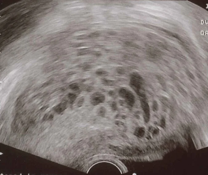
Long description
Transvaginal ultrasonography of a molar pregnancy with multiple small fluid-filled vesicles producing a bunch-of-grapes / snowstorm pattern.

- Pelvic ultrasound (transvaginal where feasible) for uterine size, myometrial invasion, adnexal masses and, in a twin pregnancy, viability.
- Chest radiograph to count lung metastases for staging. Chest CT, pelvic or abdominal CT or MRI, and brain MRI according to risk, as set out below.
| Test or measure | Threshold or expected finding |
|---|---|
| Total serum beta-hCG assay | Detects all hCG isoforms; persistently low values need continued monitoring because some rise later. [1] |
| Post-molar diagnostic plateau | hCG unchanged, i.e. a change of less than 10% (rise or fall), across four measurements over 3 weeks or more (days 1, 7, 14, 21). [2] |
| Post-molar diagnostic rise | An increase of over 10% across three weekly measurements over 2 weeks or more (days 1, 7, 14). [2] |
| Histological diagnosis | Confirmed choriocarcinoma, PSTT or ETT. [2] |
| Lung metastasis counted for scoring | Only lesions measuring 1 cm or more in diameter; micrometastases do not change management. [2] |
| Lymph node defined as metastasis | Short-axis diameter 10 mm or more on imaging. [2] |
| PSTT/ETT pre-treatment hCG | Not a sensitive marker; diagnosis rests on histology and imaging rather than hCG trend. [1] |
The "hCG elevated for 6 months" criterion was removed from the 2026 revision of the FIGO diagnosis because total assay quality made it redundant; the modern definitions are the plateau and rise criteria above, or histology.
The current standard is the FIGO system, which combines an anatomical stage with a modified WHO prognostic score. Classifying properly before starting treatment is what lets low-risk disease be treated gently and high-risk disease treated thoroughly.
- plateau of hCG of less than 10% change over four weekly measurements across 3 weeks or more (days 1, 7, 14 and 21), or
- rise of over 10% over three weekly measurements across 2 weeks or more (days 1, 7 and 14), or
- a histological diagnosis of choriocarcinoma, PSTT or ETT. [1][2]
| Stage | Description |
|---|---|
| I | Tumour confined to the uterus and cervix |
| II | Extension to adnexa, vagina or broad ligament, but limited to the genital tract |
| III | Lung metastases, with or without genital tract involvement; lymph node metastasis below the diaphragm |
| IV | All other metastatic sites, including lymph node metastasis above the diaphragm |
Eight prognostic factors are each given 0, 1, 2 or 4 points. The total places the patient in a risk group.
| Factor | 0 | 1 | 2 | 4 |
|---|---|---|---|---|
| Age at diagnosis (years) | Under 40 | 40 or older | — | — |
| Antecedent pregnancy | Mole | Abortion, miscarriage or ectopic | Term pregnancy | — |
| Interval from index pregnancy (months) | Under 4 | 4 to 6 | 7 to 12 | Over 12 [2] |
| Pre-treatment hCG (mIU/mL) | Under 1000 | 1000 to 9999 | 10000 to 99999 | 100000 or more [2] |
| Largest tumour size, uterine or metastatic (cm) | Under 3 | 3 to 5 | Over 5 | — [2] |
| Site of metastases | Lung or vagina | Spleen or kidney | Gastrointestinal tract | Brain or liver |
| Number of metastases | — | 1 to 4 | 5 to 8 | More than 8 [2] |
| Previous failed chemotherapy | — | — | Single drug | Two or more drugs |
Risk groups: total score 6 or below is low risk, above 6 is high risk. Low risk is usually treated with single-agent chemotherapy; high risk with multi-agent chemotherapy. The 2026 revision removed previous failed chemotherapy as a factor and, because no robust data support re-weighting, kept the same scoring format. Patients with scores of 5 or 6 deserve attention: metastatic disease (odds ratio 1.9, 95% confidence interval 1.1 to 3.2), choriocarcinoma histology (odds ratio 3.7) and very high pre-treatment hCG predict single-agent failure. [1][2]
Imaging for staging should be done as close as possible to the start of treatment, because GTN can double in volume every 24 to 48 hours. The minimum in low-risk disease is pelvic ultrasound and chest radiograph. In high-risk disease or suspected choriocarcinoma, contrast-enhanced brain MRI and CT of the chest, abdomen and pelvis are needed. PET/CT is not routine. [2] Brain MRI is preferred to CT for brain assessment; liver MRI is reserved for problem-solving.
- Medical stabilisation: treat anaemia, pre-eclampsia, hyperthyroidism and heavy bleeding. Measure hCG before and after treatment.
- Uterine evacuation: conventional suction evacuation, ideally ultrasound-guided, is the preferred method, using a 7 to 12 mm suction cannula; manual vacuum aspiration is acceptable where electronic suction is unavailable. Intravenous oxytocin may be started at the time of the procedure and continued for a few hours afterwards to help the uterus contract. Medical induction, hysterotomy and routine repeat curettage are not recommended; a second evacuation is reserved for selected cases with bleeding plus ultrasound evidence of retained molar tissue. [1]
- Hysterectomy with ovarian preservation is a reasonable alternative for women who have completed their family, but post-operative hCG surveillance (and chemotherapy if needed) are still required; hysterectomy is not more cost-effective than single-agent chemotherapy for low-risk disease. [1]
- Oxytocin infusion is not started before evacuation because of concern about trophoblastic dissemination.
- Rh prophylaxis: Rhesus-negative women should receive Rh immune globulin at the time of evacuation. [1][6]
- Prophylactic chemotherapy at or immediately after evacuation reduces the incidence of post-molar GTN by about 3% to 8%, but should be reserved for special situations where the risk of progression is much greater than normal or follow-up hCG is not possible. [1]
- Register the woman with the regional trophoblast service and arrange hCG monitoring.
- Do not promise patients that chemotherapy has already started; it is reserved for those who develop GTN.
Management is by regional trophoblast centres. Women with a confirmed or suspected mole are registered, cases of GTN are discussed, and most low-risk disease can be managed locally with remote specialist supervision. Patients with choriocarcinoma or metastatic disease are referred urgently to the specialist centre.
Treatment is by chemotherapy, chosen by the risk score.
Low risk (FIGO stage I to III and score 6 or less). Single-agent methotrexate or actinomycin D. Standard first-line regimens include:
- methotrexate 50 mg intramuscularly on days 1, 3, 5 and 7 with folinic acid 15 mg orally 24 hours later (days 2, 4, 6 and 8), repeated every 2 weeks;
- methotrexate 0.4 mg/kg intravenously or intramuscularly for 5 days every 2 weeks, to a maximum of 25 mg per dose;
- actinomycin D pulse 1.25 mg/m2 intravenously every 2 weeks; and
- actinomycin D 0.5 mg intravenously for 5 days every 2 weeks. [1]
Other methotrexate schedules used include 30 to 50 mg/m2 intramuscularly weekly and 300 mg/m2 by infusion every 2 weeks. Actinomycin D is more effective in head-to-head comparisons (complete remission around 80.2% versus 65.1% for methotrexate) but carries a little more nausea, vomiting and hair thinning. [1]
About 30% of low-risk patients develop resistance after a single agent. If the hCG plateaus or rises, or dosing is limited by toxicity, switch to the other single agent: the response to actinomycin D after methotrexate failure is 76% to 87%, particularly at relatively low hCG levels. [1] Women with FIGO scores of 5 or 6, particularly with metastatic disease, choriocarcinoma histology or very high pre-treatment hCG, are more likely to fail single-agent treatment and should start multi-agent therapy from the outset. [1]
After hCG normalises, a further 2 to 3 chemotherapy cycles are given to reduce relapse; complete remission rates approach 100%. Hysterectomy can be offered to selected low-risk women who have completed their family, but surveillance and any post-operative chemotherapy are still required, and chemotherapy alone is usually simpler and cheaper. [1]
High risk (FIGO stage II to III with score 7 or more, or stage IV). Multi-agent chemotherapy. The most common regimen is EMA-CO: etoposide, methotrexate, actinomycin D, cyclophosphamide and vincristine; there is no consensus from clinical trials that one combination is clearly superior. About 20% fail EMA-CO, but most are rescued with further therapy, so survival exceeds 96%. Interval disease beyond about 2.8 years from the index pregnancy, and liver or brain metastases, predict a worse outcome. Gentle induction chemotherapy for ultra-high-risk disease with a heavy tumour burden reduces early deaths from haemorrhage and tumour lysis, although late deaths from resistant tumours still occur. Choriocarcinoma in the brain is treated with radiotherapy in addition to chemotherapy; resistant deposits at one site are sometimes removed surgically. Salvage regimens in resistant disease include high-dose ifosfamide-etoposide and cisplatin or taxane-based therapy, and immunotherapy (for example, avelumab, which has produced complete remission in only about 53% of methotrexate-resistant low-risk cases and is not standard salvage) is being evaluated. [1]
PSTT and ETT are treated primarily surgically, with chemotherapy reserved for metastatic or recurrent disease; they are not scored on the FIGO risk score. [1][2]
This is the part of GTD care that must run like clockwork.
- Register the diagnosis with the trophoblast centre.
- Measure serum or urine hCG every 1 to 2 weeks after evacuation. If hCG is measured every 2 weeks and a rise or plateau is detected, sample again after 1 week. [1]
- GTN is diagnosed on the plateau or rise criteria above, or on histology; by about 2 weeks after evacuation a rising trend is plain.
- Partial mole: follow-up is concluded once hCG has returned to normal on two samples taken at least 4 weeks apart. [3]
- Complete mole: if hCG normalises within 56 days of evacuation and a confirmatory normal hCG 4 weeks later, prolonged 6-month monitoring is unnecessary and the woman can be discharged after that confirmation. If the 56-day window is missed, the standard 6 months of monthly monitoring apply from normalisation. [3][6] [1]
- NICE acknowledges molar pregnancy outside its own early-pregnancy scope and requires histological examination of all tissue from a non-continuing pregnancy, so a mole is not mistaken for an ordinary miscarriage. [7]
- After GTN treated with chemotherapy: monthly hCG for at least 12 months. [4]
Persistent but falling low hCG for up to 6 months does not necessarily need treatment; such women need continued monitoring rather than empiric chemotherapy. [6]
Until hCG has returned to normal, effective contraception is essential because a rising hCG in pregnancy could be mistaken for persistent disease. General principles are:
- Progestogen-only, combined hormonal and barrier methods are suitable after evacuation, including before hCG returns to normal. [6]
- The levonorgestrel intra-uterine system should not be fitted immediately after evacuation; it can be used once hCG has normalised. [6]
- Women who have had chemotherapy should avoid conception for 1 year after completion of treatment, as a precaution. [3]
- Hormone replacement therapy can be used once hCG has normalised. [6]
- An early pregnancy after a mole (particularly after 6 months) should be registered with the trophoblast centre; women with two or more moles or who received chemotherapy need follow-up in a subsequent pregnancy. [6]
- Subsequent pregnancies after 12 months are not at increased risk of miscarriage or fetal abnormality. After a single mole the chance of another mole is about 1%; after two, follow-up is needed. [6]
The risk of GTN in a future pregnancy is low, and the risk of recurrence in any later pregnancy after a single mole is low (about 0.6% to 2% after a single mole, increasing after consecutive moles). [1]
- Heavy bleeding at evacuation, and disseminated intravascular coagulation in longer-standing disease.
- Anaemia, infection and uterine perforation at evacuation.
- Progression to GTN as above.
- Metastatic disease: lung, vagina, brain, liver, gastrointestinal tract, spleen and kidney, producing breathlessness, bleeding, neurological deficits or abdominal symptoms.
- Ovarian theca-lutein cysts take months to regress.
- Hyperemesis, pre-eclampsia and hyperthyroidism are now uncommon because of early diagnosis.
With modern management, survival is excellent. In low-risk disease complete remission rates approach 100%, and in high-risk disease survival exceeds 90% even though some patients relapse and need further therapy. [1] Almost all women can expect cure if managed properly in a trophoblast centre, and fertility is generally preserved. Relapse after chemotherapy can occur and usually needs further chemotherapy; brain and liver metastases can relapse late. Site of metastasis, interval from the index pregnancy and treatment history are the variables that matter most after staging. [1][4]
There is no primary prevention for molar pregnancy. Practical secondary prevention is:
- histological examination of all products of conception after a non-continuing pregnancy, with routine trophoblast assessment, because not all moles are suspected before evacuation;
- periconceptional folate supplementation through a standard antenatal care programme (not specific to GTD, but part of good early pregnancy care);
- registering a mole early with the trophoblast centre and completing hCG follow-up; and
- offering genetic counselling and NLRP7/KHDC3L testing to women with recurrent mole or a family history.
Screening is not offered to the general population because incidence is low and follow-up is effective once a diagnosis is made.
Pregnancy with an existing mole. A complete or partial mole may co-exist with a normal twin pregnancy. Between 40% and 60% of these result in a live birth, although the risk of miscarriage and of GTN is higher (GTN risk compared with a singleton mole rises from 15% to 20% to 27% to 46%). In the absence of complications and typical genetic and ultrasound findings, these pregnancies can be managed expectantly in a specialist unit. [1]
Age. Older women need the same management but have a higher risk of GTN and are more likely to be high-risk; fertility counselling is different if hysterectomy is contemplated. Young women with a previous mole should be reassured that a later normal pregnancy is expected.
Men and their partners. Familial recurrent mole is an autosomal recessive disorder of the oocyte driven by NLRP7 and KHDC3L; partners do not carry the risk alleles in the usual case, and oocyte donation is an eventual option in recurrent cases.
- Suction curettage can be done with a manual vacuum aspirator.
- Keep to ultrasound guidance if available, but a clear clinical indication plus histology is better than no diagnosis.
- Start oxytocin after the cavity is cleared rather than before.
- Rhesus-negative women still need immune globulin.
- Send a fixed percentage of tissue for histology; in a low-resource setting even a single slice identifies many moles.
- Follow hCG with a total assay if possible; where a quantitative assay is unavailable, serial urine hCG or the shortest practical follow-up plus pregnancy avoidance gives some protection.
- Telemedicine and remote hCG submission have been shown to maintain follow-up compliance in women who live far from care. [1]
- Prophylactic chemotherapy is considered where hCG follow-up cannot be delivered at all.
Approach to a suspected or confirmed molar pregnancy
- First-trimester scan shows a suspected mole, or non-viable pregnancy tissue is obtainedUSS: cystic placenta with no fetus (CHM) or abnormal fetus and placenta (PHM)
- Measure serum quantitative beta-hCG before treatment; stabilise the patientTreat heavy bleeding, anaemia, pre-eclampsia, hyperthyroidism
- Suction evacuation under ultrasound guidanceCannula 7-12 mm; oxytocin only after evacuation; Rh immune globulin if Rhesus negative
- Send all tissue for histology with genotyping if equivocalp57 staining and STR genotyping separate CHM, PHM and non-molar pregnancy
- Register with the trophoblast centre and start hCG surveillanceEvery 1-2 weeks
- Question: hCG trend after evacuation?
Falls and normalises
- Partial mole: discharge when normal on two samples at least 4 weeks apartComplete mole: if normal within 56 days plus a confirmatory normal value, no 6-month follow-up is needed
Plateaus for 3 weeks or rises for 2 weeks
- Diagnose GTN and stageFIGO anatomical stage plus modified WHO score; CT, MRI or chest X-ray as indicated
- Low risk (score 6 or less): single-agent methotrexate or actinomycin D
- High risk (score above 6, or stage IV): EMA-CO multi-agent chemotherapy
- Continue monthly hCG for at least 12 months after normalisation; 2-3 extra cycles
- Complete and partial moles are both pre-malignant but differ in genetics, histology and progression risk (13% to 20% vs 0.5% to 5%).
- hCG is the monitor: GTN is defined by a plateau or rise in hCG, or by histology of the malignant forms.
- Diagnosis is first-trimester: ultrasound plus histology; p57 and STR genotyping resolve difficult cases.
- Evacuation is by suction, with anti-D at the time; medical induction is avoided.
- Follow-up is strict: 1 to 2 weekly hCG, with a 56-day rule that shortens care for complete mole when values normalise early.
- Chemotherapy is risk-based: single-agent (methotrexate or actinomycin D, with set doses) for low risk; EMA-CO for high risk; survival above 90% even in high-risk disease.
- Contraception changes with the hCG, and pregnancy is usually deferred for 12 months after chemotherapy.
- Low-resource practice is workable: manual suction, anti-D, histology and remote hCG follow-up cover most needs.
Answer each one in your head first, then open the answer.
What is the most common presenting feature of a complete hydatidiform mole in the first trimester, and which test confirms rather than just suggests the diagnosis?
Show answerHide answer
Vaginal bleeding is the commonest presenting feature, although many moles are found incidentally on ultrasound. The diagnosis is confirmed by histological examination of the evacuated tissue, with p57 immunohistochemistry and, where needed, STR genotyping to distinguish a complete mole from a partial or non-molar pregnancy.
According to the current combined FIGO threshold, when may post-molar GTN be diagnosed on hCG grounds alone?
Show answerHide answer
When the hCG (total assay) plateaus, defined as less than 10% change, across four measurements over 3 weeks or more, or when it rises by more than 10% across three weekly measurements over 2 weeks or more. A histological diagnosis of choriocarcinoma, PSTT or ETT is the third route. The older criterion of hCG remaining elevated for 6 months has been dropped.
A woman's complete mole hCG normalises 42 days after evacuation and is still normal on a confirmatory sample 4 weeks later. What follow-up is needed?
Show answerHide answer
Prolonged 6-month monthly monitoring is not required in that situation; she can be discharged from central follow-up after the confirmatory normal value, with advice not to conceive until follow-up is complete.
Give one first-line single-agent methotrexate regimen for low-risk GTN, with dose, route and frequency.
Show answerHide answer
Methotrexate 50 mg intramuscularly on days 1, 3, 5 and 7, with folinic acid 15 mg orally 24 hours after each methotrexate dose (days 2, 4, 6 and 8), repeated every 2 weeks. Alternatives are methotrexate 0.4 mg/kg, maximum 25 mg, intravenously or intramuscularly for 5 days every 2 weeks, or actinomycin D 1.25 mg/m2 intravenously every 2 weeks.
A woman with complete mole has a history of two previous moles and starts GTN treatment. Why would her disease be at higher risk of failing single-agent therapy?
Show answerHide answer
Because prior molar pregnancies (particularly consecutive ones) raise the risk of a further mole and more aggressive disease, and the additional factors that predict failure of single-agent therapy, such as metastatic disease, choriocarcinoma histology and very high pre-treatment hCG, may also be present. The practical point is to stage and score before choosing a regimen and to start multi-agent therapy when the score is above 6.
After evacuation of a mole, which contraceptive methods are acceptable before hCG returns to normal, and which device should be deferred?
Show answerHide answer
Progestogen-only, combined hormonal and barrier methods may be used. The levonorgestrel intra-uterine system should not be fitted until hCG has normalised. [6]
Gestational trophoblastic disease links to early pregnancy assessment, miscarriage and ectopic pregnancy, anaemia, hyperthyroidism and, in high-risk disease, to the oncology sections on chemotherapy and metastatic cancer. Those topics are described elsewhere in the library.
- Ngan HYS, Seckl MJ, Berkowitz RS, Xiang Y, Golfier F, Sekharan PK, Braga A, Garrett A. Diagnosis and management of gestational trophoblastic disease: 2025 update. International Journal of Gynaecology and Obstetrics 2025;171(Suppl 1):78-86. Accessed .
- Ngan HYS, Seckl MJ, Horowitz N, Cheung ANY, Braga A, Sekharan PK, Fridsten S, Garrett A, Yanaihara N, Wilailak S, Schmalfeldt B. FIGO staging for gestational trophoblastic neoplasia: 2026. International Journal of Gynaecology and Obstetrics 2026;174:1172-1180. Accessed .
- Tidy J, Seckl M, Hancock BW, on behalf of the Royal College of Obstetricians and Gynaecologists. Management of Gestational Trophoblastic Disease (Green-top Guideline No. 38, 4th edition, 2020; reviewed 2023). BJOG 2021;128:e1-e27. Accessed .
- Soper JT. Gestational trophoblastic disease: current evaluation and management. Obstetrics and Gynecology 2021;137(2):355-370. Accessed .
- Committee on Practice Bulletins-Gynecology, American College of Obstetricians and Gynecologists. Diagnosis and treatment of gestational trophoblastic disease (ACOG Practice Bulletin No. 53). Obstetrics and Gynecology 2004;103(6):1365-1377. Accessed .
- NHS Greater Glasgow and Clyde (Right Decision Service). Gestational Trophoblastic Disease, (Molar Pregnancy) (guideline 423). Right Decisions for Health and Care, last reviewed 2026-03-25. Accessed .
- National Institute for Health and Care Excellence (NICE). Ectopic pregnancy and miscarriage: diagnosis and initial management (NG126). NICE guideline NG126. Accessed .
- National Cancer Institute. Gestational Trophoblastic Disease Treatment (PDQ): Health Professional Version. NCI PDQ, updated 2026-10-28. Accessed .
Figure credits
- Figure 1: Mikael Häggström. When using this image in external works, it may be cited as: Häggström, Mikael (2014). "Medical gallery of Mikael Häggström 2014". WikiJournal of Medicine 1 (2). DOI:10.15347/wjm/2014.008. ISSN 2002-4436. Public Domain. or By Mikael Häggström, used with permission., via Wikimedia Commons, CC0, CC0.
Educational content for medical students and doctors. It is not a substitute for clinical judgement or local guidelines.