Congenital malformations of the newborn
Congenital structural defects present at birth: abdominal wall, neural tube, GI, diaphragmatic, craniofacial and genital anomalies.
- A congenital malformation is a structural (and sometimes functional) abnormality present at birth, most often arising during organ formation in the first weeks of pregnancy.
- Congenital anomalies span single defects, multiple defects and whole syndromes; some are lethal in utero, while others are compatible with a normal life after repair.
- Antenatal ultrasound and, where indicated, maternal blood tests can find many anomalies before birth, allowing delivery at a centre equipped for the newborn.
- Folic acid 400 micrograms daily, started before conception and continued to 12 weeks of gestation, prevents many neural tube defects.
- Many malformations are treated surgically in the newborn period or in infancy; survival is excellent for several once fatal conditions, but lifelong follow-up is often needed.
By the end of this page you should be able to:
- Define a congenital malformation and classify malformations as isolated, multiple, or syndromic.
- Explain why early gestation is the vulnerable window, giving examples of failed closure, fusion, recanalisation and rotation.
- Interpret an antenatal ultrasound report, including the markers that raise suspicion for a neural tube defect, an abdominal wall defect or a diaphragmatic hernia.
- Compare gastroschisis and omphalocele on examination and management at birth.
- Recognise the clinical presentation, investigation and surgical principles for oesophageal atresia with tracheo-oesophageal fistula, duodenal atresia, anorectal malformations and congenital diaphragmatic hernia.
- Describe the types and treatment timeline of cleft lip and palate and of craniosynostosis.
- Plan the investigation and surgical management of hypospadias.
- Counsel parents on folic acid prevention and recurrence risk.
A congenital malformation is a structural abnormality of the body present at birth, resulting from an error in development between fertilisation and birth. The term includes defects of organs, limbs, the neural tube, the abdominal wall, the diaphragm, the face and the external genitalia. Most are isolated (one defect in an otherwise normal infant); others occur as multiple defects or as part of a syndrome or an association. The effects range from a minor variant requiring no treatment to a life-threatening emergency in the first minutes of life. Congenital malformations are a leading contributor to neonatal mortality and to long-term disability worldwide.
This page provides a single overview of the major structural defects that present in the newborn, grouped by system. It then sets out antenatal diagnosis, folic acid prevention, the syndromes most often associated with these defects, low-resource practice and key revision points. Each malformation would, in clinical practice, be managed by a team that includes obstetricians, neonatologists, paediatric surgeons and, where relevant, urologists and craniofacial surgeons.
A useful first distinction is between a malformation (an error in morphogenesis, such as failure of a structure to form), a deformation (an abnormal shape produced by external mechanical forces on a normally formed part), a disruption (destruction of a previously normal structure, for example by vascular compromise) and a dysplasia (abnormal cellular organisation within a tissue). This page deals mainly with malformations. Most structural malformations arise during organogenesis, when tissues are rapidly proliferating, migrating and fusing; the vulnerable period runs through early gestation, before many women know they are pregnant.
Malformations are also classified clinically:
- Isolated: a single defect (for example an isolated cleft lip).
- Multiple: two or more defects without an obvious unifying diagnosis (for example an infant with both a cardiac and a renal anomaly).
- Syndromic: a recognised pattern of anomalies from one underlying cause (genetic or teratogenic), such as Down syndrome (trisomy 21) or a VACTERL association.
An association is a non-random collection of anomalies that occur together more often than chance, without (yet) a single known cause. The classic example used throughout this page is VACTERL: at least three of vertebral defects, anal atresia, cardiac defects, tracheo-oesophageal fistula, oesophageal atresia, renal anomalies and limb defects. [6] A second association is CHARGE: coloboma, heart defect, atresia of the choanae, growth retardation, genital abnormality and ear abnormalities. Recognising an association matters because finding one defect obliges a systematic search for the others.[4][6]
A further clinically vital distinction is between an open and a closed (or covered) defect. In an open neural tube defect or a gastroschisis the abnormal tissue is exposed to amniotic fluid, with consequences for both prognosis and in-utero therapy; in a closed defect (such as a meningocele covered by skin, or an omphalocele covered by a membrane) the surface is intact.
The baseline risk of a major structural malformation varies widely between populations and registries, and reported rates depend on how carefully minor anomalies are ascertained as well as on geography. Most individual malformations in this page are uncommon, of the order of 1 in a few thousand births, and several are genuinely rare. Because the rate of each defect is low, many clinicians will meet some of them only a handful of times in a career; the key is to recognise the pattern quickly and to know where to send the infant.
Specific incidences cited in this page are given with each defect and with references: gastroschisis about 2 to 4 per 10,000 live births (about 1 in 4000 to 1 in 2500, depending on era and population) [1]; omphalocele 3.38 per 10,000 pregnancies (about 2 to 3 per 10,000 pregnancies or births depending on ascertainment) [2]; duodenal atresia a rare cause of neonatal high intestinal obstruction (no figure given here); anorectal malformations 1 in 5000 births in the United States [6]; congenital diaphragmatic hernia 2.3 per 10,000 live births [7]; cleft lip with or without cleft palate 0.1% overall risk, with racial variation (2 per 1000 in Asian, 1 per 1000 in Caucasian and 0.5 per 1000 in African-American populations) [8]; and craniosynostosis and hypospadias discussed below.
The incidence of several defects differs by region, ethnicity, maternal age and geography; neural tube defects in particular vary widely between and within countries, which is partly why folic acid policy (fortification and supplementation) differs around the world.
The cause of most individual malformations is unknown and is best accepted as multifactorial: a genetic susceptibility meeting an environmental trigger at a critical moment in development. Recognised contributors include the following.
Genetic. Chromosomal abnormalities (trisomies 13, 18 and 21; sex-chromosome aneuploidy) and single-gene mutations (for example FGFR2/3 and TWIST1 in craniosynostosis syndromes, RET in Hirschsprung disease) account for a minority of malformations overall but an important minority of syndromic ones. A recurrence in a sibling is high when a mutation is found, and low but not zero for isolated, sporadic defects.
Nutritional. Periconceptional folate deficiency is the best established nutritional cause of neural tube defects, and is the basis of prevention.
Clinical pearlTiming
Teratogens and maternal disease. Antiseizure medications (notably valproate), retinoids, folate antagonists, poorly controlled maternal diabetes, maternal obesity and hyperthermia are associated with increased rates of several malformations. Specific associations mentioned in the source literature for individual defects in this page include maternal tobacco, nitrosamine exposure, cyclo-oxygenase inhibitor use and decongestant exposure for gastroschisis[1]. Recreational drugs and alcohol have well described but less quantifiable effects.
Maternal age. Extreme reproductive age (under 20 and over 40) is associated with omphalocele[2]; advanced maternal age is a classic risk factor for Down syndrome and hence duodenal atresia.
Environmental and mechanical. Mechanical constraints on the fetus (oligohydramnios, multiple pregnancy, uterine malformation) account for deformations rather than true malformations, but these can be difficult to separate on examination.
Where a teratogen is identified, the practical message is to avoid it in any woman who could become pregnant and to review medications before conception. Regional advice on valproate and pregnancy is a good example of how guidelines differ between regulators.
Most structural defects in this page can be traced to one of a small number of embryological errors.
- Failure of closure. The neural tube must close along its entire length in the first month. Failure at the cranial end gives anencephaly or encephalocele; failure at the caudal end gives spina bifida. The same principle of failed fusion of body-wall folds explains gastroschisis, and premature fusion of a cranial suture explains craniosynostosis.
- Failure of return and rotation. The midgut normally herniates into the umbilical cord early in gestation and returns later, rotating as it does so. Failure of return gives omphalocele; failure of recanalisation after the lumen is temporarily occluded gives atresias of the oesophagus, duodenum and lower gut.
- Failure of separation of a common tube. The trachea and oesophagus share a common foregut tube that must separate in early gestation; failure gives oesophageal atresia and tracheo-oesophageal fistula.
- Failure of development of a partition. The diaphragm forms from several embryonic components that must fuse in the first trimester; failure gives congenital diaphragmatic hernia.
- Failure of fusion of facial or palatal processes. Cleft lip and cleft palate result when the maxillary and medial nasal prominences, or the palatine shelves, fail to meet and fuse.
Each defect's mechanism tells you what to expect clinically: the tissue that should have developed is absent or misplaced, while surrounding structures may be hypoplastic from lack of normal functional stimulus (most importantly, the lungs in congenital diaphragmatic hernia). It also tells you why an early (sometimes fetal) repair can preserve function, for example closing a spina bifida lesion before amniotic fluid damages the exposed cord.
Modern obstetric ultrasound has transformed the recognition of congenital malformations. A detailed anomaly scan in the second trimester can detect most of the defects on this page, although sensitivity varies greatly by defect, operator and maternal habitus. Detection matters for three reasons: it allows parents to make informed choices, it allows delivery to be planned at a centre with paediatric surgery and neonatal intensive care, and it can improve outcome (for example delivery of an infant with gastroschisis at 37 to 38 weeks rather than prolonged gestation).
Antenatal markers and findings worth remembering:
- Neural tube defects: a second-trimester maternal serum alpha-fetoprotein that is higher than expected raises strong suspicion; ultrasound looks for open cranial and spinal defects and for cranial signs (the "lemon" and "banana" signs). Anencephaly can be seen early. [3]
- Gastroschisis: free-floating loops of bowel in the liquor, usually to the right of the cord insertion, often associated with restricted fetal growth; maternal serum alpha-fetoprotein is elevated. Diagnosis is often made at the 20-week scan. [1]
- Omphalocele: a midline abdominal mass covered by a membrane at the base of the cord; detectable by the end of the first trimester, with elevated maternal serum alpha-fetoprotein. The suspected presence of liver within the sac is an important prognostic detail. [2]
- Oesophageal atresia: polyhydramnios in the third trimester (because the fetus cannot swallow), with a small or absent stomach bubble. Sensitivity of prenatal ultrasound is reported at 42% with a positive predictive value of 56%. [4]
- Duodenal atresia: polyhydramnios, with the classic "double bubble" of two fluid-filled structures (stomach and proximal duodenum) and no distal bowel gas; often associated with Down syndrome.
- Anorectal malformations: difficult to detect before birth; a dilated distal colon or calcified meconium may be the only clue, so most are diagnosed after delivery.
- Congenital diaphragmatic hernia: detection is reported in up to 60% of cases at 18 to 22 weeks. Findings include abdominal organs in the chest, mediastinal shift, polyhydramnios, and absence of the stomach bubble on the left side. The liver position and lung measurements are used to predict severity. [7]
- Cleft lip: detectable by ultrasound in the second trimester. Isolated cleft palate is much harder to see and is often only found after birth.
- Craniosynostosis: the head shape and, by the third trimester, sutural ridges may be visible; suspected on ultrasound and confirmed after birth.
- Hypospadias: may be seen in the third trimester as an abnormal penile configuration; in practice many are first noticed at the newborn check.
Where a defect is suspected, further assessment usually includes a fetal echocardiogram and magnetic resonance imaging in a specialist centre, chromosomal testing where appropriate, and a multidisciplinary counselling appointment so that parents understand both the defect and the expected pathway. Unless a specific reason exists to deliver earlier, many defects are managed with delivery at (or after) 37 to 39 weeks in a high-volume centre; congenital diaphragmatic hernia management, for example, specifically favours planned delivery after 39 weeks. [7] For conditions where the stomach and bowel are involved, an orogastric tube should be passed immediately after birth rather than enteral feeding.
Antenatal suspicion to postnatal care of a major congenital malformation
- Antenatal suspicion of a structural anomalySecond-trimester anomaly scan; maternal serum alpha-fetoprotein where indicated
- Question: Is the defect isolated or part of a syndrome or association?
Isolated
- Detailed fetal ultrasound and echocardiogramPlan delivery at a centre with paediatric surgery and neonatal intensive care
- Defect-specific perinatal planTiming of delivery, mode of delivery and neonatal resuscitation agreed before labour
Multiple or syndromic features
- Fetal medicine and genetics reviewChromosomal microarray, targeted testing, and counselling for recurrence
- Multidisciplinary counsellingDiscuss prognosis, treatment options including fetal therapy where available, and palliative pathways
- Delivery at a high-volume centreObstetric, neonatal and paediatric surgical teams ready
- Immediate newborn care follows the defect-specific planAirway protection, thermoregulation, vascular access, and only then the planned investigations or surgery
Neural tube defects (anencephaly, spina bifida and encephalocele) result from failure of the neural tube to close between roughly 21 and 28 days after conception (rostral closure around days 23 to 26, caudal closure around days 26 to 28). [3] Because that closure happens before many pregnancies are recognised, prevention cannot wait for a positive pregnancy test. Folic acid, the synthetic form of vitamin B9, taken in the periconceptional period, has the strongest evidence base of any measure in this page.
- Who: all women capable of becoming pregnant, including those not planning a pregnancy.
- Standard dose: 400 micrograms (0.4 mg) of folic acid daily. The World Health Organization recommends 400 micrograms daily from the moment a woman begins trying to conceive until 12 weeks of gestation. [12]
- High-risk dose: the World Health Organization recommends 5 mg daily for women who have previously had a pregnancy affected by a neural tube defect (or who have a personal or strong family history), together with advice on the protective effect and on increasing dietary folate. [12] Guidelines differ on this point: the United States commonly advises 4 mg daily after a previous affected pregnancy, while the WHO advises 5 mg. [13]
- Regional point: clinical practice guidelines in the United States recommend 0.4 to 0.8 mg (400 to 800 micrograms) daily for women planning or capable of pregnancy, and higher doses for selected high-risk situations. [13]
United States
In the United States, folic acid fortification of cereal grains is mandatory and the recommendation is 0.4 to 0.8 mg daily, consistent with the US Preventive Services Task Force. -
World Health Organization
The WHO recommendation of 400 micrograms daily from preconception to 12 weeks applies worldwide, including in settings without food fortification, and 5 mg daily is reserved for women with a previous neural tube defect pregnancy. - Food fortification with folic acid is the population strategy in many countries and lowers the incidence of neural tube defects; where fortification is not in place, supplementation carries the whole burden.
- Folate comes from diet (leafy greens, legumes, fortified foods) but most non-pregnant women do not reach the protective intake from diet alone, which is why a daily supplement is recommended.
- Harm note: total folic acid intake from supplements and fortified foods above 1000 micrograms daily is not recommended for the general population without medical indication, because of the possibility of masking vitamin B12 deficiency.
Figure 1Newborn with an omphalocele
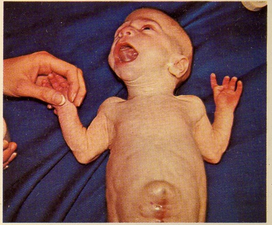
Long description
Photograph of a young newborn lying on its back on a blue cloth, mouth open and crying. At the umbilicus a rounded, translucent sac bulges from the abdominal wall, covered by a thin membrane through which the contents show. The skin of the rest of the abdomen is intact.

These are two distinct defects that look similar from a distance but differ in anatomy, association pattern, prognosis and management.
Gastroschisis is a full-thickness paraumbilical (almost always right-sided) abdominal wall defect, typically about 4 cm across, through which bowel herniates into the liquor without any covering membrane. The exposed loops become matted, thickened and inflamed; infants are often growth-restricted. Additional extraintestinal malformations occur in about 10% of cases, while additional gastrointestinal problems (atresia, stenosis, perforation, necrosis, malrotation, volvulus) occur in up to a quarter. The karyotype is abnormal in roughly 1% of cases, usually in the setting of other anomalies. Gestation is characteristically short; spontaneous delivery usually occurs at 37 to 38 weeks. Diagnosis is usually made on the 20-week ultrasound, with elevated maternal serum alpha-fetoprotein. Overall survival is greater than 90%. Cryptorchidism accompanies gastroschisis in 15% to 30% of cases. [1]
Management at birth is resuscitative first: protect the exposed bowel (saline dressings and a plastic wrap or bowel bag), insert an orogastric tube and peripheral intravenous access, and stabilise the airway. Fluid needs are high (2 to 3 fold maintenance) because of evaporative losses from the exposed bowel, but over-resuscitation worsens oedema and delays closure. Formal repair is either primary closure or delayed closure with a spring-loaded silo and slow serial reduction over days, followed by fascial closure; outcomes are similar between the two approaches. Vaginal delivery is preferred for most, and there is no contraindication to labour on the basis of gastroschisis alone, although very large liver herniation may prompt caesarean section. Infants can be classified as "simple" or "complex" according to the presence of bowel atresia, stenosis, perforation, necrosis, malrotation or volvulus; complex disease carries a significantly worse course. [1]
Omphalocele (exomphalos) is a midline defect at the base of the umbilical cord, through which abdominal contents herniate and are covered by a membrane of peritoneum (and, later, amnion). It occurs in 3.38 per 10,000 pregnancies. The defect ranges from small (a few loops of bowel) to "giant" (5 cm or more, with liver partly protruding), the latter causing restrictive lung problems. Unlike gastroschisis, omphalocele is frequently accompanied by other anomalies or syndromes, including Beckwith-Wiedemann syndrome, trisomies 13, 18 and 21, pentalogy of Cantrell, cloacal exstrophy (OEIS) syndrome, CHARGE syndrome, Shprintzen-Goldberg syndrome, Carpenter syndrome, Meckel-Gruber syndrome and Marshall-Smith syndrome. Survival is close to 80% overall and closer to 90% when the omphalocele is isolated; liver herniation carries a poorer prognosis. Detection is possible by the end of the first trimester on ultrasound, and maternal serum alpha-fetoprotein is elevated. Extreme maternal age (under 20 or over 40), multiple pregnancy and male sex are associated. Infants with omphalocele tolerate oral feeding after closure, unlike the more protracted course common with gastroschisis. [2]
| Feature | Gastroschisis | Omphalocele |
|---|---|---|
| Defect | Paraumbilical, usually to the right of the cord | Midline, at the cord insertion |
| Covering | No membrane; bowel free in liquor | Membrane of peritoneum (and amnion) |
| Cord insertion | Adjacent to, but separate from, the defect | Inserts on the apex of the sac |
| Typical herniated contents | Small bowel (often matted and inflamed) | Bowel and commonly liver |
| Extraintestinal anomalies | About 10% of cases [1] | Frequent; often part of a syndrome |
| Typical associated syndromes | Rare | Beckwith-Wiedemann, trisomies 13/18/21, pentalogy of Cantrell, CHARGE, others |
| Fetal growth | Frequent growth restriction | Less consistent |
| Delivery | Trial of labour usually; 37 to 38 weeks [1] | Depends on severity and associated anomalies |
| Postnatal management | Protect bowel, silo or primary closure; delayed enteral feeding | Sterile wrap, staged or primary closure; lung support if giant |
| Survival | Greater than 90% [1] | Close to 80% overall; about 90% if isolated [2] |
Neural tube defects are malformations of the brain and spinal cord arising from failure of the neural tube to close in early gestation. [3] They are the most common severe congenital malformations of the central nervous system after cardiac defects, and a leading cause of stillbirth and lifelong disability. There are two classic open forms: anencephaly (failure of the cranial (rostral) neuropore to close, appearing between days 23 and 26 of gestation; incompatible with postnatal life) [3] and spina bifida (failure of the caudal neuropore to close, anywhere along the spine). Between these extremes lie encephalocele and the spectrum of spina bifida from a tiny, skin-covered spina bifida occulta to an open myelomeningocele in which the spinal cord and meninges are exposed.
Clinical points:
- Anencephaly presents with absence of the cranial vault and cerebral hemispheres; most cases are detected on first-trimester ultrasound and are lethal.
- Meningocele (meninges only, usually skin-covered and neurologically intact) is less damaging than myelomeningocele (meninges plus spinal cord), which carries variable paralysis, hydrocephalus and an Arnold-Chiari type II malformation.
- Encephalocele is a protrusion of brain and meninges through a skull defect, often occipital, and commonly accompanies hydrocephalus.
- Spina bifida occulta affects about 10% of people and is usually asymptomatic, marked by a tuft of hair or a skin dimple over the lower spine; it does not need surgical treatment unless tethering produces symptoms. [3]
Diagnosis before birth relies on a raised maternal serum alpha-fetoprotein in open defects and on the characteristic ultrasound appearances (the "lemon" and "banana" signs for spinal lesions). Women with a prior affected pregnancy have a recurrence risk that justifies high-dose folate supplementation as set out above. Postnatal management of an open spinal lesion consists of covering the defect with a sterile, non-adherent dressing, nursing prone, avoiding pressure on the sac, arranging early surgical closure, and managing the complications: hydrocephalus (frequently needing a shunt), neurogenic bladder and bowel, orthopaedic deformity and tethered cord. Selected fetuses are candidates for open or minimally invasive fetal repair in the second trimester, available only in specialist centres. [3]
Oesophageal atresia (EA) and tracheo-oesophageal fistula (TEF) are congenital discontinuity or abnormal communication between the oesophagus and the trachea, resulting from failed separation of the common foregut tube in early gestation. They occur together in about 90% of cases [4] and, taken together with pure atresia, at roughly 1 in 3000 live births. [4] Around 84% to 87% are Type C: oesophageal atresia with a distal tracheo-oesophageal fistula; the remaining types (type A, isolated atresia, 7% to 8%; type B, proximal fistula, 1% to 2%; type D, both fistulas, under 1% to 3%; type E, "H-type" fistula without atresia, about 4%) are important to recognise on imaging. [4]
Presentation. In utero, polyhydramnios is common (two-thirds of pregnancies), especially in the third trimester, because the fetus cannot swallow. After birth the infant has excessive oral secretions, drooling, choking, cyanosis and respiratory distress with feeds, and a nasogastric or orogastric tube cannot reach the stomach or coils in the upper pouch. A distal fistula produces gastric distension. H-type fistulas may be subtle and present late, even years later, with recurrent aspiration or pneumonia.
Associated anomalies. Roughly half of affected infants have another congenital anomaly, classically within the VACTERL spectrum [4]; among a large single-centre series the reported rates were congenital heart disease 59.1%, vertebral 25.5%, renal 21.8%, anorectal 11.6% and limb 7.1%. [4] Trisomies 18 and 21 and CHARGE syndrome are also associated. The risk of recurrence in a subsequent pregnancy for nonsyndromic disease is 1%. [4]
Diagnosis. Failure to pass an orogastric tube beyond 10 to 15 cm, with the tube coiled in the upper pouch on a chest radiograph, supports the diagnosis; water-soluble contrast may outline the pouch. A right-sided aortic arch (2.5%) mandates left-sided surgical access. [4] Pre-operative evaluation includes echocardiography, renal ultrasound, spinal ultrasound and limb radiographs to complete the VACTERL work-up.
Management. Stabilise first: airway control if needed, suction of the upper pouch, nil by mouth, intravenous fluids and antibiotics, with consideration of total parenteral nutrition. Very low birth weight infants (under 1500 g) [4] are treated in stages (fistula ligation first, repair later); infants over 2 kg [4] generally proceed to primary repair once cardiac issues are addressed. Repair is by open thoracotomy or thoracoscopic division of the fistula and primary oesophageal anastomosis; the Foker technique may be used for a "long gap" where the ends do not meet. Proton pump inhibition is routinely recommended for at least one year because gastro-oesophageal reflux is almost universal after repair. Prognosis is relatively good, with overall survival around 85% to 90% [4], determined more by associated cardiac and chromosomal abnormalities than by the atresia itself. [4]
Figure 2Double bubble sign in duodenal atresia
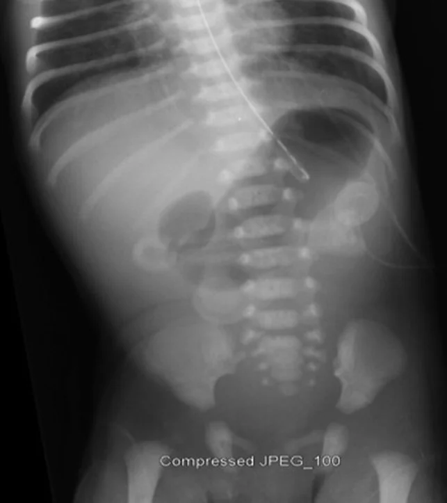
Long description
Frontal abdominal radiograph of a newborn with a nasogastric tube in place. A large gas-filled stomach lies to the left and a second gas-filled bubble, the dilated proximal duodenum, lies beside it, with little or no gas in the rest of the bowel.

Duodenal atresia is a congenital intestinal obstruction, usually of the descending duodenum, producing bilious or non-bilious vomiting within the first 24 to 48 hours of life (most often after the first feed; StatPearls cites 24 to 38 hours), typically after the first feed. [5] It is strongly associated with Down syndrome and with other duodenal anomalies (annular pancreas, duodenal web, stenosis), which share a common mechanism of failed recanalisation of the intestinal lumen during the early first trimester. Polyhydramnios is the usual antenatal clue.
The double bubble sign on a plain abdominal radiograph is the classic finding: two rounded air-filled shadows representing the dilated stomach and the dilated first part of the duodenum, with no distal intestinal gas. Confirmatory imaging (a limited water-soluble contrast study or ultrasound) may be used, but the plain film is usually sufficient. Care must be taken that air in the stomach is not mistaken for perforation, and to exclude midgut volvulus, which is a surgical emergency requiring entirely different management.
Management begins with correction of fluid and electrolyte deficits and nasogastric decompression, followed by surgical bypass (duodenoduodenostomy) once the infant is stable. Chromosomal and cardiac evaluation should accompany repair, and transient low calcium or associated congenital heart disease may complicate postoperative care. Feeding resumes after recovery; long-term outcomes are generally good. [5]
Anorectal malformations (ARMs) are a spectrum of defects in which the anus and rectum are malformed, ranging from a simple membranous covering to high (supralevator) lesions with fistulas to the urinary or genital tracts. They occur in about 1 in 5000 births in the United States, with males affected slightly more often. Rectourethral fistula is the most common defect in males and rectovestibular fistula in females. About half to 60% of infants have at least one additional anomaly, so a systematic search is mandatory; the commonest associated systems are genitourinary, vertebral/spinal cord and cardiac. Anorectal malformation is the "A" in VACTERL, in which at least three of the named defects must be present. [6]
Diagnosis and evaluation.
- At birth, inspect the perineum: look for an anal opening in the correct position, a fistulous opening on the perineum or in the vestibule, meconium from the urethra or vagina, and the "flat bottom" (absent midline gluteal fold) that predicts a high lesion and poor continence.
- A newborn who has no stool in the first 24 hours needs explanation; examination after 24 hours can reveal the defect once pressure forces meconium through a fistula. Delayed diagnosis occurs in about one in five neonates and increases morbidity. [6]
- Examination should include the heart and limbs; a passage of a nasogastric tube excludes oesophageal atresia, and a urine dipstick looks for meconium in male infants.
- Prone cross-table radiography with a marker at the anal dimple measures the distance between the distal rectal air bubble and the perineum: under 1 cm suggests a low defect, over 1 cm a high one. [6] Ultrasound and pelvic imaging further define the anatomy. Plain radiographs of the spine and echocardiography exclude associated vertebral and cardiac disease.
- On physical examination, normal anal size is a 10 to 12 Hegar dilator in a term newborn and about 15 by 12 months. [6]
Management.
- Stabilise and resuscitate, then complete the diagnostic work-up in the first 24 to 36 hours. [6] Primary anoplasty suits low, simple lesions.
- High or complex lesions require an initial diverting (descending or sigmoid) colostomy, with definitive posterior sagittal anorectoplasty 4 to 8 weeks later once the infant is growing and well. [6] Many infants are suitable for one-stage repair in experienced hands.
- After repair, anal dilatation begins 2 weeks later to prevent stricture [6], and the colostomy is closed once adequate dilation is achieved (usually over 6 to 12 months). [6] Feeding begins as soon as the bowel is working.
- Long term, prognosis depends on lesion level, sacral ratio and spinal cord quality. Most patients develop some constipation or incontinence and are managed with laxatives or a structured bowel-management programme. [6]
Congenital diaphragmatic hernia (CDH) is a developmental defect of the diaphragm that allows abdominal viscera to enter the chest, occurring in 2.3 per 10,000 live births. [7] The result is a triad of pulmonary hypoplasia (fewer airway generations and alveoli), pulmonary vascular remodelling with persistent pulmonary hypertension, and cardiac dysfunction. The most common type is a left-sided posterolateral (Bochdalek) hernia, accounting for roughly 70% to 85% of cases (Bochdalek hernias overall are about 80% to 90%, and most of these are left-sided); about half of affected newborns have an additional anomaly. [7] Morgagni (anterior, retrosternal) hernias make up about 2% to 5% of cases [7] and are less symptomatic, often diagnosed later because pulmonary hypoplasia is uncommon.
Presentation and diagnosis. Neonates present with immediate respiratory distress in the first hours of life, variable severity, sometimes life-threatening. Antenatal detection occurs in up to 60% of cases at 18 to 22 weeks: on the left side a heterogeneous intrathoracic mass with mediastinal shift and an absent stomach bubble; on the right side a homogeneous mass (the herniated liver is sonographically similar to lung). Polyhydramnios is common. Postnatal diagnosis is on chest radiograph: air- or fluid-filled bowel loops in the hemithorax with little aerated lung, mediastinal shift and reduced abdominal girth. A nasogastric tube may be seen coiled in the chest. Associated pulmonary hypertension develops in about 70% of infants and, of these, 38% ultimately require extracorporeal life support. [7]
Management. Delivery is planned for after 39 weeks [7] at a high-volume tertiary centre, with no proven difference between vaginal and caesarean birth. The immediate postnatal approach is gentle: intubation for high-risk infants, nasogastric decompression, avoidance of mask ventilation (which distends the herniated bowel), pressure-limited ventilation, optimisation of oxygenation, correction of acid-base status and, where indicated, inhaled nitric oxide; ECMO is reserved for suitable candidates. Surgery (reduction and repair of the defect, with patch if necessary) is performed only after stabilisation. Survivors require long-term follow-up for chronic lung disease, pulmonary hypertension, feeding difficulty and neurodevelopmental outcome. Overall survival is about 65% to 80% in many series (international population-based estimates are nearer 62%), and falls to around 50% in the most severe, high-risk cases; isolated disease treated in specialist centres has survival around 70% to 90%. [7]
Cleft lip and cleft palate are among the most common congenital craniofacial anomalies, arising from failure of fusion of the facial prominences or palatine shelves in early gestation. The overall risk of cleft lip with or without cleft palate is about 0.1%, with reported rates of about 2 per 1000 births in Asian, 1 per 1000 in Caucasian and 0.5 per 1000 in African-American populations; reported rates vary by ancestry and by ascertainment. The defect may be unilateral or bilateral, complete or incomplete, and may involve lip only, palate only, or both. Isolated cleft palate is a distinct entity and also needs assessment for submucous defects that present late. [8][9]
Presentation and complications. Beyond the visible split, the defect impairs feeding (poor nipple seal, nasal regurgitation) and speech (hypernasality), and predisposes to recurrent otitis media with effusion and hearing loss. Dental development and facial growth are affected. Palate repair must balance speech (early closure) against midfacial growth (delayed closure). About half of cleft palates are nonsyndromic; the remainder occur in the context of a genetic syndrome. [9]
Associated syndromes. Cleft lip/palate may be part of van der Woude syndrome (lip pits), 22q11.2 deletion (DiGeorge/velocardiofacial) syndrome, Pierre Robin sequence (U-shaped cleft with micrognathia and glossoptosis, which can obstruct the airway), Stickler syndrome and ectrodactyly-ectodermal dysplasia-clefting. Teratogenic and environmental factors reported with palatal clefts include maternal smoking, alcohol, anticonvulsant therapy and retinoid intake.
Management. Care is multidisciplinary from the outset: feeding support (specialised bottles, lactation advice), audiology, speech and language therapy, dentistry/orthodontics and, ultimately, surgery. Lip repair follows the "rule of 10s" and is usually complete in early infancy; nasoalveolar moulding may be used pre-operatively. Palatoplasty is most commonly performed between 9 and 15 months [9], with centres differing on exact timing; a staged approach (lip plus hard palate early, soft palate later) is one of several accepted strategies. Further surgery for velopharyngeal insufficiency, nasal symmetry or residual fistula is common during childhood and adolescence. [8][9]
Recurrence risk rises with affected parents or siblings: about 4% for a second child after one affected child in otherwise unaffected families, 9% after two affected children, and about 17% when one parent and one sibling are affected. [9] Counselling families requires care, since each of these figures is a guideline rather than an individual prediction.
Craniosynostosis is the premature fusion of one or more of the cranial sutures. It alters head shape, can restrict brain growth and may, in multi-suture or syndromic disease, raise intracranial pressure and impair neurodevelopment. Isolated (nonsyndromic) craniosynostosis accounts for roughly 75% of cases; the remaining are syndromic. The pattern by suture is characteristic and informs the operation: sagittal 55% to 60%, unilateral coronal 20% to 25%, metopic about 15%, and lambdoid (the rarest) 3% to 5%. [10]
The key clinical distinction is between craniosynostosis and deformational (positional) plagiocephaly, which is far more common, results from external moulding of the growing skull, and is managed with repositioning and, in selected cases, a helmet rather than surgery. Craniosynostosis produces a hard, palpable suture ridge and a typical skull shape for each suture (for example the long, narrow "scaphocephaly" of sagittal fusion; the triangular "trigonocephaly" of metopic fusion).
Syndromic forms (Apert, Crouzon, Pfeiffer, Saethre-Chotzen, Muenke) involve the coronal sutures with midface hypoplasia, shallow orbits, hand and foot anomalies and high rates of airway and dental problems. These children need coordinated craniofacial, airway, ophthalmological and neurosurgical teams rather than an isolated skull operation. Raised intracranial pressure, obstructive sleep apnoea and hydrocephalus are the principal serious complications.
Investigation and treatment. Clinical examination is usually enough to raise suspicion; skull radiography or computed tomography with three-dimensional reconstruction confirms sutural closure and bony detail. For young infants an endoscopic strip craniectomy with postoperative helmet therapy is effective; older infants and multi-suture disease require open cranial vault remodelling. Surgery is ideally timed in infancy for the best cosmetic and developmental result, balancing brain protection against operative risk. Airway management deserves specific attention in syndromic and multi-suture disease. [10]
Hypospadias is one of the most common urogenital anomalies in newborn males and is, after undescended testis, the second most common congenital anomaly in boys. It consists of an abnormally positioned urethral meatus on the ventral aspect of the penis, often with an abnormal ventral curvature (chordee) and a dorsally hooded prepuce. A meatus in the glans or coronal region is "distal"; one on the penile shaft, penoscrotal junction or perineum is "proximal", and proximal disease is the form most likely to be syndromic or to need endocrine evaluation.
Evaluation at birth should include inspection of the meatal position, corporal curvature on erection, scrotal contents and the anal region; circumcision should not be performed before paediatric urology assessment because the foreskin is required for repair. Isolated distal hypospadias is usually a sporadic, treatable defect with no recurrence risk for siblings. Proximal forms, or hypospadias with undescended testis, severe chordee, ambiguous genitalia or a disorder of sex development, warrant karyotype and endocrine investigation and full evaluation for associated anomalies.
| Aspect | Distal hypospadias (glanular, coronal, distal shaft) | Proximal hypospadias (mid or proximal shaft, penoscrotal, perineal) |
|---|---|---|
| Presentation | Ventral meatus; mild or no chordee; hooded foreskin | Ventral meatus far from tip; marked chordee; abnormal scrotal or perineal anatomy |
| Diagnosis | Clinical examination at birth; severity classified by meatal position and chordee | Clinical; plus karyotype, endocrine studies and renal/genital ultrasound if proximal or complex |
| Associated problems | Usually isolated | Undescended testis, inguinal hernia, disorders of sex development, renal anomalies |
| Treatment | Single-stage urethroplasty (for example tubularised incised plate) after infant calm enough for safe anaesthesia; cosmesis and straight forward stream as goals | Often staged repair; correction of chordee first, urethroplasty later; advanced techniques reserved for specialist centres |
| Postoperative care | Catheter drainage; analgesia; prevention of infection; urethral stenting | As for distal, but higher rates of urethrocutaneous fistula, meatal stenosis, stricture and chordee recurrence; long-term follow-up for voiding and sexual function |
| Prognosis | Excellent in most | Good in experienced hands; higher complication and reoperation rates |
Surgical repair is generally undertaken in infancy or early childhood once anaesthetic safety is established, and should be concentrated in centres with paediatric urology volume. Parents should be told that a "top-up" operation for fistula, stenosis or persistent curvature is not a failure but a recognised part of the spectrum of outcomes. [11]
A single obvious malformation should prompt a search for others, because syndromes multiply problems and change both prognosis and counselling. The pairings most often encountered are:
- Abdominal wall: omphalocele with Beckwith-Wiedemann syndrome, trisomies 13, 18 and 21, CHARGE, pentalogy of Cantrell, OEIS, Meckel-Gruber and Shprintzen-Goldberg. [2]
- Neural tube: open spina bifida with Arnold-Chiari II, hydrocephalus and clubfoot; anencephaly is lethal.
- Oesophageal atresia: VACTERL components and CHARGE syndrome. [4]
- Anorectal malformations: VACTERL, especially renal and vertebral anomalies. [6]
- Diaphragmatic hernia: roughly half have another defect, including cardiac, genitourinary and central nervous system abnormalities and chromosomal/genetic syndromes. [7]
- Craniofacial: cleft palate with 22q11.2 deletion and Pierre Robin sequence; craniosynostosis with Apert, Crouzon, Pfeiffer, Saethre-Chotzen and Muenke syndromes.
- Urogenital: hypospadias, in its proximal forms, with disorders of sex development, renal anomalies and (in selected syndromes) midline defects.
- Whole-chromosome and single-gene: Down syndrome (notably duodenal atresia and congenital heart disease), Edwards and Patau syndromes, Turner syndrome and a wide range of single-gene overgrowth and malformation syndromes.
Genetic counselling and, where indicated, chromosomal microarray and targeted gene testing should be offered for any infant with multiple or suspiciously patterned anomalies.
Across the malformations in this page, the common serious complications cluster into four groups:
- Respiratory (diaphragmatic hernia, oesophageal atresia, craniosynostosis with airway compromise, cleft palate with aspiration) including chronic lung disease, pulmonary hypertension and airway obstruction.
- Nutritional and gastrointestinal (gastroschisis, duodenal and anorectal anomalies) including short bowel, dependence on parenteral nutrition, cholestasis, constipation and incontinence.
- Neurological and developmental (neural tube defects, craniosynostosis, hypoxic injury during neonatal transport or from the defect itself) including hydrocephalus, paralysis, intellectual disability and epilepsy.
- Genitourinary (hypospadias, anorectal and spinal lesions) including renal compromise, urinary tract infection and sexual or continence problems in later life.
- Pregnancy with a known fetal malformation: management rests with fetal-medicine, neonatal and surgical teams; birth at term or near term at a centre able to care for the infant beats premature delivery in most situations.
- Infants born in district facilities without paediatric surgery: the priorities are recognition, stabilisation (airway, warmth, glucose, vascular access) and safe transport to a referral centre, with the specific "do not" points for each defect (for example, do not attempt bag-mask ventilation in a newborn with known diaphragmatic hernia if it can be avoided, and do not feed an infant suspected of oesophageal atresia).
- Families: recurrent risk counselling should be based on an established diagnosis; most are reassured to hear that an isolated, sporadic malformation carries a low (but not zero) recurrence risk.
- Immunocompromised and premature infants: any of these malformations is more dangerous in preterm infants; gestational age and birth weight inform surgical timing (for example, oesophageal atresia repair in infants under 1500 g).
- Look before you treat. A careful newborn physical examination at birth and 24 hours later detects most abdominal wall, spinal and anorectal defects without any technology. Palpate the anal dimple, pass an orogastric tube when intestinal obstruction is suspected, and test urine for meconium in male infants.
- Folic acid is cheap and is the single most effective prevention. Where food is not fortified, a daily 400 microgram supplement started before conception, taken by every woman capable of pregnancy, prevents most preventable neural tube defects. [12]
- Ultrasound where it exists (even point-of-care) changes plans: late-presenting omphaloceles, gastroschisis, diaphragmatic hernia and duodenal atresia are all easier to manage when identified before delivery.
- Nasogastric decompression, fluid and electrolyte support, warmth and infection control carry infants with atresias and abdominal wall defects safely to referral centres; expensive interventions matter less than these basics.
- Surgical capacity (neonatal anaesthesia, intensive care) is the rate-limiting step for many repairs. Early referral and transport are preferable to attempting complex repair in a centre without the expertise.
- Follow-up is often possible in primary care: anal dilatation schedules, bowel-management programmes, colostomy care, hearing surveillance after cleft repair and developmental monitoring.
Prognosis depends more on whether a defect is isolated and on the quality of care than on the defect alone. Gastroschisis (>90% survival), oesophageal atresia (85% to 90%), isolated omphalocele (about 90%) and distal hypospadias (excellent) have outcomes that would have been unthinkable a generation ago. Congenital diaphragmatic hernia (about 65% to 80% overall, lower in high-risk disease) and proximal anorectal or hypospadias malformations still carry substantial morbidity. Neural tube defects occupy a spectrum from asymptomatic occulta to lethal anencephaly; functional outcome in surviving children is determined by level of lesion and by access to hydrocephalus management, urological care and rehabilitation. Craniosynostosis outcomes are generally good when treated in infancy but depend on whether the disease is syndromic. Long-term, even "simple" repairs can leave sequelae (reflux and dysmotility after oesophageal atresia, constipation after anorectal repair, speech after cleft repair), which is why transition and lifelong follow-up matter.
- A congenital malformation is a structural defect present at birth; most arise during early gestation, before pregnancy is recognised.
- Classify as isolated, multiple or syndromic; one obvious defect obliges a search for others, especially along the VACTERL spectrum.
- Antenatal ultrasound at 18 to 22 weeks, with maternal serum alpha-fetoprotein for neural tube and abdominal wall defects, detects most major anomalies.
- Folic acid 400 micrograms daily from preconception to 12 weeks prevents many neural tube defects; 5 mg daily is used after a previously affected pregnancy. [12]
- Gastroschisis (no membrane, right paraumbilical, about 2 to 4 per 10,000) and omphalocele (membrane, midline, about 3 per 10,000 pregnancies) differ in associations, delivery and outcome. [1][2]
- Oesophageal atresia with tracheo-oesophageal fistula: Type C in 84% to 87% of cases; associated anomalies in about half; survival 85% to 90%. [4]
- Duodenal atresia presents at 24 to 48 hours with vomiting and the "double bubble" sign on plain film; it is commonly associated with Down syndrome. [5]
- Anorectal malformations: 1 in 5000 births; low lesions get primary anoplasty, high lesions a colostomy then posterior sagittal anorectoplasty 4 to 8 weeks later. [6]
- Congenital diaphragmatic hernia: 2.3 per 10,000 births; pulmonary hypoplasia and pulmonary hypertension define severity; survival about 65% to 80% (higher for isolated disease, lower for high-risk), planned delivery after 39 weeks. [7]
- Cleft lip/palate: repair the lip early in infancy and the palate 9 to 15 months; multidisciplinary lifelong care. [9]
- Craniosynostosis: sagittal suture most commonly involved (55% to 60%); distinguish from positional plagiocephaly; treat in infancy. [10]
- Hypospadias: second most common male congenital anomaly; do not circumcise before urology review; treatment differs by meatal position. [11]
- In low-resource settings, newborn examination, folic acid, basic supportive care and timely referral matter more than technology.
Answer each one in your head first, then open the answer.
A newborn is born with loops of small bowel outside the abdomen at the right side of the umbilicus, with no covering membrane. Which diagnosis is this, and why is preservation of body temperature and careful fluid management important?
Show answerHide answer
Gastroschisis. The exposed bowel loses fluid and heat rapidly, and fluid requirements are 2 to 3 times maintenance; evaporative losses from the exposed bowel can be 2.5 times those of a healthy newborn. Over-resuscitation worsens bowel oedema and delays closure. [1]
A newborn vomits bilious fluid within the first two days after birth and a plain abdominal film shows two adjacent air bubbles with no distal gas. What is the diagnosis, and what chromosomal association should you consider?
Show answerHide answer
Duodenal atresia, with the classic double bubble sign. Down syndrome is the classic association. After assessment of hydration and nasogastric decompression, surgical correction and cardiac and chromosomal evaluation should follow.
A newborn has immediate severe respiratory distress, a scaphoid abdomen and bowel sounds heard over the chest. What is the diagnosis, and what immediate respiratory measures are sensible while preparing for surgery?
Show answerHide answer
Congenital diaphragmatic hernia. Intubate high-risk infants early, decompress the stomach and bowel with a nasogastric tube, and avoid bag-mask ventilation; use pressure-limited ventilation, optimise oxygenation, correct acid-base status and consider inhaled nitric oxide for pulmonary hypertension. Surgery follows stabilisation.
A 3-month-old infant has an unusually shaped long narrow skull with a palpable bony ridge along the sagittal suture. What is the diagnosis, how does it differ from positional plagiocephaly, and what is the treatment window?
Show answerHide answer
Sagittal craniosynostosis (scaphocephaly). Positional plagiocephaly is common, soft and moulded, with no suture ridge, and is treated with repositioning. Craniosynostosis needs imaging (skull radiograph or CT) and surgical correction, optimally in infancy.
A male newborn has a urethral opening on the ventral surface of the penis. Why should circumcision be deferred, and what does the position of the meatus tell you?
Show answerHide answer
This is hypospadias. Circumcision removes the foreskin needed for urethroplasty. A distal meatus usually predicts a straightforward single-stage repair; a proximal meatus raises the chance of chordee, associated anomalies and a staged operation.
A woman asks when to start folic acid before pregnancy and how much to take. How would your answer differ for a woman who has already had a pregnancy affected by a neural tube defect?
Show answerHide answer
For any woman capable of pregnancy, 400 micrograms daily starting before conception and continuing to 12 weeks. [12] After a previously affected pregnancy, the dose is raised to 5 mg daily (WHO) [12] alongside advice on dietary folate. In the United States clinical guidelines cite 0.4 to 0.8 mg daily for standard use and 4 mg after a previous affected pregnancy [13].
Atrial myxoma is unrelated in content but shares the MedGrasp teaching style; topics that would naturally follow include paediatric surgery and anaesthesia, birth asphyxia and neonatal resuscitation, and high-risk obstetrics. No MedGrasp topic links are inserted here.
- Rentea, Rebecca M.; Gupta, Vikas. Gastroschisis. StatPearls [Internet]. Treasure Island (FL): StatPearls Publishing; 2026 Jan-.. Accessed .
- Zahouani, Tarik; Mendez, Magda D. Omphalocele. StatPearls [Internet]. Treasure Island (FL): StatPearls Publishing; 2026 Jan-.. Accessed .
- Karsonovich, Torin; Munakomi, Sunil. Spina bifida. StatPearls [Internet]. Treasure Island (FL): StatPearls Publishing; 2026 Jan-.. Accessed .
- Baldwin, Dustin L.; Yadav, Deepak. Esophageal atresia. StatPearls [Internet]. Treasure Island (FL): StatPearls Publishing; 2026 Jan-.. Accessed .
- Sigmon, David F.; Eovaldi, Benjamin J.; Cohen, Harris L. Duodenal atresia and stenosis. StatPearls [Internet]. Treasure Island (FL): StatPearls Publishing; 2026 Jan-.. Accessed .
- Singh, Meenakshi; Mehra, Kashish. Imperforate anus. StatPearls [Internet]. Treasure Island (FL): StatPearls Publishing; 2026 Jan-.. Accessed .
- Spellar, Kayleigh; Sharma, Sanjeev; Gupta, Nagendra. Diaphragmatic hernia. StatPearls [Internet]. Treasure Island (FL): StatPearls Publishing; 2026 Jan-.. Accessed .
- Walker, Nicholas J.; Anand, Sachit; Podda, Silvio. Cleft lip. StatPearls [Internet]. Treasure Island (FL): StatPearls Publishing; 2026 Jan-.. Accessed .
- Phalke, Neelam; Goldman, Joshua J. Cleft palate. StatPearls [Internet]. Treasure Island (FL): StatPearls Publishing; 2026 Jan-.. Accessed .
- Karsonovich, Torin; Das, Joe M. Craniosynostosis. StatPearls [Internet]. Treasure Island (FL): StatPearls Publishing; 2026 Jan-.. Accessed .
- Anand, Sachit; Lotfollahzadeh, Saran. Hypospadias urogenital reconstruction. StatPearls [Internet]. Treasure Island (FL): StatPearls Publishing; 2026 Jan-.. Accessed .
- World Health Organization. Periconceptional folic acid supplementation to prevent neural tube defects. eLENA, last updated 9 August 2023.. Accessed .
- US Preventive Services Task Force. Folic acid supplementation to prevent neural tube defects: preventive medication. Final Recommendation Statement, 1 August 2023.. Accessed .
Figure credits
Educational content for medical students and doctors. It is not a substitute for clinical judgement or local guidelines.