Croup, epiglottitis and bacterial tracheitis
How to recognise and manage the three big causes of acute stridor in children: viral croup, bacterial epiglottitis and bacterial tracheitis.
- Acute stridor in a child is an airway problem until proven otherwise. The three diagnoses in this title, together with foreign-body aspiration, make up most life-threatening causes, and delay kills.
- Viral croup (laryngotracheobronchitis) is common, self-limited and usually harmless: a barking cough after a cold, managed with dexamethasone and, if needed, nebulised adrenaline. It peaks between 6 and 36 months of age.
- Epiglottitis is a bacterial airway emergency in the post-vaccine era: it declares itself with drooling, a muffled voice and the tripod posture, and is caused by organisms including Haemophilus influenzae type b. Do not examine the throat; secure the airway.
- Bacterial tracheitis (membranous laryngotracheobronchitis) often follows viral croup, most often involves Staphylococcus aureus, and produces thick purulent secretions that obstruct and often need intubation and suction.
- The Westley croup score grades severity and guides admission; dexamethasone is the disease-modifying drug, while nebulised adrenaline is the rescue drug that buys the 4-6 hours dexamethasone needs to work.
By the end of this page you should be able to:
- Explain why a child's airway obstructs more easily than an adult's, and describe the mechanism that produces stridor.
- Recognise viral croup and grade its severity using the Westley croup score, and outline its medical and supportive treatment.
- Tell epiglottitis from croup and from bacterial tracheitis on history and examination, and state the immediate priorities including airway, antibiotics and imaging.
- Recognise bacterial tracheitis and foreign-body aspiration as the two conditions most often mistaken for severe croup, and describe how they are managed.
- Apply an algorithm for acute stridor in children, including red flags, investigations and when to refer.
- State the indications, doses and monitoring for dexamethasone, nebulised adrenaline and other agents, and describe the role of heliox and airway securing.
DefinitionStridor
A high-pitched, harsh respiratory sound produced when airflow becomes turbulent through a partially narrowed upper airway, most often at or above the thoracic inlet. It is a sign, not a diagnosis. Inspiratory stridor points to a lesion at or above the glottis; biphasic stridor suggests subglottic disease; expiratory stridor points to lower airway disease and is more properly called wheeze.
DefinitionCroup (viral laryngotracheobronchitis)
An acute viral inflammation of the larynx, trachea and bronchi. The pathological process is subglottic mucosal oedema and inflammation; the subglottis is the narrowest fixed part of the paediatric airway, so small amounts of swelling cause large resistance to airflow.
DefinitionEpiglottitis (supraglottitis)
A rapidly progressive bacterial infection of the epiglottis and adjacent supraglottic structures (aryepiglottic folds, arytenoids, vallecula). The swollen tissues fold down like a ball valve on inspiration and can completely obstruct the airway within minutes.
DefinitionBacterial tracheitis (membranous laryngotracheobronchitis)
A bacterial infection of the tracheal mucosa, usually secondary to a viral croup, with mucosal ulceration, thick mucopurulent secretions and pseudomembrane formation that can detach and obstruct the trachea or bronchi.
These conditions share the final common pathway of acute upper airway obstruction, but their causes, pace, treatment and outcome are different. The rest of this page treats them together, because that is how a child presents: as "stridor".
Croup is one of the commonest respiratory illnesses of early childhood. It affects mainly children from about 6 months to 3 years of age, with a peak in the second year of life, and is rare in adults whose airways are wider and whose cartilage is stiffer. Reports from the United States put annual paediatric emergency visits for croup at more than 350,000, about 1-2% of all paediatric encounters. [1] It occurs more often in boys, runs with the autumn and winter virus season, and more than 95% of cases are viral. [3][1] Around 85% of cases are mild and self-limited, while severe disease develops in 5-15% of children. [3]
Epiglottitis has changed dramatically since the Hib conjugate vaccine. Before vaccination, Haemophilus influenzae type b (Hib) was the most frequent cause and affected mainly children under 5 years of age. Since the widespread introduction of the Hib conjugate vaccine in the early 1990s there has been a dramatic decline in invasive Hib disease, including epiglottitis. The 2023 Active Bacterial Core Surveillance Report by the CDC estimated annual rates of Hib infection of 0.22 cases per 100,000 among preschool children, compared with rates that could exceed 20 per 100,000 before vaccination. [6] Today the disease is rare in children but still present in older children, adolescents and adults, who present with different organisms and a more indolent course. In adults the incidence has been quoted at 1-4 per 100,000, with a male-to-female ratio of about 2:1 and presentation from 19 to 96 years of age. [5] Hib vaccination coverage exceeds 95% in some countries and contributes to herd immunity. [6] [2]
Bacterial tracheitis is rare, with an estimated annual incidence of 0.1 cases per 100,000 children. [4] It usually follows a viral croup in toddlers, clustering in the same winter season, and MRSA is an important and increasing isolate. [4]
Foreign-body aspiration is a leading cause of acute airway events in young children. The peak age is the same broad toddler age as for croup, because toddlers explore objects with their mouths and have immature chewing and swallowing coordination.
Viral croup is most often caused by parainfluenza virus type 1; other agents include influenza A and B, respiratory syncytial virus, human rhinovirus, adenovirus and, recently, SARS-CoV-2. [1] Atopy, a history of croup and a narrow subglottis predispose to worse obstruction. Spasmodic croup, a separate non-infectious variant with sudden nocturnal stridor, low fever and prompt resolution, is not covered here. Recurrent croup suggests an underlying condition (subglottic stenosis, vascular ring, gastro-oesophageal reflux, allergy) and needs evaluation.
Epiglottitis is a bacterial infection arising from direct invasion by organisms from the posterior nasopharynx or from bacteraemia. [6] Haemophilus influenzae type b remains the classic pathogen, but non-type b serotypes and non-typable strains now predominate in the post-vaccine era; other causes are Streptococcus pneumoniae, Staphylococcus aureus, Streptococcus pyogenes, and viral agents (influenza, parainfluenza, EBV, SARS-CoV-2). [6] Risk rises with immunocompromise, incomplete vaccination, crowding, and, in adults, diabetes, hypertension, obesity (body mass index over 25 kg/m2) and older age. [5]
Bacterial tracheitis usually follows a viral tracheitis (classically parainfluenza or influenza) by a few days: the damaged mucosa is overrun by bacteria, most often Staphylococcus aureus, sometimes including MRSA, Streptococcus pyogenes, Streptococcus pneumoniae, Moraxella catarrhalis and Haemophilus influenzae. [4]
Foreign bodies are usually food (nuts, seeds, peas, sweets) in toddlers and small objects (beads, buttons, small toys or their parts) in young children. The right main bronchus is the usual destination because it is wider and more vertical.
In croup the virus infects the laryngeal and tracheal mucosa, producing oedema and inflammatory infiltrate in the loose submucosa between the cartilage and the basement membrane. Because the subglottis is a rigid ring in the young child, any swelling reduces its lumen. The child's larynx is only 4-5 mm wide; the review by Hou and colleagues notes that even 1 mm of mucosal swelling can reduce cross-sectional area by over 60%. [3] Air flowing through this narrowed tube becomes turbulent, producing the barking cough and inspiratory stridor. Symptoms peak at night because lying down increases mucosal blood flow and pooling; crying and agitation worsen them. Fever is low grade and the child often sounds worse than they look.
In epiglottitis the bacterial infection leads to intense cellulitis of the supraglottic mucosa. The epiglottis loses its normal leaf-like shape and becomes a swollen red "thumb" that obstructs, first swallowing (drooling, odynophagia), then phonation (muffled "hot-potato" voice), and finally breathing (stridor, tripod, retractions). Because the lesion is above the glottis, expiration is easier than inspiration, so inspiratory stridor predominates early. The case by Ferreira and colleagues describes the child avoiding the supine position, sticking the tongue out and drooling as an attempt to keep the airway open. [6] The airway can close without warning when the child lies flat or is agitated, which is why the examination stops at a distance.
In bacterial tracheitis the viral damage to the ciliated tracheal mucosa is followed by a secondary bacterial infection. The mucosa ulcerates and secretes thick, yellow-green, mucopurulent material that forms crusts and pseudomembranes. Pieces of membrane can detach and obstruct the trachea or a main bronchus, and the surrounding oedema adds fixed narrowing. The disease is essentially a "medical tracheostomy": the child is toxic, febrile and stridorous, and usually needs an artificial airway to clear the debris and support breathing. [4]
In foreign-body aspiration the object lodges at the narrowest point appropriate to its size: large objects occlude the larynx or trachea completely (sudden collapse, the cafe coronary), while smaller ones pass to a main or lobar bronchus. The classic history is a choking episode followed by cough, sometimes a silent interval, then unilateral wheeze, decreased breath sounds and mediastinal shift on the chest radiograph. Fever appears later if infection supervenes.
Take the history at the bedside without distressing the child, and listen before touching.
- Onset and pace. Croup follows a cold by 1-2 days and builds over hours, worse at night. [1] Epiglottitis has a sudden onset over a few hours. [6] Bacterial tracheitis usually develops 2-5 days after a viral illness and then deteriorates rapidly. [4] Foreign body is abrupt, often witnessed, sometimes with gagging or vomiting.
- Fever. Low or modest in croup, high and toxic in bacterial tracheitis and epiglottitis. [4][6]
- Voice. Hoarse in croup. Muffled "hot potato" in epiglottitis (dysphonia rather than hoarseness). Normal or hoarse in tracheitis.
- Cough. The barking, seal-like cough is the signature of croup. Its median duration is roughly two to three days, and persistence beyond the typical 24-72 hour course, or poor response to standard treatment, should make you reconsider the diagnosis. [1] A dry or paroxysmal cough in croup; tracheitis produces a brassy cough with thick sputum; epiglottitis patients cough little but drool because swallowing hurts.
- Sore throat and drooling. In a prospective study comparing croup and epiglottitis in children, nearly all patients with croup presented with cough and inspiratory stridor, whereas sore throat and drooling were observed in fewer than 10% of croup cases. Sore throat and drooling are suggestive of epiglottitis. [1]
- Dysphagia and odynophagia point to supraglottic disease (epiglottitis, abscess) rather than croup.
- General appearance. Most children with croup look relatively well between paroxysms; children with bacterial tracheitis and epiglottitis look toxic and frightened.
- Atopic or reflux history, recurrent episodes, and the nature of the last meal (foreign body) matter for differentials.
Watch the child breathing. Look for:
- Stridor: timing (inspiratory versus biphasic), loudness, and change with position.
- Retractions (suprasternal, intercostal, subcostal), nasal flaring, tracheal tug.
- Respiratory rate, oxygen saturation, level of consciousness. Tachypnoea with normal or slightly low oxygen saturation is common; cyanosis, bradypnoea or drowsiness are late and ominous.
- Voice and cry: hoarse, absent, muffled.
- Drooling and reluctance to swallow; the child sitting forward in the tripod posture with the neck extended and chin thrust out. [6]
- A "sniffing" posture: neck extended, nose up, trying to open the airway.
- Chest auscultation for unilateral decrease (foreign body, membrane) versus bilateral changes.
- Fever and toxic appearance.
- Hydration.
Examination traps:
- Do not try to visualise the epiglottis with a tongue depressor or supine positioning in a child with suspected epiglottitis; it can precipitate complete obstruction. This is a true red flag.
- Do not agitate the child: crying narrows the airway and can convert partial into total obstruction. Sit the child with the parent, examine from a distance, and minimise interventions.
- In croup, the absence of fever and a harsh cough with good colour means severity is low; do not over-treat.
- Stridor that is loudest on expiration or associated with wheeze suggests lower airway or a foreign body lower down rather than croup.
| Feature | Viral croup | Epiglottitis | Bacterial tracheitis |
|---|---|---|---|
| Usual age | 6 months to 3 years | Previously 2 to 5 years, now older children and adults | 6 months to 8 years |
| Usual cause | Parainfluenza and other viruses [1][3] | Haemophilus influenzae type b and other bacteria [6] | Staphylococcus aureus (often MRSA) and other bacteria [4] |
| Onset | Follows a cold by 1 to 2 days | Sudden, over hours | 2 to 5 days after a viral illness |
| Main symptoms | Barking cough, hoarse voice, inspiratory stridor, low-grade fever | Toxic, drooling, muffled voice, dysphagia, tripod posture, high fever, little cough | Toxic, high fever, harsh cough, stridor unresponsive to nebulised adrenaline, thick purulent secretions |
| Radiograph | Anteroposterior neck film may show the steeple sign; supports but does not diagnose | Lateral neck film may show the thumbprint sign; must not delay treatment [6] | Film not diagnostic; bronchoscopy shows purulent secretions and pseudomembranes [4] |
Stridor in a child has a broad but manageable differential. The table compares the big three with foreign body and other causes.
| Condition | Age and onset | Hallmarks | Usual cause |
|---|---|---|---|
| Viral croup | 6 months-3 years; follows a cold by 1-2 days | Barking cough, hoarse voice, inspiratory stridor, low-grade fever | Parainfluenza and other viruses [1][3] |
| Epiglottitis | Previously 2-5 years, now older children and adults; sudden over hours | Toxic, drooling, muffled voice, dysphagia, tripod posture; high fever; few cough | Haemophilus influenzae type b and other bacteria [6] |
| Bacterial tracheitis | 6 months-8 years; 2-5 days after a viral illness | Toxic, high fever, harsh/brassy cough, stridor unresponsive to nebulised adrenaline, thick purulent secretions | Staphylococcus aureus (often MRSA) and other bacteria [4] |
| Foreign-body aspiration | Peak toddler and preschool age; abrupt, often witnessed | Choking episode, sudden cough, unilateral wheeze or decreased breath sounds, no fever initially | Food or small objects |
| Retropharyngeal or peritonsillar abscess | Any age; often follows tonsillopharyngitis | Neck stiffness, drooling, torticollis, muffled voice, unilateral swelling | Polymicrobial; Group A Streptococcus |
| Laryngomalacia or subglottic stenosis | From birth to weeks; recurrent or persistent | Positional inspiratory stridor, otherwise well | Anatomical; congenital or post-intubation |
| Diphtheria, anaphylaxis, thermal or caustic injury | Variable | Sudden onset, erythema or oedema, exposure history | Corynebacterium, allergen, burn |
Two clinical clues from comparative work: in children, nearly all with croup cough and have inspiratory stridor, whereas fewer than 10% have sore throat or drooling, which point towards epiglottitis. The median duration of croup cough is about two to three days, so a course that drags beyond 24-72 hours or fails to respond to standard therapy should prompt reconsideration. [1]
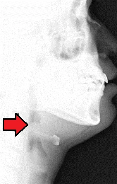
Figure 1Epiglottitis
Long description
Lateral soft-tissue neck radiograph of acute epiglottitis showing the thumb sign of a swollen epiglottis.

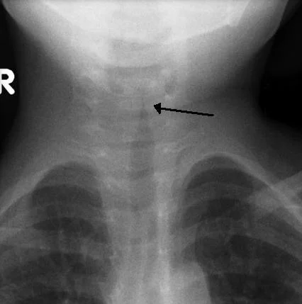
Figure 2Croup steeple sign
Long description
Anteroposterior neck radiograph of a child with croup showing the steeple sign, narrowing of the upper trachea.

Where the diagnosis is clear croup in a mild case, no investigation is needed. Investigate when the picture is atypical, severe, or deteriorating, when a foreign body is suspected, or when epiglottitis or bacterial tracheitis is possible. [4]
- Clinical (bedside). Pulse oximetry, respiratory rate, heart rate, temperature, work of breathing. The primary assessment (airway, breathing, circulation) comes first; security of the airway before any test.
- Laboratory. No blood test diagnoses croup. Full blood count and CRP are non-specific but may be markedly elevated in bacterial disease; take blood cultures in toxic children before antibiotics and whenever epiglottitis or tracheitis is suspected. [6]
- Imaging. Lateral neck radiograph shows the "thumbprint" sign in epiglottitis - a thickened, rounded, radiopaque shadow resembling a thumb - when there is diagnostic doubt, and should not delay treatment. [6] In croup, anteroposterior neck films may show subglottic narrowing, the "steeple sign", named after the steeple shape of the narrowed subglottis; this supports but does not diagnose croup, since it appears in other subglottic narrowings, and it is unnecessary when the clinical picture is typical. Chest X-ray looks for aspiration, mediastinal shift, a radiopaque or radiolucent-gap foreign body, and complications. Dynamic imaging or bronchoscopy is needed when foreign body is strongly suspected.
- Laryngoscopy and bronchoscopy. Indirect or flexible laryngoscopy confirms epiglottitis by showing a swollen, erythematous epiglottis and supraglottic oedema; it should only be performed where airway control is immediately available. In bacterial tracheitis, bronchoscopy is both diagnostic and therapeutic, showing circumferential purulent secretions and pseudomembranes adhering to the tracheal mucosa. [4]
- ECG and monitoring. Continuous cardiorespiratory monitoring in moderate or severe disease; nebulised adrenaline can produce tachycardia and hypertension, which usually resolve within 60-90 minutes. [3]
There is no laboratory test for croup; diagnosis is clinical. Severity grading uses the Westley croup score, the standard clinical tool (score range 0-17).
| Feature | 0 points | 1 point | 2 points | 3 points | 4-5 points |
|---|---|---|---|---|---|
| Stridor | None | Only with agitation or crying | Mild at rest | Moderate at rest | Severe at rest, with minimal air exchange (5 points) |
| Retractions | None | Mild | Moderate | Severe | - |
| Air entry | Normal | Decreased | Markedly decreased | - | - |
| Colour | Normal | - | Cyanotic on crying or exertion | Cyanotic at rest | - |
| Level of consciousness | Normal | - | - | Disoriented or drowsy | - |
The score is approximate and most useful as a trend. Two validated interventions change it: nebulised adrenaline improves the score by 2-3 points within 30 minutes; dexamethasone takes 4-6 hours to show its effect. [3] Severe scores mandate admission and continuous monitoring; any fall in level of consciousness, exhaustion or a rising score despite treatment marks impending respiratory failure.
Epiglottitis is a clinical diagnosis; confirmation by laryngoscopy is only where airway control is available. Bacterial tracheitis is confirmed at bronchoscopy. Foreign-body aspiration is clinical, with bronchoscopy both diagnostic and therapeutic.
Acute stridor in a child: assessment and first actions
- First look: ABCDEAirway patent? Work of breathing? Colour, tone, level of consciousness. Give oxygen and keep the child with the parent
- Position and keep calmSit the child upright, avoid supine positioning and unnecessary procedures, do not agitate
- Question: Is this a life threat?
Apnoea, cyanosis, exhaustion, confusion, or stridor at rest with severe obstruction
- Call for senior help and an anaesthetist or airway team nowPrepare for difficult intubation and tracheostomy; do not leave the child
- Secure the airway in theatre if possibleGas induction with experienced operators; orotracheal intubation or surgical airway if needed
Stable but significant symptoms
- Question: What is the most likely diagnosis?
Viral croup (barking cough, hoarse, follows a cold, mild fever)
- Give dexamethasone (oral or parenteral) for all severitiesUnder 0.15 mg/kg underdose, over 0.6 mg/kg overdose; typical 0.15-0.6 mg/kg as a single dose
- If moderate-severe: nebulised adrenaline and observe 2-4 hoursImproves Westley score by 2-3 points within 30 minutes; wears off in 90-120 minutes
- Reassess and admit if not improvingDischarge with clear return advice if mild and improved
Epiglottitis (toxic, drooling, muffled voice, tripod, high fever)
- Do NOT examine the throat or lie the child flatKeep NPO, avoid agitating, keep an experienced airway team on standby
- Admit, start intravenous antibiotics (third-generation cephalosporin plus anti-staphylococcal cover)Typically ceftriaxone plus clindamycin; add steroids as adjunct
- Secure airway early if any deteriorationOrotracheal intubation by the most experienced operator, or tracheostomy
Bacterial tracheitis (toxic, high fever, brassy cough, unresponsive stridor after viral croup)
- Intubate and suction purulent secretions70-90% require intubation; short-term intubation 2-4 days usual
- Start broad-spectrum IV antibiotics covering Staphylococcus aureus (including MRSA)Switch to directed therapy when culture results return
Foreign body (abrupt choking, unilateral findings)
- Rigid or flexible bronchoscopy by ENT or anaestheticsDiagnostic and therapeutic; imaging to help, but do not delay if the history is strong
- Red flags throughout: apnoea, cyanosis, exhaustion, altered consciousness, stridor at rest, recurrent or multiple episodes, age under 6 months or immunocompromiseThese patients need admission, monitoring and senior review
The goals are to relieve obstruction and reduce its duration, and to avoid unnecessary admission.
Corticosteroids. Systemic dexamethasone is the drug of choice for all severities of croup, including mild disease, because even mild cases improve in symptom scores, need less adrenaline, and have fewer return visits. [2] Corticosteroids reduce upper airway obstruction within 2-6 hours, cutting hospital admissions, length of stay and the need for intubation compared with placebo. [2] Typical total dose is a single dose of 0.15-0.6 mg/kg; less than 0.15 mg/kg is underdosing and more than 0.6 mg/kg is overdosing. [2] Dexamethasone can be given orally, intramuscularly or intravenously; the oral route is preferred when the child can swallow and is not vomiting, and intramuscular is a useful alternative when oral intake is unreliable. The inhaled route is an alternative in some guidelines: nebulised budesonide has evidence, but oral dexamethasone is at least as effective and cheaper, with allowance for the 4-6 hours onset. An equivalent corticosteroid such as prednisolone is an alternative where dexamethasone is unavailable; check your local formulary. Repeat doses are occasionally needed in severe disease; for mild cases in primary care a single outpatient dose is standard. [3]
Nebulised adrenaline. For moderate to severe croup (stridor at rest, significant retractions), nebulised adrenaline is the rescue drug because it works in minutes by alpha-1 vasoconstriction of submucosal vessels, shrinking oedema, and by beta-2 bronchodilation. According to Hou and colleagues it improves the Westley croup score by 2-3 points within 30 minutes, and the effect lasts about 90-120 minutes, the "rebound window". [3] Administration is by nebuliser with oxygen, keeping the child quiet while the drug acts (vasoconstriction within 10-15 minutes), with reduction of crying and agitation. Both racemic and L-epinephrine (the pure R-isomer used in anaphylaxis) are equally effective and guideline-equivalent; the choice is dictated by availability and local formulary. [3] Systemic effects are transient tachycardia (15-20% rise in heart rate) and a 10-15 mmHg systolic blood pressure rise, usually resolving in 60-90 minutes. [3] Adrenaline at this route and dose is generally well tolerated; avoid it only in situations where severe tachycardia or hypertension would be dangerous, and always in children who look unstable, who need admission and monitoring. Because the effect wears off in 1-2 hours, observe for at least 2-4 hours after a dose; many systems admit a child who has required more than two doses. [2]
Oxygen and supportive care. Give oxygen for hypoxaemia, keep the child upright with a parent, avoid unnecessary interventions (radiography, blood tests, intravenous lines) and aerosol-generating distress. Keep the child nil by mouth only if unwell; hydration otherwise orally. Low threshold for intravenous fluids in tachypnoea.
Heliox. A helium-oxygen mixture has lower density than air and reduces the work of breathing through the narrowed glottis. Evidence in croup is limited and inconsistent; it is used as an adjunct in severe obstruction when intubation is not yet needed, but it delays definitive therapy and requires tight-fitting delivery.
When to refer/admit. Admit moderate-severe croup, any need for adrenaline, age under 6 months, pre-existing airway disease or immunocompromise, poor social circumstances, or clinical uncertainty. Discharge only if mild, observations normal for at least 2-4 hours after any adrenaline, and clear safety-net advice is given.
Epiglottitis is an airway emergency first and an infection second.
- Do not examine the throat, do not lie the child flat, do not distress. Allow the position of comfort (often sitting forward, tongue out, drooling), keep nothing by mouth, and keep an experienced airway team (senior emergency clinician, anaesthetist, ENT surgeon) immediately available. [5][6]
- Secure the airway early. Many patients can be observed safely if breathing is stable, but rapid deterioration is characteristic. Definitive airway (orotracheal intubation with gas induction by the most experienced operator present, or tracheostomy) is needed when there is stridor at rest, respiratory distress, drooling with dysphagia, cyanosis, rising CO2 or any decline. [5] A child already intubated has a much easier course; delaying risks complete obstruction.
- Antibiotics intravenously. Start empirically to cover the expected organisms; typical regimen is a third-generation cephalosporin (ceftriaxone) combined with an anti-staphylococcal agent with MRSA coverage (clindamycin) [6] Switch to directed therapy when cultures return, and always take blood cultures when feasible before starting antibiotics (culture carries a risk of airway compromise and is usually reserved for intubated patients). [6]
- Steroids as adjunct. Intravenous steroids (hydrocortisone or methylprednisolone) reduce oedema and are given together with antibiotics; oral or nebulised steroids are added as the child improves, followed by step-down. [5]
- Supportive care. Oxygen, hydration, monitoring, and an interdisciplinary plan involving paediatrics, ENT, anaesthetics and infectious diseases. [5]
- Imaging is not essential for diagnosis and should not delay treatment, but may help when the diagnosis is uncertain: a lateral neck radiograph shows a swollen epiglottis, the thumbprint sign. [6]
Bacterial tracheitis behaves like a medical emergency with high risk of obstruction from detached membranes.
- Airway: 70-90% of patients need intubation to relieve obstruction and to allow repeated suctioning of the purulent secretions. Following initial intubation to relieve obstruction, patients often show dramatic improvement. [4]
- Bronchoscopy (flexible or rigid as appropriate) clears the pseudomembranes and secretions and establishes the microbiological diagnosis; repeated inspection guides progress (mucosal healing usually allows extubation in 2-4 days). [4]
- Antibiotics: start broad-spectrum intravenous therapy covering Staphylococcus aureus, including MRSA, and other possible pathogens (Streptococcus pyogenes, Streptococcus pneumoniae, Haemophilus influenzae); common first-line in reported practice includes meropenem, vancomycin and clindamycin for severe cases, with de-escalation by culture, guided by clinical and microbiological response.
- Supportive care: mechanical ventilation (often high inspiratory pressures and PEEP), oxygenation targets, suctioning every few hours, avoidance of premature extubation, rehabilitation of swallowing once extubated.
- Severe disease can progress to pneumothorax (1.3%) or ARDS (1.0%) and may require advanced support such as veno-venous ECMO. [4]
Suspected foreign body needs airway control. Rigid bronchoscopy (or flexible bronchoscopy in selected older children) by an anaesthetist and ENT team is both diagnostic and therapeutic. Imaging helps localise the object and assess complications but should not delay removal when the history is convincing. Post-procedure observe for laryngeal oedema and secondary infection.
- Reassess croup with the Westley score after each adrenaline dose and at 2-4 hours; declining score is the trigger for downgrading care.
- Bacterial tracheitis patients need serial bronchoscopic inspection until mucosal healing.
- Epiglottitis patients need close follow-up laryngoscopy and completion of antibiotics; consider underlying immunodeficiency in atypical or recurrent cases.
- All patients with suspected epiglottitis who do not require intubation still need admission and observation with an airway plan.
- Croup: secondary bacterial infection, prolonged respiratory failure, hypoxic brain injury in severe cases, hospitalisation; recurrence.
- Epiglottitis: airway obstruction, hypoxia, aspiration, death; the mortality is quoted at 1-20%. [5]
- Bacterial tracheitis: pneumothorax (1.3%), ARDS (1.0%), septic shock, prolonged mechanical ventilation, tracheal stenosis after healing, death. [4]
- Foreign body: complete obstruction, pneumonia, atelectasis, bronchiectasis if retained, and iatrogenic complications during removal.
- All of these conditions in combination can cause family distress, repeat attendances and overuse of antibiotics.
Most children with croup recover within days with treatment, and serious outcomes are rare. Epiglottitis in the modern era still carries mortality (1-20%), mostly from airway events before or during intubation, which is why airway readiness is the single most important intervention. [5] Bacterial tracheitis usually has a favourable prognosis with intubation, bronchoscopic suction and appropriate antibiotics, although a minority become critically ill and need prolonged support. [4] Foreign-body aspiration has excellent prognosis if the object is removed promptly.
- Vaccination against Haemophilus influenzae type b has transformed epiglottitis; it remains the single most effective preventive measure, with coverage exceeding 95% contributing to herd immunity. [6]
- Continuous microbiological surveillance is needed because non-type b and non-typable strains are not covered by the Hib vaccine. [6]
- Seasonal influenza and parainfluenza prevention (hygiene, staying home when ill, annual influenza vaccination when recommended) reduces the triggers for croup and secondary bacterial infections.
- Foreign-body prevention: keep small objects away from children under 3 years, supervise eating (avoid whole nuts, hot dogs, grapes; cut food small), and follow age guidance on toys.
- Recurrent croup should trigger evaluation for atopy, gastro-oesophageal reflux and airway lesions rather than repeated steroid courses.
- Under 6 months. Croup is uncommon; consider congenital anomalies, subglottic stenosis, vascular rings, and admission.
- Immunocompromised or unvaccinated children. Higher risk of severe epiglottitis and uncommon organisms; broader antibiotic cover and lower threshold for airway intervention.
- Adults. Epiglottitis increasingly affects adults with a more insidious onset; stridor may be absent initially. Bacterial tracheitis and croup are rare. Foreign body is common in older adults with poor dentition or neurological swallowing problems.
- The diagnosis is clinical; no investigation should delay treatment in a toxic, stridorous child. The steeple and thumbprint signs need X-rays, which are often unavailable and unnecessary.
- A transthoracic echocardiogram is not relevant here, but bedside oxygen, nebulisation and careful positioning are universal.
- Dexamethasone is cheap, heat-stable and oral and is the single most effective intervention; nebulise with simple air-driven nebulisers if oxygen is scarce.
- If nebulised adrenaline is unavailable, intramuscular or subcutaneous adrenaline at paediatric doses is not a substitute and should not be extrapolated; use dexamethasone early and refer promptly.
- Airway securing requires trained staff and equipment (appropriate endotracheal tubes, laryngoscopes, suction, tracheostomy set); if these are absent, transfer early before decompensation.
- Antibiotics for epiglottitis and tracheitis: any third-generation cephalosporin plus anti-staphylococcal agent is acceptable if ceftriaxone and clindamycin are unavailable, directed at local resistance patterns.
- Continuous observation after adrenaline can be done on a paediatric ward with a nurse, pulse oximeter and clear escalation criteria if an ICU is unavailable.
- Stridor in a child is an airway problem first: stabilise, keep calm, position upright, and reassess continuously.
- Viral croup dominates: a barking cough after a cold, managed with a single dose of dexamethasone (typical 0.15-0.6 mg/kg) for all severities. [2]
- Dexamethasone needs 4-6 hours; nebulised adrenaline covers the gap, improving the Westley score by 2-3 points within 30 minutes and lasting 90-120 minutes. [3]
- Adrenaline works by vasoconstriction of submucosal vessels, shrinking oedema within 10-15 minutes, and lasts 1-2 hours; observe at least 2-4 hours afterwards. [3]
- Epiglottitis is now rare after Hib vaccination but still an emergency: drooling, muffled voice, tripod posture, high fever; do not examine the throat; secure the airway and give intravenous antibiotics. [5][6] [1]
- Thumbprint sign on the lateral neck film supports epiglottitis but should not delay treatment; management is clinical. [6]
- Bacterial tracheitis follows viral croup, is often Staphylococcus aureus, produces obstructing mucopurulent secretions and pseudomembranes; 70-90% need intubation and bronchoscopic suction, with intubation typically 2-4 days. [4]
- Age 6 months-3 years is the high-risk band for croup and stridor, and about 1 mm of subglottic oedema can cut cross-sectional area by over 60%. [3] [1]
- Westley score grades croup severity; nebulised epinephrine improves it by 2-3 points within 30 minutes and steroid onset is 4-6 hours. [3]
- Roughly 85% of croup is mild and self-limited; severe disease occurs in 5-15%. [3]
- About 350,000 paediatric ED visits for croup occur annually in the USA, about 1-2% of paediatric encounters; croup is a common cause of acute upper airway obstruction. [1]
- Influenza and SARS-CoV-2 remain important precipitants of croup and of secondary bacterial tracheitis.
- Recurrent croup or croup in an infant should raise suspicion of an underlying anomaly rather than another viral episode.
- Safety-netting advice and a low threshold to return are essential after discharge, especially in the first 24 hours.
Answer each one in your head first, then open the answer.
A 3-year-old has had a barking cough and hoarse voice for two days after a cold. He is playing happily between coughs, has a temperature of 37.5 °C and mild inspiratory stridor when distressed. What is the diagnosis and what drug does he need now?
Show answerHide answer
Viral croup (laryngotracheobronchitis). He needs a single dose of oral dexamethasone; even mild croup benefits. Antibiotics and nebulised adrenaline are not indicated in this mild case. Reassess and give safety-net advice.
A 5-year-old sits upright, leaning forward with the neck extended, drooling and refusing to swallow. He is febrile to 39 °C and has a muffled voice. His oxygen saturation is normal. What is the diagnosis, what should you NOT do, and what is your first priority?
Show answerHide answer
Acute epiglottitis. Do not examine the throat with a tongue depressor or lay him flat, because airway obstruction can be provoked. Keep him calm and nil by mouth, keep an experienced airway team available, and be ready for early intubation or tracheostomy. Start intravenous antibiotics (third-generation cephalosporin plus anti-staphylococcal cover) and steroids.
A child has been treated for croup with dexamethasone and nebulised adrenaline two hours ago and now looks well, with no stridor. Can he go home?
Show answerHide answer
Only if he is mild, observations are normal, and at least 2-4 hours have passed since the last dose of nebulised adrenaline; the effect of adrenaline lasts about 90-120 minutes and rebound can occur. Give clear written return advice, especially for the first 24 hours.
A 2-year-old returns on day 4 of a viral croup with worsening stridor, a temperature of 39.5 °C and a toxic appearance. Nebulised adrenaline barely helps. What do you suspect and how do you manage it?
Show answerHide answer
Bacterial tracheitis (membranous laryngotracheobronchitis). Suspect it because of the toxic course, high fever and failure to respond to adrenaline. Intubate and suction the purulent secretions, and start broad-spectrum intravenous antibiotics with cover for Staphylococcus aureus including MRSA. Expect to ventilate for 2-4 days while mucosal healing occurs.
A 2-year-old suddenly clutches his throat while eating nuts. He then starts coughing and has reduced air entry on the right. What is the priority and why is the history more important than the chest film?
Show answerHide answer
Foreign-body aspiration. The priority is urgent bronchoscopy to remove the object. Imaging may be normal or non-specific in the first hours, so a strong history of choking mandates bronchoscopy without waiting for a chest film.
How do you grade a child with croup, and what is the score that signals severe disease?
Show answerHide answer
Use the Westley croup score (0-17) based on stridor, retractions, air entry, colour and level of consciousness. Higher scores mean more severe obstruction and impending respiratory failure; because severity thresholds vary by protocol, use your local guideline and treat the trend after each treatment.
Severe acute respiratory illness in children; asthma and wheeze in children; pneumonia and bronchiolitis; airway management in children; foreign body ingestion; anaphylaxis and airway obstruction. (Topic links will be added when those pages are published.)
Guidelines and regions differ on the dose of dexamethasone (single 0.15-0.6 mg/kg with underdosing below 0.15 mg/kg and overdosing above 0.6 mg/kg), on the need for post-adrenaline observation (2-4 hours versus less than 2 hours in some systems), and on whether nebulised adrenaline is restricted to severe cases or allowed when any stridor is present. [2] Where sources disagree, follow your national paediatric guidance.
- Taniguchi S, Kanno K, Ito Y. When Should the Differential Diagnosis of Croup Be Reconsidered?. Cureus 2025;17:e99177. doi:10.7759/cureus.99177. Accessed .
- Jirawong J, Anuntasaree W, Ruangnapa K, Saelim K, Prasertsan P. A Decade of Croup Management: Guideline Adherence, Practice Variation, and Opportunities for Improvement. Children (Basel) 2026;13:1046. doi:10.3390/children13081046. Accessed .
- Hou YT, Shi Q, Zhang L, Cheng Q. Clinical advances in racemic epinephrine for pediatric croup: a mini-review of evidence and practice. Frontiers in Pediatrics 2025;13:1616521. doi:10.3389/fped.2025.1616521. Accessed .
- Kano K, Obara T, Ninomiya R, Honda K, Yogo N, Muto Y, Hirai K. Severe respiratory failure caused by bacterial tracheitis in a two-year-old girl: ECMO and bronchoscopic management. Pediatrics International 2025;67:e70185. doi:10.1111/ped.70185. Accessed .
- Correa EJ, Caballero AS, Ordonez CA, Formichelli AC, Conti DM. Acute epiglottitis in an older patient: a case report and review of the literature. Journal of Medical Case Reports 2025;19:539. doi:10.1186/s13256-025-05363-3. Accessed .
- Ferreira M, Condessa L, Roquette M, Antao R, Cardoso C, Chaves M. Haemophilus influenzae Epiglottitis: A Rare Disease Not to Be Forgotten. Cureus 2026;18:e101680. doi:10.7759/cureus.101680. Accessed .
Figure credits
Educational content for medical students and doctors. It is not a substitute for clinical judgement or local guidelines.