Kawasaki disease
Acute medium-vessel vasculitis of childhood: prolonged fever with mucocutaneous signs and risk of coronary aneurysm; treated with IVIG and aspirin.
- Kawasaki disease is an acute, self-limited medium-vessel vasculitis that usually occurs in children under 5 years of age and has a striking tendency to involve the coronary arteries. [1]
- The diagnosis is clinical: at least 5 days of fever at 38.0 °C or higher together with at least 4 of 5 mucocutaneous and lymph-node features. [1][2]
- Children who meet only 2 or 3 criteria, especially infants, may have incomplete Kawasaki disease and still develop coronary aneurysms, so they must not be missed. [1]
- Untreated, about a quarter of children develop coronary artery abnormalities; with intravenous immunoglobulin (IVIG) and aspirin started within 10 days, that falls to under 5%. [1]
- Live vaccines such as measles-mumps-rubella and varicella are deferred for about 11 months after IVIG, and low-dose aspirin is continued at least 8 weeks or longer if coronary disease is present. [1]
By the end of this page you should be able to:
- Define Kawasaki disease as a medium-vessel vasculitis and state why it matters in paediatrics.
- List the diagnostic criteria and apply the "4-day" modification, including incomplete disease.
- Explain the acute, subacute and convalescent phases from mechanism to signs.
- Interpret the laboratory and echocardiographic findings.
- Plan acute treatment with IVIG and aspirin, and recognise when to intensify therapy.
- State the vaccine delay, aspirin follow-up and prognosis rules.
DefinitionKawasaki disease
Kawasaki disease is an acute, self-limited vasculitis of medium-sized arteries, with a special predilection for the coronary arteries. It is the leading cause of acquired heart disease in children in developed countries. [1]
It was first described in 1967 as "acute febrile mucocutaneous lymph node syndrome", a name that still matches the clinical picture well. [2] Other names include Kawasaki's disease and mucocutaneous lymph node syndrome (MCLS).
There is no staging by size of vessel other than "medium-vessel vasculitis", but it helps to think of three temporal phases rather than stages of disease:
- Acute phase: fever and mucocutaneous signs, ending with resolution of fever. [1]
- Subacute phase: desquamation and thrombocytosis, roughly days 10 to 25.
- Convalescent phase: silent recovery until 6 to 8 weeks after onset. [1]
Incomplete (atypical) Kawasaki disease is the same disease with fewer of the classical features, most often in infants under 1 year of age; these children carry a higher risk of coronary complications. [1]
Kawasaki disease is uncommon but important. In the United States an estimated 4,000 to 5,500 cases occur each year. [1] Worldwide it occurs in all ethnic groups, with higher incidence in children of Japanese and wider Asian descent. [1]
- Age. Eighty percent of patients are under 5 years of age, with a peak between 6 and 24 months. [1] Cases in adolescents, adults and infants under 4 months are rare. [1]
- Sex. Boys are affected about 1.5 times as often as girls. [1]
- Recurrence. About 2% of children have a recurrence, usually months to years later. [1]
- Seasonality. Cases occur year round but cluster in spring and winter. [1]
The cause is unknown. The illness behaves like an infection acting on a genetically predisposed child, or an abnormal immune response to an infectious trigger in a susceptible host. [1] There is no proven preventive measure, no person-to-person transmission and no known modifiable risk factor. [1] Family history is a clue to susceptibility rather than to a specific exposure.
The whole clinical picture follows from one process: an acute inflammation of the wall of medium-sized arteries, most dangerously the coronary arteries, with endothelial activation, mural oedema and aneurysm formation in the subacute phase.
- Fever and acute-phase response. The systemic inflammatory signals produce persistent fever, irritability, acute-phase proteins and sometimes hepatitis, meningism or gallbladder hydrops. [1]
- Mucous membranes. Inflammation explains the bilateral non-purulent conjunctivitis, fissured lips, strawberry tongue and injected pharynx. [1]
- Skin and extremities. A polymorphous rash and early erythema/oedema of the palms and soles reflect vasculitis of small vessels in the skin, followed by periungual and palm/sole desquamation as recovery begins. [1]
- Lymph nodes. Enlargement of a single cervical node reflects regional immune activation rather than primary infection. [1]
- Coronary arteries. Inflammation weakens the arterial wall, so dilation and aneurysm form in the second and third weeks, when the surface illness is often settling. [1] This is why a child can look better while the most dangerous complication is developing. [1]
- Thrombosis. Aneurysms provide stagnant blood flow and a damaged endothelium, so thrombus can form on acute and chronic time scales; giant aneurysms remain at risk long after the fever has gone. [3]
Figure 1Kawasaki symptoms B
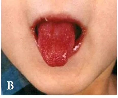
Long description
Strawberry tongue and bright red, swollen, cracked lips in a child with Kawasaki disease.

The illness classically runs in three phases. [1]
- Fever. At least 5 days of unremitting fever, usually 38.0 °C or higher, often with irritability or lethargy. [1][2]
- Eyes. Bilateral bulbar conjunctival injection without discharge, appearing within a day or two of fever onset. [1]
- Mouth. Injected pharynx, reddened cracked lips, and a red "strawberry" tongue. [1]
- Rash. A polymorphous erythematous macular rash, mainly truncal, often accentuated in the perineal region. [1]
- Extremities. Erythema or purple-red discolouration with variable oedema of the palms and soles, usually days 3 to 5; the oedema can be tense and non-pitting. [1]
- Nodes. Tender non-suppurative cervical lymphadenopathy, at least one node 1.5 cm or more in diameter. [1][2]
- Desquamation. Periungual, palmar, plantar and perineal peeling beginning around day 10; the superficial skin may come off in sheets. [1]
- Joints. Arthralgia or arthritis, mainly large joints, in about one third of patients (Merck Manual) and 7.5% to 25% in one Kuo 2023 cohort. [1][2]
- Heart. Cardiac manifestations become apparent as the rash and fever settle, usually 1 to 4 weeks after onset. [1]
- Platelets. Thrombocytosis appears in the second or third week. [1]
Clinical signs disappear and the child returns to normal; coronary changes may still be evolving. [1]
Children, especially those under 1 year of age, may have fewer than four criteria but still develop coronary aneurysms. [1] Incomplete Kawasaki disease should be considered when there are at least 5 days of fever at 38.0 °C or higher plus at least 2 of the 5 criteria. [1] These children are often the ones in whom the diagnosis is delayed, so repeated examination every few days and a low threshold for specialist review and echocardiography are essential. [2]
Diagnosis is clinical. There is no single blood test for Kawasaki disease. [1]
| Element | Detail |
|---|---|
| Fever | At least 38.0 °C (about 100.4 °F) for 5 or more days. [1][2] |
| Conjunctivitis | Bilateral, non-exudative bulbar injection. [1][2] |
| Oral changes | Fissured lips, strawberry tongue, or erythematous pharynx. [1][2] |
| Rash | Polymorphous truncal exanthem. [1][2] |
| Extremity changes | Erythema/oedema of hands and feet in the acute phase and/or periungual desquamation later. [1][2] |
| Cervical nodes | At least 1 node 1.5 cm or more in diameter, usually unilateral. [1][2] |
| Atraumatic exit rule | If only 4 days of fever but 4 or more major criteria, and redness or swelling of the hands/feet is present, the diagnosis may be made earlier. [2] |
Approach to a child with suspected Kawasaki disease
- Child with 5 or more days of fever at 38.0 C or higherPlus mucocutaneous features; consider Kawasaki disease
- Count the 5 major criteriaConjunctivitis, oral changes, rash, extremity changes, cervical node 1.5 cm or more
- Question: How many criteria are present?
4 or 5 criteria
- Kawasaki disease (complete)Start IVIG 2 g/kg and high-dose aspirin once diagnosis is made
2 or 3 criteria
- Question: Age under 1 year, or coronary changes on echo?
Yes
- Incomplete Kawasaki diseaseTreat and monitor as complete disease; repeat examination
No
- Incomplete or unlikelyReassess every 1-2 days; involve cardiology; labs and baseline echo
0 or 1 criterion, but disease suspected
- Reassess and investigateRepeat examination; infection work-up; echocardiography; avoid delaying IVIG if evolution becomes obvious
- ECG and echocardiogram at diagnosisRepeat imaging at 2-3 weeks, 6-8 weeks, and later if coronary disease is found
Many febrile exanthems can mimic Kawasaki disease. The table separates the common mimics by the feature that helps to tell them apart. [1]
| Condition | Feature that separates it from Kawasaki disease |
|---|---|
| Scarlet fever | Sandpaper rash, circumoral pallor, Pastia's lines, and group A streptococcal pharyngitis; conjunctivitis and extremity oedema are not typical. [1] |
| Measles | Cough, coryza, Koplik spots, and a cephalocaudal rash; lymphadenopathy is generalised rather than a single neck node. [1] |
| Staphylococcal toxic shock / scalded skin | Severe sepsis, hypotension, multi-organ shock, desquamation; blood cultures may be positive. [1] |
| Drug reaction | Temporal relation to a new drug; usually no prolonged high fever with coronary disease. [1] |
| Systemic juvenile idiopathic arthritis | Quotidian fever, evanescent rash, arthritis, with only modest conjunctival or oral features. [1] |
| Multisystem inflammatory syndrome in children (MIS-C) | Follows SARS-CoV-2; older children, more gastrointestinal and neurological symptoms, shock, and different cardiac phenotype. [1] |
| Leptospirosis / Rocky Mountain spotted fever | Relevant exposure, specific rash distribution, laboratory confirmation. [1] |
Kawasaki disease is a medium-vessel vasculitis of young children with coronary risk; Henoch-Schönlein purpura (IgA vasculitis) is a small-vessel vasculitis seen more often in slightly older children, presenting with palpable purpura on the legs and buttocks. [1]
| Feature | Kawasaki disease | Henoch-Schönlein purpura |
|---|---|---|
| Typical age | Under 5 years, peak 6-24 months | Older than Kawasaki disease (school-aged children) |
| Vasculitis level | Medium-sized arteries | Small vessels (capillaries, venules, arterioles) |
| Signature rash | Polymorphous trunk rash, extremity and perineal change; no purpura | Palpable purpura on lower limbs and buttocks |
| Fever | Prolonged, 5 or more days, central to diagnosis | Variable; not prolonged in the same way |
| Coronary involvement | Common if untreated; aneurysm risk | Not a feature |
| Joint involvement | Arthralgia/arthritis in about one third (Merck) or 7.5-25% (Kuo), mainly large joints | Arthritis/arthralgia, often knees and ankles |
| Renal involvement | Sterile pyuria or proteinuria less often | IgA nephritis with haematuria/proteinuria common |
| Gastrointestinal | Vomiting, diarrhoea, gallbladder hydrops | Colicky abdominal pain, bleeding, intussusception risk |
| Diagnostic anchor | Clinical (fever plus mucocutaneous/lymph-node criteria) | Palpable purpura with IgA on biopsy and extra-cutaneous features |
There is no diagnostic blood test, but the pattern supports the diagnosis and helps exclude infections. [1]
- White cell count. Leukocytosis with immature forms is common in the acute phase. [1]
- Haemoglobin. A mild normocytic anaemia is typical. [1]
- Platelets. Thrombocytosis, often 450,000 per microlitre or more, appears in the second or third week. [1]
- Inflammatory markers. Erythrocyte sedimentation rate and C-reactive protein are raised. [1]
- Other. Sterile pyuria, elevated liver enzymes, proteinuria, reduced serum albumin and cerebrospinal fluid pleocytosis occur depending on organs involved. [1]
- Exclusions. Cultures and serology are negative; antinuclear antibody and rheumatoid factor are generally negative. [1]
A baseline C-reactive protein of 13 mg/dL or more is one of the North American factors that mark a child at higher risk of coronary aneurysm. [3]
Transthoracic echocardiography is the essential imaging test: it shows coronary artery size, aneurysm, valvular regurgitation, pericarditis and myocarditis. [1][3]
- ECG and echocardiography are performed at diagnosis. [1]
- Because coronary abnormalities may appear later, imaging is repeated at 2 to 3 weeks, 6 to 8 weeks, and 6 to 12 months after onset if needed. [1]
- An initial echocardiogram can be normal; if suspicion remains, repeat the study 2 to 3 days later, especially in high-risk children. [3]
- Giant coronary aneurysms are defined as more than 8 mm internal diameter on echocardiography. [1]
Treatment should be started by, or in close consultation with, an experienced paediatric cardiologist, infectious disease specialist or rheumatologist. [1] It should not be delayed, including in children with atypical disease, because those infants are at high risk. [1]
Therapy is most effective when started within the first 10 days of illness, optimally as soon as the diagnosis is made. [1] The standard is a single dose of IVIG, 2 g/kg, given by intravenous infusion over 10 to 12 hours, together with high-dose oral aspirin. [1]
- IVIG dampens the systemic and vascular inflammation and reduces coronary complications. [1]
- Aspirin is used at an anti-inflammatory dose during the acute febrile phase; its metabolism is erratic in acute disease, which partly explains the high dose requirement. [1]
- The aspirin dose is reduced once the child has been afebrile for 4 to 5 days; some authorities continue high-dose aspirin until day 14. [1]
- If the child has cardiac dysfunction, IVIG may need a slower infusion or splitting over 2 days because of its large volume. [1]
- IVIG may be given after day 10 if diagnosis is late, although efficacy is less certain. [1]
A minority remain febrile after the initial infusion and are considered IVIG-resistant. [3] Escalation options include a second dose of IVIG, corticosteroids, infliximab, etanercept, anakinra or cyclosporine, guided by a cardiologist and rheumatologist. [1][3]
Children at greatest risk of coronary aneurysm are those aged 6 months or younger, or with a baseline coronary artery z-score of 2.5 or more, and North American models also flag age under 6 months, Asian race, baseline left anterior descending or right coronary artery z-score of 2 or more, and C-reactive protein of 13 mg/dL or more. [1][3] High-risk children may benefit from intensification of initial therapy with an adjunctive anti-inflammatory agent; both corticosteroids and infliximab reduce progression of coronary artery size compared with IVIG alone. [3]
- After symptoms settle for 4 to 5 days, low-dose aspirin is continued for at least 8 weeks, until repeat echocardiography is complete and inflammation has resolved. [1]
- If no aneurysm is found and inflammatory markers have normalised, aspirin can be stopped. [1]
- If coronary abnormalities are present, low-dose aspirin is continued indefinitely for its antiplatelet effect. [1]
- Children with giant coronary aneurysms may need additional anticoagulation with warfarin or low-molecular-weight heparin; direct oral anticoagulants are emerging as an alternative in this setting. [1][3]
- Giant aneurysms are least likely to regress and require the most intensive follow-up. [1]
Live viral vaccines are less effective after IVIG, so the measles-mumps-rubella vaccine should generally be delayed for 11 months after IVIG and the varicella vaccine for 11 months or more. [1] If measles exposure risk is high, vaccination proceeds but is repeated (or serology checked) 11 months later. [1]
Children on long-term aspirin, especially during influenza or varicella outbreaks, carry a small risk of Reye syndrome. Annual influenza vaccination is therefore important for children 6 months or older on long-term aspirin, varicella vaccination should be kept up to date, and parents should seek prompt advice after exposure or symptom onset. [1] Temporary interruption of aspirin with substitution of dipyridamole may be considered in selected children with documented aneurysms. [1]
- Coronary artery aneurysm, including giant aneurysm with thrombosis, myocardial infarction or, rarely, rupture. [1]
- Myocarditis, arrhythmia, pericarditis and heart failure in the acute phase. [1]
- Desquamating rash, arthritis and sterile pyuria. [1]
- IVIG-related haemolysis (more common in A, B or AB blood groups) and occasionally aseptic meningitis. [3]
- Recurrence in about 2% of children. [1]
With adequate therapy, mortality in the United States is reported as approximately 0.17% in older in-hospital data. [1] The single most important risk modifier is duration of fever: longer fever means greater cardiac risk. [1]
Effective treatment relieves symptoms and lowers the incidence of coronary artery aneurysm, from about 20% to under 5% in the Merck Manual and from about 25% to about 3-5% in other sources. [1] Untreated, coronary artery abnormalities develop in about 25%. [1]
In the absence of coronary disease, the outlook for full recovery is excellent. About two-thirds of coronary aneurysms regress within a year, but whether residual stenosis remains is uncertain. [1] Giant aneurysms are least likely to regress. [1]
- Infants under 1 year. They are the most likely to have incomplete Kawasaki disease and a higher aneurysm rate; investigation and treatment should not wait for a full picture. [1][3]
- Older children and adolescents. Recognised but rare; the diagnosis is more often missed. [1]
- Pregnancy. Not a disease of pregnancy, but women with residual coronary disease from childhood Kawasaki disease need cardiology follow-up. (No firm pregnancy-specific guideline figure is given here.) [3]
A careful clinical diagnosis and bedside pieces of equipment matter more than tests.
- A transthoracic echocardiogram, even a basic one, is the test that confirms or excludes coronary disease; repeat imaging can be performed per local capacity. [1][3]
- IVIG is expensive and may be unavailable; work with local specialists to source it and to use adjunctive corticosteroids or infliximab where supported. [1][3]
- Where laboratory tests are limited, serial clinical examination and temperature charts still define the illness and its course. [1]
- Ensure the follow-up echocardiogram is not lost: in many settings coronary disease is only discovered late. [1]
East Africa
In settings where IVIG is scarce, early high-dose aspirin and close follow-up, plus urgent referral, remain the cornerstone while sourcing immunoglobulin.
- Kawasaki disease is an acute medium-vessel vasculitis of young children, the leading cause of acquired heart disease in children, with a dangerous tendency to form coronary aneurysms. [1]
- Diagnosis rests on 5 or more days of fever at 38.0 °C or higher plus at least 4 of 5 mucocutaneous/lymph-node criteria; an early 4-day modification exists when 4 criteria are present with extremity changes. [1][2]
- Infants under 1 year can have incomplete disease with only 2 criteria and still develop aneurysms; reassess frequently. [1]
- Coronary involvement occurs in about 25% of untreated children but falls to under 5% with IVIG and aspirin started within 10 days. [1]
- IVIG is given as 2 g/kg over 10 to 12 hours with high-dose aspirin; aspirin is later reduced and given as low-dose for at least 8 weeks, indefinitely if coronary disease is present. [1]
- High-risk children (young age, coronary z-score 2.5 or more, raised C-reactive protein) may benefit from adjunctive corticosteroids or infliximab. [3]
- Live vaccines are deferred for about 11 months after IVIG; long-term aspirin requires influenza vaccination and varicella precautions. [1]
- Echocardiography is performed at diagnosis and repeated at 2-3 weeks and 6-8 weeks; giant aneurysms are over 8 mm. [1]
- Differential includes scarlet fever, measles, toxic shock, drug reactions, systemic juvenile arthritis and MIS-C; Henoch-Schönlein purpura is a small-vessel vasculitis with palpable purpura and different age distribution. [1]
- Prognosis is excellent without coronary disease; about two-thirds of aneurysms regress within a year. [1]
Answer each one in your head first, then open the answer.
A 3-year-old has had 6 days of fever at 39 °C, bilateral non-purulent conjunctivitis, a truncal rash, cracked lips with strawberry tongue, and a cervical node measuring 2 cm. What is the most likely diagnosis and the first investigations?
Show answerHide answer
Kawasaki disease: at least 5 days of fever at 38 °C or higher plus 4 of the 5 criteria. The first tests are an ECG and echocardiogram, with blood tests (full blood count, ESR/CRP, liver enzymes, cultures) mainly to exclude other conditions. [1]
A 4-month-old has fever for 5 days, a rash and conjunctivitis only. The echo is normal. How should this be managed?
Why is the echocardiogram often normal at first even when Kawasaki disease is diagnosed correctly, and why must it be repeated?
Show answerHide answer
Coronary dilatation and aneurysm develop in the second and third weeks, when the fever and rash are often settling, so the first study can be normal. Repeat imaging at 2-3 weeks and 6-8 weeks detects late changes. [1]
What does IVIG and aspirin achieve, and within what time should treatment start?
Show answerHide answer
Treatment within 10 days of onset lowers coronary aneurysms (about 20% to under 5% per the Merck Manual; about 25% to about 3-5% in other sources). IVIG is 2 g/kg over 10-12 hours plus high-dose aspirin initially, then low-dose aspirin for at least 8 weeks. [1]
A child needs IVIG for Kawasaki disease two weeks before a scheduled measles-mumps-rubella booster. What do you advise?
Show answerHide answer
Defer the live vaccine for 11 months after IVIG (11 months or more for varicella), because passively transferred antibody can blunt the response; if exposure risk is high, vaccinate now and repeat or check serology later. [1]
How does Henoch-Schönlein purpura differ from Kawasaki disease in age, vessel size and coronary risk?
Show answerHide answer
Henoch-Schönlein purpura is a small-vessel vasculitis of slightly older children (typically 3-10 years) with palpable purpura on the legs and buttocks, arthralgia, abdominal pain and IgA nephritis, whereas Kawasaki disease is a medium-vessel vasculitis of children under 5 with prolonged fever and mucocutaneous signs, carrying coronary aneurysm risk. [1]
- Henoch-Schönlein purpura (IgA vasculitis)
- Multisystem inflammatory syndrome in children (MIS-C)
- Scarlet fever and other streptococcal infections
- Measles and viral exanthems
- Toxic shock syndrome
- Congenital and acquired heart disease in children
- Mehta J. Kawasaki disease. Merck Manual Professional Version, updated June 2025 (full review February 2025). Accessed .
- Kuo HC. Diagnosis, Progress, and Treatment Update of Kawasaki Disease. International Journal of Molecular Sciences 2023;24(18):13948. Accessed .
- Elias MD, Jone PN, Harahsheh AS. AHA Scientific Statement Update on Kawasaki Disease: Identifying and Managing Patients at High Risk. American College of Cardiology Expert Analysis, 28 April 2025. Accessed .
Figure credits
Educational content for medical students and doctors. It is not a substitute for clinical judgement or local guidelines.