Lead poisoning
Paediatric lead poisoning: sources, screening, BLL thresholds, neurological and abdominal features, heme mechanism, chelation, public health.
- Lead poisoning in children usually happens silently: there are often no obvious symptoms while the blood lead level rises, and the harm falls on the developing brain.
- Exposure is nearly always environmental, most often deteriorating lead-based paint in homes built before 1978, together with dust, contaminated soil, certain water pipes and some imported toys, jewellery, candies and remedies. [3]
- A blood lead test is the only way to know whether exposure has occurred; treatment and follow-up are decided by the blood lead level in micrograms per decilitre.
- The mechanism that the blood count and chemistry reveal is inhibition of two enzymes (delta-aminolevulinic acid dehydratase and ferrochelatase) in the heme pathway, producing basophilic stippling of red cells and a characteristic anaemia.
- Children with very high levels (45 micrograms per decilitre or above in the CDC thresholds used here) need removal from the source plus chelation; lead encephalopathy needs dimercaprol as well as calcium disodium EDTA.
- Prevention is the cure: no blood lead level is known to be safe, so primary prevention (removing lead before exposure) plus screening and follow-up protect the next birth cohort.
By the end of this page you should be able to:
- Explain where childhood lead comes from and why toddlers are at greatest risk.
- Describe the neurological and abdominal presentation and recognise the red flags of lead encephalopathy.
- Interpret a blood lead level and say what action each range triggers.
- Explain the haem pathway mechanism and why basophilic stippling and Burton line appear.
- Choose and monitor the right chelator for a given blood lead level, route and duration.
- Outline the public-health response (source removal, screening, notification) and place lead among other heavy-metal toxicities.
DefinitionLead poisoning
Lead poisoning is the clinical condition produced by absorption of the heavy metal lead (Pb) into the body, in amounts that disturb the haematopoietic, nervous, gastrointestinal, renal and other systems. In children it is most often chronic, low-dose exposure from the environment; severe acute exposure also occurs. An elevated blood lead level is the laboratory definition of exposure, and clinical lead poisoning is present when that exposure causes symptoms or organ dysfunction.
The terminology matters for communication. An elevated blood lead level is a biomarker of exposure; it is not the same as symptomatic poisoning, and even levels once considered acceptable are now recognised to harm development. No level of lead exposure is known to be safe for children, and CDC uses a Blood Lead Reference Value of 3.5 micrograms per decilitre to mark the level above which a child's result deserves follow-up and source investigation.[1]
Lead poisoning is one of the most common and preventable environmental diseases of childhood, but it is unevenly distributed. Children under six years are the most vulnerable, because their nervous systems are developing rapidly, their gut absorbs a larger fraction of ingested lead than an adult gut, and their normal hand-to-mouth behaviour puts contaminated dust into the mouth.[2] Most children with an elevated blood lead level have no obvious symptoms, which is why case-finding by screening is essential.[2]
No safe blood lead level in children has been identified, and even low levels have been shown to affect learning, attention and academic achievement.[3] The exposure is global wherever lead paint, lead pipes, leaded fuel residues, mining, smelting or recycling of lead-acid batteries occur; local data therefore drive each region's screening plan, and universal testing is used where local plans are absent.[1] [2]
Lead enters through three routes: the lungs (inhaled dust or fume), the gut (swallowed dust, chips, water or food) and, to a lesser extent, the skin. The respiratory route dominates in adults who work with lead; the gastrointestinal route dominates in small children, who put objects in their mouths.[7]
The sources to consider deliberately are old paint, dust, water pipes and imported products.[3]
- Old lead-based paint and dust. In the United States, interior lead-based paint was banned from 1978, so homes built before then commonly contain it. When paint peels and cracks it produces lead dust, and that dust is the main vehicle by which children are poisoned. Renovation that scrapes, sands or heats old paint creates very high exposures.[3]
- Dust and soil. Lead deposited over decades from leaded petrol, from paint weathering and from industrial emissions accumulates in household dust and in bare soil around homes; children swallow or breathe it.[3]
- Water pipes. Lead can leach from certain water pipes and from solder or brass fittings, especially in older plumbing, into drinking water.[3]
- Imported and traditional products. Lead is still found in some toys and jewellery (especially cheap metal costume jewellery), in candies imported from other countries, and in traditional home remedies and cosmetics.[3]
- Occupational take-home exposure. Parents whose jobs or hobbies involve lead (stained-glass work, certain recycling, bridge repainting) can carry dust home on clothes and hands.[3]
- Other environmental sources. Lead from air and soil near airports using leaded aviation fuel, and from living near battery recycling or smelting, also matters.[3]
Risk is highest in children under six, in children living in older housing, in children from low-income households, and in children of workers with occupational exposure.[1] Iron deficiency increases lead absorption and is itself a risk factor; children with pica or hand-to-mouth behaviour ingest more dust.[1]
Once lead is absorbed it enters the blood, where it has a half-life of about one to two months in adults and a little longer in children; most of the total body burden ends up in bone, where it can remain for decades and be mobilised again during pregnancy, lactation or osteoporosis.[2] Blood lead therefore measures recent exposure and current mobilisation, not the whole-body burden. Lead is absorbed through the lungs, the gastrointestinal tract and, to a lesser extent, the skin.[7]
Lead's best-understood biochemical action is on the haem (heme) biosynthetic pathway. It directly inhibits two enzymes in that pathway: delta-aminolevulinic acid dehydratase (ALA dehydratase), which catalyses the second step and converts two molecules of delta-aminolevulinic acid into porphobilinogen, and ferrochelatase, which inserts ferrous iron into protoporphyrin IX to make haem.[7] The consequences are:
- ALA dehydratase inhibition causes overproduction and urinary excretion of delta-aminolevulinic acid, with normal porphobilinogen levels. That pattern is one of the keys that separates lead poisoning from acute porphyria, in which both ALA and porphobilinogen rise.[7]
- Ferrochelatase inhibition causes free erythrocyte protoporphyrin to accumulate.
- Binding of lead to sulphydryl groups of other enzymes and proteins disrupts membrane function and neurotransmission.
- Lead's interference with calcium signalling and its competition with calcium in the nervous system is a major reason for its neurotoxicity.
These changes produce the anaemia (decreased haem synthesis, shortened red cell survival) and the basophilic stippling of red cells (degeneration of ribosomal RNA in erythrocytes, which then clumps and stains).[7]
The brain is the critical target in children. Lead crosses the blood-brain barrier and accumulates in astrocytes, where it disrupts calcium-dependent signalling, impairs myelination and synaptic pruning, and generates oxidative stress. Effects are greatest while the brain is developing. In adults the pattern is more often peripheral neuropathy and encephalopathy at high levels; in young children even low exposure shifts intelligence, attention and behaviour.[2]
The most important clinical fact is how quiet this condition is: most children with an elevated blood lead level look well, with no immediate symptoms, and the harm only appears later in growth, learning and behaviour.[2] Clinical suspicion based on risk factors, plus screening, is therefore more useful than waiting for symptoms.
- Abdominal pain and colic. Cramping, poorly localised abdominal pain ("lead colic") is a classic feature of adult high exposure, related to altered smooth-muscle tone and mucosal effects of lead.[7]
- Constipation and anorexia are common; nausea and vomiting occur with higher levels. Some children have mild hepatitis-like transaminase elevations.
- Pica, the eating of non-food items such as paint chips, is both a risk factor and a feature of iron deficiency occurring alongside lead exposure.
- Irritability, fatigue, headache and difficulty concentrating.
- Sleep disturbance, memory problems and slowed processing at higher loads.
- Developmental delay, learning difficulty, speech and hearing problems and behavioural dysregulation in chronic childhood exposure.[2]
- Peripheral neuropathy in adults (classically wrist or foot drop), which is uncommon in children.
- Lead encephalopathy at very high levels: vomiting, ataxia, seizures, confusion, altered consciousness and cerebral oedema. This is a life-threatening emergency, mainly seen in toddlers in very poor housing.[6]
- Burton line (Burtonian line): a blue-purplish line along the gingival margin, seen with chronic lead exposure, caused by deposition of lead sulphide in the gums. It is a useful but insensitive sign and is worth looking for in the mouth and gum margin.[7]
- Lead lines on plain radiographs: dense metaphyseal bands at the ends of growing long bones, reflecting lead deposition in calcifying cartilage. They indicate chronic heavy exposure in a child and can be seen even without clinical symptoms.
- Abdominal radiopacity: radiopaque flecks of ingested paint or other material may be visible on an abdominal X-ray in a young child who eats paint chips.
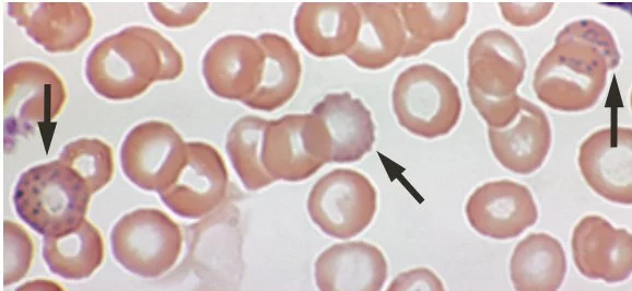
Figure 1Lead poisoning - blood film
Long description
Peripheral blood film in lead poisoning showing basophilic stippling of red cells.

Lead poisoning causes anaemia by two mechanisms: decreased haem synthesis and shortened red-cell survival. The typical picture is a normocytic or microcytic anaemia with an elevated red cell distribution width, and the characteristic finding is basophilic stippling on the peripheral blood smear, produced by degeneration of ribosomal RNA that clumps in the erythrocyte.[7] With severe exposure the white cell count may be raised and liver enzymes mildly raised; renal tubular effects produce glycosuria and aminoaciduria.
- Free erythrocyte protoporphyrin rises because ferrochelatase is blocked.
- Urinary delta-aminolevulinic acid rises and urinary porphobilinogen stays normal or near normal, the pattern that separates lead poisoning from acute porphyria.[7]
- Zinc protoporphyrin (ZPP) is often used as a screening aid in some programmes but is less specific than blood lead.
Red flagLead encephalopathy
In a toddler with irritability, vomiting, ataxia, seizures or altered consciousness, especially one living in older housing, think lead encephalopathy. Blood lead levels are usually very high (often above 70 micrograms per decilitre in adults, and the same order in severe paediatric exposure). The patient needs urgent admission, neurological management of cerebral oedema, intramuscular (not rapid intravenous) calcium disodium EDTA, and dimercaprol at a separate injection site.
The differential for lead poisoning falls into three families: conditions that look like abdominal lead colic; conditions that produce anaemia with basophilic stippling; and other heavy-metal exposures.
| Condition | Feature that separates it |
|---|---|
| Acute porphyria (acute intermittent porphyria) | Both urinary delta-aminolevulinic acid and porphobilinogen are raised; neuropsychiatric symptoms and abdominal pain without anaemia; no exposure source |
| Iron deficiency anaemia | Microcytic hypochromic anaemia without basophilic stippling; raised total iron binding capacity; low ferritin; no gum line or lead lines |
| Idiopathic thrombocytopenic or haematological disease | Cytopenias explained by marrow diagnosis; no source; confirmatory tests negative |
| Chronic abdominal pain, functional dyspepsia | No basophilic stippling, no gum line, no elevated blood lead |
| Other heavy-metal toxicity (mercury, arsenic, cadmium) | Different source, different dominant syndrome and, in several cases, different antidote; see comparison table below |
| Acute gastroenteritis | Self-limiting; vomiting and diarrhoea; no anaemia or neurodevelopmental decline |
A venous blood lead level is the diagnostic and monitoring test. Capillary finger-prick screening is acceptable when a venous sample is not immediately available, but any elevated capillary result must be confirmed with a venous sample on a schedule that shortens as the level rises.[1] Blood must be collected into lead-free tubes, and contamination at collection (dust on the skin, contaminated wipes) can falsely raise the result.
- Full blood count and blood film (basophilic stippling, anaemia).
- Zinc protoporphyrin or free erythrocyte protoporphyrin, which rise when ferrochelatase is inhibited; useful when blood lead testing is delayed but less specific.
- Iron studies, because iron deficiency commonly accompanies lead exposure and raises absorption.
- Renal function, urinalysis and liver enzymes before any chelation, and electrolytes.
- Serum creatinine and blood urea nitrogen in particular before calcium disodium EDTA, because its nephrotoxicity is dose dependent.[5]
- Abdominal X-ray for radiopaque paint chips or other foreign bodies when ingestion is suspected and in young children under environmental investigation.[1]
- Raised urinary delta-aminolevulinic acid with normal or near-normal urinary porphobilinogen points to lead rather than porphyria.[7]
- Erythrocyte protoporphyrin above 35 micrograms per decilitre of whole blood indicates the need to perform a venous blood lead determination.[5] [1]
- Long-bone radiographs may show lead lines at the metaphyses in growing children with chronic exposure, and abdominal radiographs may show radio-opaque material in the abdomen.[5]
Lead poisoning is diagnosed by an elevated blood lead level together with a credible source of exposure and, where present, compatible clinical findings. Severity is stratified by the blood lead level: any level above the reference value triggers action, and the intensity of action rises through the bands. Current CDC guidance (the values used on this page, last reviewed 2025) sets a Blood Lead Reference Value of 3.5 micrograms per decilitre; the 200 micrograms per litre figure sometimes seen in older literature is simply 20 micrograms per decilitre in different units.
| Blood lead level (micrograms per decilitre) | Tier | Key actions |
|---|---|---|
| Under 3.5 | Below reference value | Education on sources and prevention; developmental and nutritional check at well visits (iron and calcium focus); routine follow-up testing by age and risk |
| 3.5 to 19 | Elevated, below urgent | As above, plus report to state or local health department; take an environmental exposure history; arrange environmental investigation of the home; check for iron deficiency; confirmatory follow-up testing on tiered schedule |
| 20 to 44 | Higher | All of the above, plus complete history and physical examination; environmental investigation and lead hazard reduction; consider abdominal X-ray for paint chips or radiopaque foreign bodies and start bowel decontamination if indicated; consult a paediatric environmental health unit or poison centre |
| 45 or above | Highest | All of the above with a detailed neurological examination; abdominal X-ray and bowel decontamination if needed; admit if symptomatic (confusion, weakness, seizures, coma, vomiting, abdominal pain) or if the home is not lead-safe; arrange chelation in consultation with a medical toxicologist or paediatrician experienced in lead treatment, and consult a paediatric environmental health unit or poison centre |
Approach to a child with suspected or confirmed lead exposure
- Identify riskAge under 6, pre-1978 housing, parental occupational exposure, pica, iron deficiency, migrant or low-resource setting
- Screen blood lead levelCapillary or venous sample; confirm raised capillary results with venous blood
- Question: Confirmed venous level?
Under 3.5 micrograms per decilitre
- Educate and preventSource counselling, handwashing, wet-wiping, check development and nutrition
- RescreenBy age, risk and local screening plan
3.5 to 19 micrograms per decilitre
- Report and investigateNotify health department; take environmental history; arrange home inspection; treat iron deficiency
- Follow upRepeat blood lead on the tiered schedule until stable
20 to 44 micrograms per decilitre
- Assess and decontaminateFull examination; environmental remediation; consider abdominal X-ray and bowel decontamination
- Manage sourceRemove from exposure; consult paediatric environmental health unit or poison centre
45 or above, or any encephalopathy
- Admit and chelateNeurological precautions; chelation as below; dimercaprol plus calcium disodium EDTA for encephalopathy
- Source controlRemove from lead source before and during treatment
- Remove the source before treating the child. Chelation while the child continues to be exposed wastes the effort, because re-exposure rebuilds the body burden immediately. Source removal, family relocation or remediation of paint and dust comes first or alongside treatment.[4]
- All poisoning is treated in context. The blood lead level, the level of symptoms, the child's age and housing, and local services determine the plan. Any complex case should be discussed with a paediatric environmental health unit or a poison control centre.[1]
- Everyone is educated. Families need clear, written guidance on sources (especially old paint and dust), handwashing, wet-wiping of windowsills, wet-mopping of floors and safe renovation, in whatever language they use.[1][3]
- Iron and calcium nutrition. Optimising iron and calcium intake reduces lead absorption.[1]
No medical treatment for lead itself. Provide education, review development and nutrition, and rescreen according to local policy (at least at 12 and 24 months for children on Medicaid in the United States, or using the local risk-based plan).[1]
As above, plus report the result to the local health department, take a careful environmental exposure history and arrange an environmental investigation of the home where required. Treat iron deficiency, discuss calcium- and iron-rich foods, refer to supportive services such as nutrition programmes, and arrange follow-up blood lead testing on the tiered schedule until the level is falling and stable.[1]
All of the above plus a complete history and physical examination, an environmental investigation and lead hazard reduction programme, consideration of an abdominal X-ray for paint chips or other radiopaque material with bowel decontamination if present, and consultation with a paediatric environmental health unit or poison control centre.[1]
All of the above, with a detailed neurological examination. Admit the child if there are signs or symptoms of poisoning, or if the home is not lead-safe and the family cannot find a lead-free living space, or if the source has not been identified. Chelation is arranged by a medical toxicologist or paediatrician experienced in lead treatment.[1]
Chelation is reserved for significant elevation or symptoms; it does not replace source removal.
Succimer is indicated for treatment of lead poisoning in paediatric patients aged 1 year and older with blood lead levels above 45 micrograms per decilitre.[4] It does not cross the blood-brain barrier, so it is not indicated for lead encephalopathy.[4] [5]
- Dose: 10 milligrams per kilogram, or 350 milligrams per square metre, by mouth every 8 hours for 5 days, then every 12 hours for a further 14 days; total 19 days.[4]
- Pretreatment checks: blood lead level, full blood count with differential and platelets (absolute neutrophil count above 1500 per microlitre), liver transaminases, and renal function with urea, creatinine and urinary protein. Ensure adequate hydration and adequate urine output.[4]
- Monitor full blood count weekly during treatment; interrupt succimer if the absolute neutrophil count falls below 1200 per microlitre until it recovers above 1500 per microlitre (or the child's baseline). Interrupt at once if signs of infection appear.[4]
- Monitor liver transaminases at baseline and at least weekly; elevations above 5 times the upper limit of normal, if confirmed, lead to dose reduction or temporary stopping.[4]
- Common adverse reactions include nausea, vomiting, diarrhoea, appetite loss, metallic taste and rash; hypersensitivity can occur.[4]
- Concomitant administration with calcium disodium EDTA is not recommended.[4]
- Blood lead usually falls during the 19-day course, stable levels under 15 micrograms per decilitre have been seen in studies of extended dosing, but rebound after stopping is common, because lead redistributes from bone to soft tissue and blood. Half of the treatment course advantage can be lost within two weeks without continued monitoring.[4] Repeat blood lead after completing the course and every week until stable; a further course may be given after at least 2 weeks off treatment if levels remain high.[4] [1]
Calcium disodium EDTA is indicated for the reduction of blood and depot (bone) stores of lead in acute and chronic lead poisoning and in lead encephalopathy, in children and adults.[5] Its route and dose differ from succimer.
- Dose: 1000 milligrams per square metre per day for 5 days, given by intravenous infusion over 8 to 12 hours (diluted in 5 per cent dextrose or normal saline) or, divided equally every 8 to 12 hours, by intramuscular injection.[5]
- Therapy is then interrupted for 2 to 4 days to allow redistribution of lead and to prevent severe zinc and essential metal depletion; two courses are usually given.[5]
- Route safety in the central nervous system. Because patients with lead encephalopathy and cerebral oedema can suffer a lethal rise in intracranial pressure after rapid intravenous infusion, the intramuscular route is preferred in that setting and in very young children; intravenous infusion must not be rapid. [5]
- Renal and hepatic safety. Edetate calcium disodium can itself produce renal damage, so renal function (urine flow, sediment, protein), liver tests and electrolytes are checked before each course and monitored during therapy; treatment stops at the first sign of renal toxicity. Adequate urine flow must be established before the first dose. Avoid rapid hydration in the presence of encephalopathy.[5]
- Combination therapy. When the blood lead level is above 70 micrograms per decilitre or clinical symptoms of lead poisoning are present, calcium disodium EDTA is used in conjunction with dimercaprol (BAL), injected at a separate intramuscular site. Published protocols and specialist references should guide the combination dosing. Adults with lead nephropathy have their own reduced schedule by serum creatinine.[5]
- Dosing caution. At no time should the recommended daily dosage be exceeded, and the drug is contraindicated with anuria, active renal disease or hepatitis. Steroids are no longer recommended because they enhance renal toxicity in animals. [5]
Dimercaprol is the third agent. Its sulphhydryl groups form complexes with certain heavy metals; it is indicated for arsenic, gold and mercury poisoning, and for acute lead poisoning when used concomitantly with calcium disodium EDTA.[6] It is expressly not indicated for iron, cadmium or selenium poisoning, because the resulting metal-dimercaprol complexes are more toxic than the metal alone, particularly to the kidneys.[6]
- For acute lead encephalopathy: 4 milligrams per kilogram by deep intramuscular injection alone in the first dose, then at 4-hourly intervals in combination with calcium disodium EDTA injected at a separate site; for less severe poisoning the dose can be reduced to 3 milligrams per kilogram after the first dose. Treatment is maintained for 2 to 7 days according to clinical response. [6]
- Dimercaprol is given by intramuscular injection only, in oil; adverse effects include a rise in blood pressure with tachycardia, nausea, vomiting, headache, oral burning, lacrimation, salivation, sweating, and fever in roughly one third of children. Dosage above 5 milligrams per kilogram usually causes vomiting, convulsions and stupor.[6]
- It should not be given to patients with hepatic insufficiency except post-arsenical jaundice, and acute renal insufficiency during therapy requires withdrawal. Because the dimercaprol-metal complex breaks down in acid medium, an alkaline urine is maintained to protect the kidney; medicinal iron must not be given concurrently. The product is formulated in peanut oil, so caution is needed in peanut-sensitive patients.[6]
Children newly treated for elevated blood lead should have a repeat blood lead level within about a month, and the level is followed on the tiered schedule until it is stable and falling.[1] Because lead redistributes from bone after chelation stops, rebounds are expected and should be explained to families.
- Neurodevelopmental impairment: lower IQ, decreased ability to pay attention, speech and hearing problems, behavioural dysregulation and underperformance at school, which can be permanent even at low exposure.[2][3]
- Anaemia with basophilic stippling and associated fatigue.
- Neurological damage: peripheral neuropathy in adults, encephalopathy, seizures and cerebral oedema at high levels.[6]
- Gastrointestinal morbidity: colic, constipation, abdominal pain.
- Renal injury: proximal tubular dysfunction and, with chelation, drug-induced nephrotoxicity.
- Growth slowing and other systemic effects in chronically exposed children.[2]
Outcome depends on the level, duration and age of exposure and on how quickly the source is removed. Removing a child from exposure usually produces a falling blood lead level within weeks, but the total body burden (mainly bone) falls slowly and can mobilise later in life. Neurodevelopmental deficits caused in early childhood are usually not fully reversible; this is why primary prevention and screening are the most important interventions. Prognosis after severe encephalopathy is guarded.[2]
Primary prevention means removing lead hazards before a child is exposed. It is the most effective way to prevent the lifelong effects of lead exposure.[3]
- Remove or encapsulate deteriorating lead paint using certified contractors following approved methods; do not scrape or sand it without protection.
- Wet-wipe windows and windowsills and wet-mop floors regularly in older homes.
- Cover chipping or peeling paint to keep dust from spreading.
- Flush pipework and address lead plumbing where required.
- Wash children's hands frequently, especially before meals, and wipe toys.
- Avoid occupational take-home contamination (change clothes, wash hands before going home).
- Ensure iron- and calcium-rich diets to reduce absorption.[1]
Secondary prevention is blood lead testing plus follow-up care and referral; it is the safety net for children who may already be exposed.[3] CDC recommends that screening plans be built on local data; in the absence of a local plan, universal blood lead testing is recommended.[1] Children enrolled in Medicaid should be tested at 12 and 24 months, or once between 24 and 72 months if never screened.[1] High-risk neighbourhoods and children (by housing age and social or demographic factors) are targeted where universal testing is not used.[1] Blood lead levels change seasonally, with greater exposure in summer months, and may require more frequent follow-up in colder climates.[1]
When a raised blood lead level is confirmed, public health and clinical steps run in parallel:
- Notify the local or state health department (for the 3.5 to 19 micrograms per decilitre band and above in current CDC guidance).[1]
- Investigate the home environment to identify and remediate the source, with certified contractors where required.[1]
- Arrange relocation if the home cannot be made lead-safe.
- Report results and track outcomes so that cohort and neighbourhood trends are visible.
- Provide education, nutritional support and developmental follow-up to the child.
- In the United States, elimination of lead hazards from homes, schools and play areas has been estimated to generate approximately 84 billion dollars in long-term benefits per birth cohort.[3]
- Infants and toddlers under 6 are the priority population, because the developing brain is most sensitive and hand-to-mouth behaviour is greatest.[2]
- Pregnancy and breastfeeding. Lead crosses the placenta and is excreted in breast milk; lead in bones mobilises during pregnancy and lactation. Women with confirmed blood lead levels of 40 micrograms per decilitre or more should not initiate breast-feeding; pumping and discarding milk is recommended until levels fall below 40 micrograms per decilitre. Calcium supplementation may reduce the amount of lead in milk. Chelation in pregnancy is a specialist decision because of fetal risk. Where such levels occur, local obstetric, toxicology and paediatric input is needed.[4]
- Adults. Occupational exposure is the usual source; abdominal colic, anaemia and neuropathy predominate. Blood lead values above 20 micrograms per decilitre are considered toxic in adults, and immediate chelation is strongly recommended when a patient is symptomatic or has a blood lead level of 70 micrograms per decilitre or more.>The commonly used agents are calcium disodium EDTA (with or without dimercaprol) for symptomatic or high-level disease.[7]
- Renal impairment. Chelators need dose reduction and close renal monitoring, and calcium disodium EDTA is contraindicated with anuria or active renal disease.
Where laboratory testing, environmental assessment or chelators are limited:
- Use all available environmental clues: peeling paint in older homes, bare contaminated soil, probable lead pipes, local battery recycling or smelting, traditional remedies.
- Teach simple prevention: handwashing, wet-wiping and wet-mopping, covering peeling paint, not letting children play in bare soil near smelters, and keeping occupational dust out of the home.
- In the absence of venous testing, use the clinical picture (developmental delay, abdominal pain, anaemia with basophilic stippling) as a prompt to obtain whatever blood lead testing is available.
- The simpler single-source removal steps save more children than chelation: relocate or temporarily remove the child from the highest-exposure environment.
- Keep a low threshold for treating anaemia and iron deficiency, because both reduce lead absorption and improve development regardless of chelation.
The table places lead among the other heavy metals that also present with neurological, abdominal or haematological features. The details of non-lead metals are summarised from general clinical knowledge and from the prescribing information for the chelators, which is why the agents listed are cross-referenced rather than claimed as primary therapy for every metal.
| Exposure | Typical source | Dominant clinical picture | Key investigations | Antidote or first-line approach |
|---|---|---|---|---|
| Lead | Old paint and dust, water pipes, imported products | Often silent; developmental and behaviour problems; colic, constipation; anaemia with basophilic stippling; lead lines; encephalopathy at very high levels | Venous blood lead level; FEP or ZPP; iron studies | Remove source; chelate with succimer (above 45) or calcium disodium EDTA (plus dimercaprol for encephalopathy or above 70) [3] |
| Mercury (inorganic / elemental) | Contaminated seafood, occupational exposure, broken thermometers | Tremor, erethism, gingivitis, proteinuria, neuropathy; acrodynia in children | Blood and urine mercury; renal function | Remove exposure; dimercaprol or succimer depending on form; consult toxicology |
| Arsenic | Contaminated water, traditional medicines, pesticides | Gastroenteritis, "rice-water" diarrhoea, neuropathy, skin changes, cancer | Blood and urine arsenic | Fluids; dimercaprol in acute poisoning |
| Cadmium | Industrial exposure, contaminated crops, tobacco | Renal tubular dysfunction, bone disease, pneumonitis (inhaled) | Blood and urine cadmium, renal tubular markers | No specific chelation that is safe; remove from exposure and support; dimercaprol is contraindicated because complexes are more toxic[6] |
| Iron (overdose) | Accidental ingestion of tablets by children | Vomiting, abdominal pain, shock, hepatic injury | Serum iron, TIBC | Deferoxamine for significant overdose |
| Copper (acute) | Ingestion of copper salts | Corrosive gastroenteritis; haemolysis | Serum copper | Penicillamine or dimercaprol in consultation |
- Lead poisoning in children is often silent while it damages the developing brain; screening, not symptoms, finds it.
- No safe blood lead level in children has been identified; CDC's current Blood Lead Reference Value is 3.5 micrograms per decilitre.[1]
- The classic sources are old lead paint and dust, contaminated soil, certain water pipes, imported toys and jewellery, and some traditional remedies and candies.[3]
- The neurological triad in chronic childhood exposure is developmental delay, behavioural change and learning difficulty; at high levels there is encephalopathy with vomiting, ataxia, seizures and cerebral oedema.[2][6]
- Abdominal colic, constipation and anorexia characterise adult high-dose and some paediatric exposure.[7]
- Anaemia is normocytic or microcytic with basophilic stippling; free erythrocyte protoporphyrin rises because ferrochelatase is inhibited.
- Burton line and lead lines (metaphyseal densities on radiograph) are chronic-exposure clues.[7]
- The mechanism is direct inhibition of ALA dehydratase and ferrochelatase in heme synthesis, giving raised urinary ALA with normal porphobilinogen and raised FEP.[7]
- A venous blood lead level is the diagnostic and monitoring test; confirmatory timing tightens as the level rises.[1]
- Management always begins with removing the source; chelation does not compensate for ongoing exposure.
- Succimer by mouth is for paediatric patients aged 1 year and older with levels above 45 micrograms per decilitre, at 10 milligrams per kilogram or 350 milligrams per square metre every 8 hours for 5 days then every 12 hours for 14 days, with weekly blood counts and liver tests and follow-up after rebound.[4]
- Calcium disodium EDTA is 1000 milligrams per square metre per day for 5 days, intravenously or intramuscularly, interrupted 2 to 4 days, two courses; intramuscular preferred in encephalopathy; renal safety is the main caution.[5]
- Above 70 micrograms per decilitre, or when clinical symptoms are present, calcium disodium EDTA is combined with dimercaprol (4 then 3 milligrams per kilogram intramuscularly), never in the same syringe or site.[5][6]
- Prevention generates the greatest benefit: universal or risk-based screening and remediation protect whole birth cohorts.[1][3]
Answer each one in your head first, then open the answer.
A 20-month-old boy in inner-city housing has developmental delay, irritability and constipation. His blood film shows basophilic stippling. What is the single most informative test, and what will it also explain about his anaemia?
Show answerHide answer
The single most informative test is a venous blood lead level. The anaemia and basophilic stippling are explained by lead's inhibition of ferrochelatase and ALA dehydratase in the heme pathway, which raises free erythrocyte protoporphyrin and urinary ALA while porphobilinogen stays normal, and by shortened red-cell survival.
Why does the urinary pattern of raised delta-aminolevulinic acid with normal porphobilinogen point to lead rather than acute intermittent porphyria?
Show answerHide answer
Lead directly inhibits ALA dehydratase, the second step of heme synthesis, so ALA accumulates while porphobilinogen does not. In acute porphyria the relevant enzyme is blocked further down the pathway, so both ALA and porphobilinogen rise.
A toddler is found to have a venous blood lead level of 52 micrograms per decilitre but is well, with no symptoms. Outline the immediate and medium-term management.
Show answerHide answer
Remove the child from the source and refer for environmental investigation and remediation; consult paediatric environmental health or toxicology. Start oral succimer (indicated above 45 micrograms per decilitre in children aged 1 year and older) at 10 milligrams per kilogram every 8 hours for 5 days then every 12 hours for 14 days with pretreatment renal and liver tests, adequate hydration, weekly full blood count (interrupt if neutrophils below 1200 per microlitre) and liver tests (interrupt if transaminases above 5 times normal). Repeat blood lead after the course and every week until stable, treating iron deficiency and following nutrition and developmental supports. [4]
The same child, two weeks later, is drowsy, vomiting and ataxic. How does management change, and which route of calcium disodium EDTA is required?
Show answerHide answer
This is lead encephalopathy. Admit for neurological care of cerebral oedema, give dimercaprol 4 milligrams per kilogram by deep intramuscular injection alone as the first dose then 4-hourly with calcium disodium EDTA at a separate site, and use calcium disodium EDTA 1000 milligrams per square metre per day for 5 days. The intramuscular route is preferred (intravenous infusion must not be rapid) to avoid a lethal rise in intracranial pressure. [4]
A public-health nurse has to design lead screening for a district with no existing plan. What does current guidance say she should do?
Show answerHide answer
Build the plan on local data (housing age, neighbourhood risk, social and demographic factors) with targeted testing of high-risk children; where no local plan exists, recommend universal blood lead testing, including at 12 and 24 months for children on Medicaid in the United States. Every confirmed elevated result should trigger an environmental investigation, source remediation and follow-up testing.
In the comparison table, why is dimercaprol contraindicated in cadmium poisoning?
Show answerHide answer
Because the dimercaprol-cadmium complex is more toxic than cadmium alone, especially to the kidneys. Dimercaprol is indicated for arsenic, gold and mercury poisoning, and for acute lead poisoning only when used with calcium disodium EDTA. It is also contraindicated in iron and selenium poisoning for the same reason. [6]
Lead poisoning in children sits alongside other haematology and toxicology topics on this site. Reading it should be followed by study of anaemia (iron deficiency and haemolytic), porphyrias and the management of acute poisoning, all of which turn on the same haem pathway biology covered here. Low-resource prevention of environmental disease and developmental surveillance are the practical counterparts of the clinical management described above.
- Centers for Disease Control and Prevention (CDC). Recommended Actions Based on Blood Lead Level. CDC Childhood Lead Poisoning Prevention. Clinical guidance for healthcare providers; updated 21 Aug 2025. Accessed .
- Centers for Disease Control and Prevention (CDC). Lead Exposure Symptoms and Complications. CDC Childhood Lead Poisoning Prevention. Information for everyone; reviewed 27 Aug 2026. Accessed .
- Centers for Disease Control and Prevention (CDC). Preventing Childhood Lead Poisoning. CDC Childhood Lead Poisoning Prevention. Information for everyone; published 12 Jun 2024. Accessed .
- Recordati Rare Diseases, Inc. CHEMET (succimer) capsule label. DailyMed, National Library of Medicine. FDA-approved prescribing information; updated 10 Jul 2025. Accessed .
- Rising Pharma Holdings, Inc. Edetate calcium disodium injection label. DailyMed, National Library of Medicine. FDA-approved prescribing information; updated 13 Feb 2025. Accessed .
- Akorn Operating Company LLC. BAL (dimercaprol) injection label. DailyMed, National Library of Medicine. FDA-approved prescribing information; updated 20 Jun 2022. Accessed .
- Kaneko M, Kazatani T, Shikata H. Occupational Lead Poisoning in a Patient with Acute Abdomen and Normocytic Anemia. Internal Medicine 2020;59(12):1565-1570. doi:10.2169/internalmedicine.4176-19. Accessed .
Figure credits
Educational content for medical students and doctors. It is not a substitute for clinical judgement or local guidelines.