Necrotising enterocolitis
Necrotising enterocolitis in premature infants: risk factors, presentation, imaging, Bell staging, management, complications and prevention with breast milk.
- Necrotising enterocolitis (NEC) is the most common gastrointestinal emergency of premature infants and the most common cause of gastrointestinal morbidity and mortality in this group.
- It usually declares itself after a week or more of age with feeding intolerance, a swollen and often discoloured abdomen, and blood in the stool.
- The diagnosis rests on an abdominal radiograph: pneumatosis intestinalis (gas in the bowel wall) and, when seen, gas in the portal venous system are pathognomonic.
- Bell staging turns these findings into a severity score that drives management: suspected disease is observed closely, definitive disease is treated medically (fasting, decompression, intravenous antibiotics), and surgical disease is treated by laparotomy or drainage.
- Prevention begins with the mother's own breast milk and careful, non-aggressive advancement of feeds.
By the end of this page you should be able to:
- Explain why prematurity is the dominant risk factor for NEC and how immature gut barriers, colonising microbes and an excessive inflammatory response interact.
- Recognise the clinical presentation of NEC and distinguish the three forms of neonatal intestinal injury that are easily confused with it.
- Read an abdominal radiograph for NEC: pneumatosis intestinalis, portal venous gas and free air.
- Stage NEC using modified Bell criteria and choose management, including the usual duration of fasting and antibiotics.
- Describe the medical and surgical management, and explain the uncertainty about peritoneal drainage versus laparotomy.
- Outline the complications, including stricture, short bowel syndrome and later neurodevelopmental compromise.
- Describe prevention centred on mother's own milk and cautious feeding practices.
DefinitionNecrotising enterocolitis (NEC)
Necrotising enterocolitis is an inflammatory disease of the intestine, occurring most often in premature infants, in which mucosal injury and necrosis of varying depth progress to full-thickness bowel necrosis and, in the most severe cases, perforation and peritonitis. The name describes what is seen at operation or autopsy: necrotising inflammation of both small and large bowel.
Classic NEC has three overlapping neighbours that must be separated before it is diagnosed. The first is disease in term or late preterm infants, usually in the first week after birth, associated with congenital heart disease, intestinal anomalies such as aganglionosis or atresia, maternal drug use or perinatal stress affecting mesenteric flow. The second is spontaneous intestinal perforation, which usually appears in the first few days after birth, is not linked to enteral feeding, and shows only minimal inflammation and necrosis, often associated with early indomethacin or glucocorticoid use. The third is classic NEC itself: an inflammatory intestinal condition of premature infants, usually after the first week of life. [1]
Bell staging was first published in 1978 and subsequently refined, and the modified Bell criteria remain the best known severity classification for NEC. [1][2]
In the usual clinical sense, NEC is a disease of the very small premature infant. Its mean prevalence is about 7% among infants with a birth weight between 500 and 1500 g. [1][3] It is also described as the most common surgical emergency in premature infants. [3]
The mortality associated with NEC is substantial: the estimated rate of death ranges between 20% and 30%, and it is highest in infants who require surgery. [1] Older population data put the incidence at 2000 to 4000 newborns affected each year in the United States, of whom 10% to 50% die, about 1000 infant deaths per year. [2] The incidence of NEC in the neonatal intensive care unit is unchanged at best, because improved survival of the most premature infants keeps the at-risk population larger.
Roughly nine out of ten cases occur in premature infants. [2] It is rare in older children and adults. NEC-like disease in term infants is a different clinical problem.
There is also a major cost: over 5 years, care for an infant with the short-bowel syndrome that can follow NEC has been estimated to cost nearly 1.5 million dollars, and the total annual estimated cost of caring for affected infants in the United States is between 500 million and 1 billion dollars. [1]
The risk factors that are consistently described are prematurity, aberrant microbial colonisation and lack of human milk feeding. [3] Many others are epidemiological associations rather than proven causes.
Prematurity. Immaturities of the developing gastrointestinal tract include decreased secretory IgA, fewer intestinal T lymphocytes, poor antibody response in infants under 35 weeks, low gastric acid output, low proteolytic activity, a thin and compositionally different mucin blanket, higher intestinal permeability, and low, disorganised intestinal motility. Each of these removes a barrier or a defence and makes it easier for microbes and their toxins to cross the mucosa. [2]
Extremely premature infants, born under 28 weeks, remain at risk for a protracted period: the risk is high until the infant reaches a post-conceptual age of 35 to 36 weeks, sometimes later if there are other gastrointestinal problems. [2]
Enteral feeding. NEC rarely if ever occurs in utero. The fetus swallows up to 150 mL/kg/day of sterile amniotic fluid but receives most nutrition from the placenta; after birth, the intestine is colonised and stressed with relatively concentrated feeds. Although NEC occasionally occurs in infants who have never been fed, it most often occurs in premature infants on enteral feeds, especially when those feeds are being aggressively increased. Advancement of formula feeding at an aggressive rate has historically been associated with an increased incidence of NEC. More recent trials and reviews have not shown faster advancement of feeds by itself to increase the risk. [2]
Infection and colonisation. Prematurity and the presence of bacteria in the gut are the most consistent risk factors. Whether bacteria are initiators, permissive agents, or both is not settled. Specific organisms associated with NEC in clusters and outbreaks include Enterobacteriaceae such as Klebsiella and Escherichia coli, Staphylococcus epidermidis, Pseudomonas species, various Clostridium species, and several viruses. Anaerobic bacteria are specifically implicated in the production of pneumatosis. [2][3]
Other associations. These include prolonged use of intravenous antibiotics, which actually increases the incidence of NEC in recent data; H2-receptor antagonist use, which lowers gastric acid; chorioamnionitis and the fetal inflammatory response; and, in some reports, enteral feeding after a transfusion of packed red cells. None of these is established as a sole cause. [1][2]
The pathophysiology of classic NEC is incompletely understood, but the consensus is that it is multifactorial: a genetic susceptibility, intestinal immaturity, abnormal microbial colonisation, a highly immunoreactive mucosa and altered microvascular tone combine to produce necrosis. [1]
The modern pathway can be traced in stages:
- Immaturity of the bowel wall. The preterm intestine has poor motility, limited digestive and absorptive capacity, reduced protective mucus, higher permeability, and immature circulatory regulation. [2]
- Excessive inflammatory response to microbes. Preterm enterocytes, which have developed in a germ-free intrauterine environment, appear to respond to colonising bacteria with an excessive inflammatory cascade, mediated in part by increased toll-like receptor 4 signalling and NF-kappa-B activation. Postnatal bacterial colonisation therefore provokes exaggerated mucosal inflammation rather than tolerance. [1]
- Microbial ecology. NEC does not appear until after the first week, when anaerobic bacteria have colonised the gut. Experimental NEC does not occur in germ-free animals. Preliminary molecular studies associate NEC with unusual intestinal species and reduced overall microbiota diversity, especially after prolonged antibiotic therapy. [1]
- Mucosal injury and bacterial translocation. When the mucosal barrier breaks down, luminal bacteria and toxins translocate across the bowel wall, producing bacteraemia, endotoxaemia and sepsis. The local inflammatory cascade, with platelet-activating factor, tumour necrosis factor and other cytokines, generates tissue injury. [2]
- Ischaemia as a downstream event. Hypoxia-ischaemia was once thought to be the primary cause. It is now considered unlikely to initiate classic NEC, but altered microvascular tone (nitric oxide and endothelin balance) probably plays a downstream role once the inflammatory cascade is underway. [1][2]
- Necrosis and perforation. Mucosal oedema, haemorrhage, coagulation necrosis and ulceration progress to full-thickness necrosis, most often in the terminal ileum and proximal colon; perforation produces pneumoperitoneum and peritonitis. [2]
Figure 1 (request): mechanism schematic of the multifactorial pathway from prematurity and microbial colonisation to NEC necrosis.
Pathological descriptions come from the most severe cases, those who died or required resection of gangrenous bowel. Gross examination shows involvement predominantly in the terminal ileum and proximal colon, although in severe cases the bowel from the stomach to the rectum may be involved. Microscopic examination shows mucosal oedema, haemorrhage, coagulation necrosis and mucosal ulceration. [2]
History is the story of a premature infant who had seemed stable and begins, after the first week or more of life, to deteriorate. The most typical initial signs and symptoms of classic NEC in a preterm infant are feeding intolerance, abdominal distension and bloody stools after 8 to 10 days of age. [1]
Symptoms may progress rapidly, often within hours, from subtle findings to abdominal discolouration, intestinal perforation and peritonitis, to systemic hypotension requiring intensive medical and surgical support. [1]
Feeding intolerance appears first in many infants: increasing gastric residuals before feeds, a distended or tense abdomen, and vomiting, which may be bilious.
Abdominal distension becomes visible and often progressive; the abdomen may be shiny, tense and erythematous around the umbilicus. Distension without radiographic pneumatosis is still treated with suspicion, because early imaging may be normal.
Bloody stool ranges from occult blood in the stool to bright red rectal bleeding.
Systemic signs follow the intestinal injury: temperature instability, apnoea, bradycardia, lethargy, metabolic acidosis, thrombocytopenia, neutropenia, hypotension, renal failure and, in the most severe cases, disseminated intravascular coagulation and shock. A low or changing platelet and neutrophil count in a premature infant with feeding intolerance is a warning sign of progression even before pneumatosis is seen.
In extremely premature infants, the presentation can be even less specific: apnoea, bradycardia and temperature instability may be the only clues. Severe NEC also places survivors at substantially increased risk of neurodevelopmental delay, with nearly a 25% chance of microcephaly and serious neurodevelopmental problems. [1]
Two questions arise on the wards: when a premature infant deteriorates, what else looks like NEC; and when a term or near-term infant has intestinal injury, which form is it.
| Condition | Typical timing and patient | Features that distinguish it from classic NEC |
|---|---|---|
| Feeding intolerance or sepsis without NEC | Any | Gastric residuals and distension, but no pathognomonic radiographic findings; often improves with conservative measures. [1] |
| Spontaneous intestinal perforation | First few days after birth | Usually in very premature infants, not linked to enteral feeding, with minimal inflammation; often associated with early indomethacin or glucocorticoids. Pneumatosis is absent. [1] |
| NEC in term or late preterm infants | First week after birth | Associated with congenital heart disease, intestinal anomalies, maternal drug use or perinatal stress; pathogenesis differs and management is tailored to the cause. [1] |
| Infectious or viral enteritis | Variable | Organism-specific features; may present with diarrhoea, and may be associated with outbreaks; severity generally less than stage III NEC. [3] |
| Primary intestinal anomalies (atresia, aganglionosis) | Usually early | Structural problem, not an acquired inflammatory necrosis; surgery addresses the anomaly. [1] |
Figure 1Necrotising enterocolitis
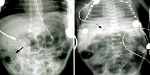
Long description
Abdominal radiograph of necrotising enterocolitis in a neonate.

The investigation of suspected NEC is clinical and radiographic; there is no single laboratory test that makes or excludes the diagnosis.
Abdominal radiograph. This is the central investigation. In the early stages the findings are non-specific: dilated bowel loops, generalised bowel distension and bowel-wall thickening. A single persistent dilated loop is suspicious but not diagnostic. Pneumatosis intestinalis, gas in the bowel wall, in the appropriate clinical setting is usually diagnostic of NEC. Portal venous gas is a further sign and is frequently associated with severe disease. Pneumoperitoneum, free air, is frequently diagnostic of intestinal perforation with NEC: in the supine infant it floats to the most non-dependent area beneath the anterior abdominal wall, often outlining the falciform ligament. [2]
Left lateral decubitus and cross-table lateral films often add information, because free air floats to the non-dependent surface of the peritoneal cavity. Careful serial review of left lateral decubitus films is also used to follow progression and to detect perforation. [2]
Laboratory monitoring. No individual laboratory feature is diagnostic. Full blood counts, differential and platelet counts, arterial blood gas, electrolytes and renal function are used to confirm the clinical impression and track severity. Progressively decreasing absolute granulocyte count and thrombocytopenia suggest increasing severity. [2]
Blood cultures should be taken because bacteraemia is common, and peritoneal cultures may grow organisms when perforation has occurred; Enterobacteriaceae dominate in perforated NEC, while Candida and coagulase-negative staphylococci are more typical of spontaneous intestinal perforation. [3]
Vermont-Oxford Network criteria provide an alternative, more specific diagnostic approach: clinical findings such as bilious gastric aspirate or emesis, abdominal distension and occult or gross blood in stool without anal fissure, plus radiographic findings such as pneumatosis intestinalis, hepatobiliary gas or pneumoperitoneum, with at least one clinical and one radiographic finding. Even this approach has the shortcomings of Bell criteria, because severe surgical NEC can occur without detected pneumatosis or portal gas. [1]
NEC is usually staged with the modified Bell criteria, which classify the illness as suspected (stage I), proven but medically treated (stage II) or advanced (stage III), including perforation. [2][1]
| Stage | Classification | Systemic signs | Intestinal signs | Radiographic signs |
|---|---|---|---|---|
| IA | Suspected NEC | Temperature instability, apnoea, bradycardia, lethargy | Increased pre-feed residuals, mid-abdominal distension, emesis, guaiac-positive stool | Normal or intestinal dilation, mild ileus |
| IB | Suspected NEC | Same as above | Bright red blood from rectum | Same as above |
| IIA | Proven NEC, mildly ill | Same as above | Same as IA plus absent bowel sounds, with or without abdominal tenderness | Intestinal dilation, ileus, pneumatosis intestinalis |
| IIB | Proven NEC, moderately ill | As above plus mild metabolic acidosis and mild thrombocytopenia | As above plus absent bowel sounds, definite tenderness, with or without abdominal cellulitis or right lower quadrant mass | As stage II, plus definite ascites |
| IIIA | Advanced NEC, severely ill, bowel intact | As stage IIB plus hypotension, bradycardia, severe apnoea, combined acidosis, disseminated intravascular coagulation and neutropenia | Signs of generalised peritonitis, marked tenderness and distension | As stage IIB, plus definite ascites |
| IIIB | Advanced NEC, severely ill, bowel perforated | As stage IIIA | As stage IIIA | As stage II, plus pneumoperitoneum |
Two limitations of Bell staging are worth stating. First, stage I findings are highly non-specific and may reflect feeding intolerance, sepsis or stress-related gastrointestinal bleeding. Second, the criteria depend on radiographic pneumatosis, which can be subtle; surgical NEC can develop in a patient whose imaging shows only a paucity of gas without pneumatosis. [1]
Speed matters in NEC. Once the diagnosis is suspected, the paediatric surgical team should be notified immediately, because the disease can become a surgical emergency within hours even when only medical management is needed at first. [2]
The management algorithm is summarised below and in the flowchart.
Approach to suspected or proven necrotising enterocolitis
- Suspected NECFeeding intolerance, distension, guaiac-positive or bloody stool; no pathognomonic radiograph
- Initial stepsNil by mouth; nasogastric or orogastric decompression; close monitoring; labs and imaging; notify surgery
- Question: Radiograph shows pneumatosis or portal venous gas?
No (stage I suspected)
- Close observationBrief feeding pause (about 24 hours in the classic review, up to about 48 hours in some pathways, around 5 days for stage 1 in the CAHS 2025 guideline); consider short course of intravenous antibiotics; repeat radiograph; reassess
Yes (definitive medical NEC)
- Medical NECBowel rest for 7 to 14 days depending on stage (7 to 10 days in the classic review; 7 to 14 days in newer guidelines); decompression; broad-spectrum intravenous antibiotics for 7 to 14 days; fluid and electrolyte support; serial radiographs and labs
- Question: Clinical or biochemical deterioration?
No
- Gradual re-introduction of feeds when stable; monitor for stricture
Yes or free air
- Surgical NECExploratory laparotomy with resection as needed; or primary peritoneal drainage in selected infants
Medical management. Medical (non-operative) treatment is appropriate for stage I and stage II disease, and it includes:
- Bowel rest and decompression. Enteral feedings are stopped. The duration of bowel rest is not standard across guidelines: approximately 7 to 10 days in the classic review [1], 7 days for stage II and 10 to 14 days for stage III in the Turkish Neonatal Society guideline [6], 7 to 14 days in the CAHS guideline [4], and 5 to 7, 7 to 10 or 7 to 14 days by stage in the Johns Hopkins pathway [5]. Bowel is decompressed through a large-bore nasogastric or orogastric tube on low intermittent suction. For suspected disease, only a brief feeding pause is used: about 24 hours in the classic review [1], up to about 48 hours in some pathways [5], and ... as above?
- Antibiotics. Broad-spectrum intravenous antibiotics are a mainstay of treatment. The classic regimen reported in reviews is ampicillin and gentamicin, with vancomycin instead of ampicillin when resistant Staphylococcus epidermidis is suspected, and clindamycin or metronidazole added when perforation or anaerobic infection is suspected. [2] Treatment duration is not standard across guidelines: 7 to 10 days in the classic review [1]; 10 to 14 days in the CAHS and Turkish guidelines [4][6]; and 7, 10 or 7 to 14 days by stage in the Johns Hopkins pathway [5]. Product choice, dosing and duration should follow the local formulary and neonatal team, because randomised trial evidence for the optimal antimicrobial regimen in premature infants is limited. [3]
- Fluids and electrolytes. Third spacing of fluids with losses of sodium and protein is considerable, and acid-base imbalance, potassium fluxes and renal failure are frequent. Careful fluid, electrolyte, parenteral nutrition and respiratory and circulatory support are required. [2]
- Monitoring. Serial abdominal radiographs (anteroposterior and left lateral decubitus), blood counts (a sudden fall suggests progression), and blood gases are needed. Pneumoperitoneum on its own is generally an indication for surgery, whereas pneumatosis without clinical deterioration or worsening of blood results is usually treated medically. [2]
Surgical management. Surgery is indicated for free intraperitoneal air, for persistent ileus, abdominal distension and absence of bowel gas on radiographs coupled with deteriorating clinical and laboratory values (for example falling neutrophil and platelet counts), and for failure to improve with medical management. [1] Two operations are described: exploratory laparotomy with resection of necrotic bowel (and creation of a stoma when needed), and primary peritoneal drainage without laparotomy. Two large multicentre studies found no significant difference in early survival or clinically important early outcomes between drainage and laparotomy, although peritoneal drainage very often required a subsequent laparotomy. A systematic review suggested mortality was increased by more than 50% with peritoneal drainage compared with laparotomy, and follow-up at 18 to 22 months in the classic review (18 to 24 months in the ERNICA guideline) showed a lower risk of death or neurodevelopmental impairment after laparotomy. [1][7] Whether drainage or laparotomy is chosen for an individual infant must be a decision by the surgical and neonatal teams.
Post-operative course and intestinal failure. Extensive resection leaves infants with short-bowel syndrome, the major cause of prolonged hospitalisation and expense. Strictures may form later in survivors and can present weeks or months afterwards as feeding intolerance, obstruction or perforation.
- Intestinal perforation and peritonitis, sometimes progressing within hours.
- Short-bowel syndrome from resection of necrotic bowel. [1]
- Intestinal stricture, often discovered after an apparently resolved episode. [2]
- Sepsis and death, with mortality highest when surgery is required. [1]
- Cholestasis and hepatic complications from prolonged parenteral nutrition.
- Neurodevelopmental impairment, with nearly a 25% chance of microcephaly and serious delays. [1]
Prognosis depends on gestational age at onset, extent of intestinal necrosis, whether perforation occurs and whether surgery is required. Mortality overall is 20% to 30%, worse in surgical disease, and long-term survivors remain at risk of strictures, intestinal failure and neurodevelopmental problems. [1]
Prevention is more likely to reduce mortality than better treatment of established disease, because once NEC is severe the outcome is often poor.
Evidence-supported measures include careful, monitored advancement of enteral feeds together with feeding with the mother's own breast milk. In an early dietary trial the incidence of NEC was 7.2% in infants fed only formula, 2.5% in those fed formula plus expressed human milk and 1.2% in those fed expressed human milk alone. [2]
The mother's expressed milk is preferred for the smallest infants; it contains bioactive factors and immunological components that formula does not, and its use has evidence of efficacy and safety. Exclusive use of human milk plus a human milk-derived fortifier may further lower the incidence of NEC. [1]
Measures with evidence of efficacy but safety questions include enteral aminoglycosides (resistance limits routine use), prophylactic probiotics (not licensed in preterm infants and require high-grade manufacture), antenatal glucocorticoids and arginine. Complete withholding of feeds may be harmful, leading to prolonged parenteral nutrition, intestinal atrophy, increased permeability and inflammation and late-onset sepsis; even delayed feeding may worsen NEC if it occurs. Prolonged empirical intravenous antibiotics also increase the incidence of NEC. [1]
Other approaches such as prebiotics, growth factors, anticytokines and toll-like receptor modulation remain investigational. [1]
Term and late preterm infants. Disease in these infants usually appears in the first week and is linked to other problems, such as congenital heart disease, intestinal anomalies or perinatal stress. Management must find and treat the underlying cause rather than apply the classic NEC pathway blindly. [1]
Infants with spontaneous intestinal perforation. These infants are often treated surgically but have much less intestinal inflammation; medical NEC protocols do not fit them well and they should not be classified as NEC if the clinical picture suggests perforation without pneumatosis. [1]
Extremely preterm infants. They remain at risk until 35 to 36 weeks post-conceptual age. [2]
- A single abdominal radiograph showing pneumatosis or portal gas is diagnostic in the right clinical setting; learn to read it before the disease presents overnight. [2]
- When radiography is unavailable, suspected perforation can be sought clinically and with a careful fluid assessment, but imaging should be obtained whenever possible.
- Feeding milk from the infant's own mother is low-cost, feasible even where donor milk banks do not exist and is a genuine preventive measure. [1]
- Surgical capacity, anaesthesia and intestinal failure support determine survival after perforation; early referral from the maternity unit matters.
- Oral antimicrobial prophylaxis during outbreaks is limited by resistance; basic infection-control measures, cohorting and interruption of the outbreak are the safe measures. [2]
- NEC is the most common gastrointestinal emergency of premature infants, with a prevalence of about 7% in infants of 500 to 1500 g birth weight. [1][3]
- Around nine in ten cases occur in premature infants; the condition is rare in term infants and adults. [2]
- After 8 to 10 days of age, feeding intolerance, abdominal distension and bloody stool are the classic presentation; deterioration can be abrupt. [1]
- Pneumatosis intestinalis, portal venous gas and free air on an abdominal radiograph are the imaging landmarks; left decubitus films are often more informative. [2]
- Modified Bell staging guides management: suspected disease is observed, definitive disease is treated with fasting, decompression and intravenous antibiotics for 7 to 10 days in the classic review and up to 10 to 14 days in newer guidelines, and surgical disease requires operation. [1][4][6]
- Surgery is indicated for free air or clinical deterioration despite medical therapy; whether to drain or operate is unresolved, and laparotomy probably reduces mortality. [1]
- Complications include perforation, sepsis, stricture, short-bowel syndrome and neurodevelopmental impairment, with nearly a 25% chance of microcephaly and serious delays after severe disease. [1]
- Mother's own milk and careful feeding practices are the measures with evidence of safety and efficacy in prevention. [1][2]
Answer each one in your head first, then open the answer.
A 10-day-old baby born at 28 weeks' gestation becomes less tolerant of feeds, vomits and passes a small amount of blood in the stool. The abdomen is distended. What is the most likely diagnosis and what radiographic finding would confirm it?
Show answerHide answer
Necrotising enterocolitis. Pneumatosis intestinalis, with or without portal venous gas, on an abdominal radiograph is pathognomonic in this clinical setting; early films may show only dilated loops and ileus.
How do the radiographic appearances of pneumatosis intestinalis, portal venous gas and free air differ in the severity and urgency of necrotising enterocolitis?
Show answerHide answer
Pneumatosis intestinalis is diagnostic of NEC and usually treated medically if the infant is stable. Portal venous gas is associated with more severe disease. Free air indicates perforation and is generally an indication for surgery.
A premature infant with pneumatosis intestinalis is made nil by mouth, given intravenous antibiotics and decompressed. After 48 hours the platelet count is falling, the abdomen is tense and the radiograph now shows no bowel gas. What should happen next?
Show answerHide answer
This is surgical NEC despite medical management: the absence of bowel gas with a fixed, dilated or gasless abdomen plus deteriorating blood results indicates progression to operation. The surgical and neonatal teams must decide between laparotomy and peritoneal drainage.
Why is an infant born at 26 weeks still at risk of NEC at a post-conceptual age of 32 weeks, while a term infant is rarely affected?
Show answerHide answer
Prematurity is the dominant risk factor because the immature gut has poor motility, reduced mucus, higher permeability and an exaggerated inflammatory response to colonising microbes. Extremely premature infants remain at risk until 35 to 36 weeks post-conceptual age.
A neonatal unit wishes to reduce its rate of necrotising enterocolitis. Which two feeding practices have the strongest evidence of efficacy and safety, and which common practice has been shown to increase it?
Show answerHide answer
Non-aggressive advancement of feeds and feeding with the mother's own breast milk have evidence of efficacy and safety; prolonged empirical intravenous antibiotics increase the incidence. Probiotics and enteral aminoglycosides have efficacy data but unresolved safety or licensing issues.
What is the difference between necrotising enterocolitis in a term infant and classic NEC in a premature infant, and why does it matter?
Show answerHide answer
Term or late preterm NEC usually arises in the first week and is linked to congenital heart disease, intestinal anomalies or perinatal stress; classic NEC arises after the first week in premature infants. The pathogenesis and required management differ, so the underlying cause must be sought in term infants.
- Pediatrics: neonatal care, feeding of the preterm infant, short bowel syndrome, intestinal failure.
- Anaemia and red-cell transfusion medicine.
- Infection control in the neonatal intensive care unit.
- Neu J, Walker WA. Necrotizing enterocolitis. N Engl J Med 2011;364:255-264. doi:10.1056/NEJMra1005408. Accessed .
- Neu J. Necrotizing enterocolitis: the search for a unifying pathogenic theory leading to prevention. Pediatr Clin North Am 1996;43:409-432. doi:10.1016/S0031-3955(05)70413-2. Accessed .
- Coggins SA, Wynn JL, Weitkamp JH. Infectious causes of necrotizing enterocolitis. Clin Perinatol 2015;42:133-154. doi:10.1016/j.clp.2014.10.012. Accessed .
- Child and Adolescent Health Service, Western Australia. Necrotising Enterocolitis. Neonatal clinical guideline, last reviewed 2025-12-23. Accessed .
- Johns Hopkins All Children's Hospital Clinical Pathway. Evaluation and Management of Suspected, Medical and Surgical Necrotizing Enterocolitis. Clinical pathway, final 2022-12-16. Accessed .
- Ergenekon E, Tayman C, Ozkan H. Turkish Neonatal Society Necrotizing Enterocolitis Diagnosis, Treatment and Prevention Guidelines. Turk Arch Pediatr 2021;56:513-524. Accessed .
- Hulscher J, et al. European Reference Network for Inherited and Congenital Anomalies Evidence-Based Guideline on Surgical Aspects of Necrotizing Enterocolitis in Premature Neonates. Neonatology 2024;122:376-384. Accessed .
Figure credits
Educational content for medical students and doctors. It is not a substitute for clinical judgement or local guidelines.