Neuroblastoma
A malignant tumour of the sympathetic nervous system in children: how it presents, how it is staged, and how treatment depends on risk group.
- Neuroblastoma is a malignant tumour that begins in immature sympathetic nervous tissue, most often the adrenal medulla or the paraspinal sympathetic chain from neck to pelvis.
- It is the most common extracranial solid tumour of childhood: more than 650 cases are diagnosed each year in the United States, about 1 case per 7,000 live births, and 90% of patients are younger than 5 years at diagnosis.[1]
- It is a tumour of infancy: 37% of patients are diagnosed as infants and the highest rate of diagnosis is in the first month of life.[1]
- The classic presentation is an abdominal mass, often with metastatic “raccoon eyes” (periorbital ecchymosis), bone pain, or hypertension; urine usually contains raised vanillylmandelic acid (VMA) and homovanillic acid (HVA).[1]
- Diagnosis rests on imaging, especially MIBG scintigraphy, together with measurement of urine catecholamine metabolites and a biopsy that establishes the histological type and genetic risk features.[1]
- Treatment is chosen by risk group: gentle surgery or observation for low-risk disease, chemotherapy with or without surgery for intermediate-risk disease, and intensive multimodal therapy for high-risk disease.[1]
By the end of this page you should be able to:
- Explain where neuroblastoma arises and why it can appear almost anywhere along the sympathetic chain.
- Link the common symptoms and signs (abdominal mass, raccoon eyes, bone pain, hypertension, opsoclonus) to the underlying biology.
- Interpret urine VMA and HVA and plan a sensible imaging work-up, including MIBG scanning.
- Distinguish neuroblastoma from Wilms tumour using clinical and imaging clues.
- Apply the International Neuroblastoma Risk Group (INRG) staging system and the COG risk groups in outline.
- Describe the role of surgery, chemotherapy, radiotherapy, stem cell transplant, retinoids and anti-GD2 immunotherapy in high-risk disease.
- Summarise how prognosis varies with age, stage and MYCN status.
DefinitionNeuroblastoma
A malignant tumour of the sympathetic nervous system arising from neural crest cells that normally form the adrenal medulla and the paravertebral sympathetic ganglia. It sits in a family with ganglioneuroblastoma (a mixed, partly mature tumour) and ganglioneuroma (a fully mature, benign tumour).
“Neuroblastoma” covers the whole family of peripheral neuroblastic tumours, which range from the very aggressive neuroblastoma through the mixed ganglioneuroblastoma (with a nodule of highly malignant cells that behaves like neuroblastoma) to the benign ganglioneuroma. The International Neuroblastoma Pathology Classification (INPC, the Shimada system) sorts these tumours into favourable and unfavourable histology using age-dependent criteria, which then feed into risk-group assignment.[1]
Classification by risk is at least as important as histological classification, because two tumours that look identical under the microscope may receive entirely different treatment depending on age, stage, MYCN amplification, chromosome 11q status and ploidy.[1]
Neuroblastoma is the most common extracranial solid tumour in childhood. More than 650 cases are diagnosed each year in the United States, a prevalence of about 1 case per 7,000 live births and an incidence of 8.3 cases per million children younger than 15 years.[1] The overall incidence has remained stable.[1] [2]
- Age. About 37% of patients are diagnosed as infants and 90% are younger than 5 years, with a median age at diagnosis of 17 months.[1] The rate of diagnosis is highest in the first month of life, and screening studies have shown that spontaneous regression without clinical detection in the first year is at least as common as clinically detected disease.[1]
- Sex. Boys are affected slightly more often than girls in most series.
- Ethnicity. Non-Hispanic White children have a higher risk of developing neuroblastoma than Hispanic, Asian or Pacific Islander, American Indian or Alaska Native and Black children; five-year relative survival is higher in White and Hispanic children than in Black children.[1]
- Family history. Only 1 to 2% of patients have a family history, and these children are usually diagnosed younger (around 9 months) and are more likely to have multifocal disease.[1]
- Genetics. About 1 to 2% of cases are familial; the main predisposition genes are ALK and PHOX2B, and other cancer predisposition syndromes also occasionally include neuroblastoma.[1]
- Late effects. Treatment-related late effects are substantial, particularly after radiotherapy in infancy, so the modern approach aims to cure the disease with the least possible toxicity.[1]
No environmental exposure has been clearly linked to neuroblastoma.[1] The aetiology is mainly genetic.
- ALK variants are the commonest cause of familial disease (about 75% of familial cases) and are also found as somatic variants in about 9% of sporadic tumours; ALK is near MYCN on chromosome 2 and may be co-amplified with it.[1]
- PHOX2B variants are seen in rare patients with familial neuroblastoma together with congenital central hypoventilation (Ondine curse) and/or Hirschsprung disease.[1]
- Germline deletions at 1p36 or 11q14-23 have been reported in familial cases, and about 14% of patients in one study carried a pathogenic germline variant in a cancer predisposition gene.[1]
- Common variants of small effect (single nucleotide polymorphisms) influence susceptibility and often associate with high-risk rather than low-risk phenotypes.[1]
Clinicians should enquire about family history, congenital anomalies and syndromic features, because a predisposition syndrome changes both screening and treatment decisions.[1]
The tumour’s behaviour follows from three facts: it derives from neural crest cells, it can secrete catecholamines, and it very commonly spreads to bone and bone marrow early.
Origin. Neural crest cells destined to become the adrenal medulla and sympathetic ganglia fail to complete their differentiation. The result is a population of small, undifferentiated neuroblasts, usually clustered into a mass with a rich stroma of Schwann cells and collagen. Grossly the tumour is grey-pink, soft and friable, and frequently calcified.[1]
Catecholamine secretion. About 90% of tumours produce excess catecholamine metabolites, which spill into the urine as VMA and HVA. This is the basis of the simple urine test that often confirms the diagnosis, and it is why hypertension can be a presenting feature.[1]
Spread. Neuroblastoma disseminates early, often before symptoms appear. The common routes are lymphatic spread to regional nodes and haematogenous spread to bone, bone marrow, liver and, in infants, subcutaneous skin nodules. About 70% of patients have metastatic disease at diagnosis.[1]
MYCN. The MYCN oncogene on chromosome 2p24 is amplified in 16 to 25% of tumours overall and in 40 to 50% of high-risk tumours. MYCN amplification drives rapid proliferation and is the single strongest biological adverse factor.[1]
Telomere maintenance and ALK. High-risk tumours often maintain telomeres through TERT rearrangement or ATRX alteration, and about 15% carry activating ALK variants. These features are used for research stratification and increasingly for targeted therapy in relapse.[1]
Why metastatic disease looks the way it does. Tumour in the retrobulbar space pushes the eye forward (proptosis) and produces the bruised “raccoon eyes”. Deposits in bone and marrow explain bone pain and pancytopenia. Massive liver infiltration in infants produces abdominal distension and respiratory compromise. Paraspinal tumours can invade the spinal canal and compress the cord.[1]
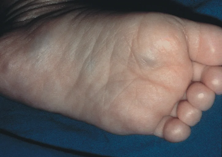
Figure 1Neuroblastoma Muffin
Long description
Stage 4S neuroblastoma in an infant with multiple bluish cutaneous metastases (blueberry muffin appearance).

The most frequent signs and symptoms are caused by tumour mass and metastases.[1]
- An abdominal mass is the most common presentation overall; the swelling is often noticed by a parent and may be painless.[1]
- Bone pain is common, particularly when metastases are present.[1]
- Fever, fatigue, weight loss and pallor are common constitutional symptoms.[1]
- Watery diarrhoea can occur when the tumour secretes vasoactive intestinal peptide, classically producing severe secretory diarrhoea with electrolyte disturbance.[1]
- Hoarseness or respiratory symptoms can occur with thoracic tumours.[1]
- Limb weakness or sensory loss occurs when a paraspinal tumour compresses the spinal cord.[1]
- An abdominal mass, often firm, irregular, crossing the midline and not moving with respiration.[1]
- Proptosis with periorbital ecchymosis (“raccoon eyes”), especially in high-risk disease, from retrobulbar metastasis.[1]
- Subcutaneous nodules with bluish discolouration of the overlying skin, seen especially in infants; these are metastases and are a hallmark of stage MS disease.[1]
- Horner syndrome (miosis, ptosis, anhidrosis) from a tumour in the stellate ganglion.[1]
- Hypertension in a minority of patients, from catecholamine secretion or renal vein compression.[1]
- Pancytopenia from extensive bone marrow involvement.[1]
- Chronic anaemia and sometimes hepatomegaly.[1]
A rare paraneoplastic presentation: about half of young children who present with opsoclonus–myoclonus syndrome are found to have neuroblastoma, usually with favourable biology and an excellent survival.[1] The expected findings are chaotic multi-directional eye movements together with myoclonic jerks, irritability and ataxia developing over days. Treatment of the neurological syndrome uses corticosteroids and, in some cases, intravenous immunoglobulin, with rituximab in selected cases.[1]
Adolescents and adults rarely develop neuroblastoma (less than 5% of cases). Their tumours run a more indolent course, show de novo chemotherapy resistance, and are markedly enriched for segmental chromosomal aberrations and ATRX loss. Five-year overall survival for adolescents and young adults is 38%.[1]
An upper abdominal mass in a child is most commonly either a neuroblastoma or a Wilms tumour (nephroblastoma); other possibilities include hepatoblastoma, renal cystic disease, retroperitoneal lymphoma, adrenal haemorrhage and, in neonates, congenital adrenal cysts. The clinical clues that favour neuroblastoma over Wilms tumour are summarised below.[1][2]
| Feature | Neuroblastoma | Wilms tumour (nephroblastoma) |
|---|---|---|
| Origin | Adrenal medulla and sympathetic chain[1] | Kidney (“renal”); embryonal renal tumour[2] |
| Typical age | Median 17 months; 90% younger than 5 years[1] | Most commonly diagnosed in early childhood[2] |
| Abdominal mass | Often crosses the midline, encases rather than displaces vessels, frequently calcified[1] | Usually arises from the kidney; often a smooth flank mass that does not cross the midline[2] |
| Haematuria | Not typical | Common presenting feature[2] |
| Catecholamine metabolites | Urine VMA and HVA usually raised; about 90% of tumours MIBG-avid[1] | Not raised; not MIBG-avid |
| Imaging hallmark | MIBG uptake in primary and metastatic sites[1] | “Claw sign” of compressed normal renal tissue; lung metastases common[2] |
| Metastatic pattern | Bone, bone marrow, liver, skin (infants), retrobulbar orbit[1] | Lung, liver; bone and marrow uncommon[2] |
| Overall survival | About 60% at 5 years for high-risk disease[1] | Over 90% at 5 years with favourable histology[2] |
Measurement of urinary VMA and HVA, expressed per milligram of creatinine, is the first-line laboratory investigation: most neuroblastomas secrete excess catecholamines and 90% are detectable this way, and a 24-hour collection is not required.[1] Serum catecholamines are not routinely used.[1]
Normal or equivocal values do not exclude the disease; conversely, elevated values together with marrow containing unequivocal tumour cells satisfy the international minimum diagnostic criteria in the absence of a biopsy.[1]
- Ultrasonography is useful for the first detection, especially of an adrenal mass in an infant.[1]
- CT or MRI with contrast of chest, abdomen and pelvis defines the primary tumour and regional nodes, and is used to assess image-defined risk factors (IDRFs) for staging.[1]
- MRI of the spine adjacent to any paraspinal tumour is mandatory to exclude spinal cord compression.[1]
- MIBG scintigraphy is the critical imaging study for staging and for response assessment: 90 to 99% of neuroblastomas are MIBG avid. Iodine-123 is preferred to iodine-131.[1]
- PET-CT is used for MIBG-non-avid tumours.[1]
- Bone marrow assessment is by bilateral iliac crest aspirates and trephine biopsies, with at least 1 cm of core.[1]
Diagnostic pathway for a child with a suspected abdominal or paraspinal mass
- Child with a palpable mass or suspicious imagingHistory, examination, bloods (FBC, renal, LDH, ferritin)
- Urine VMA and HVANormalised to creatinine; 24-hour collection not needed
- Staging imagingCT or MRI of chest, abdomen, pelvis plus spine MRI if paraspinal; MIBG scintigraphy (I-123)
- Biopsy of primary tumourCore or open biopsy for histology, MYCN, ALK, DNA index, segmental chromosomal aberrations
- Bone marrow aspirates and trephinesBilateral; required for staging and for diagnosis when biopsy is not possible
- Question: Risk group and stage assigned?
Low risk
- Surgery, often curative; observe if incidental cystic adrenal mass
Intermediate risk
- Chemotherapy with or without delayed surgery; radiotherapy rarely
High risk
- Induction, surgery, tandem myeloablative HSCT, radiotherapy, immunotherapy, retinoid
Tumour tissue is required for modern risk-group assignment; tissue from several core biopsies or about 1 cm³ from an open biopsy is usually needed, and both give similar diagnostic yield.[1] In patients older than 18 months with stage 4 (metastatic) disease, extensive bone marrow involvement together with raised catecholamine metabolites may be adequate for diagnosis, but an attempt should still be made to biopsy the primary tumour.[1]
The international minimum criterion for a diagnosis of neuroblastoma is either an unequivocal pathological diagnosis on tumour tissue, or bone marrow containing unequivocal tumour cells together with raised urinary catecholamine metabolites.[1]
INRG is a pre-surgical staging system based on image-defined risk factors (IDRFs), so that staging does not depend on how much of the tumour a surgeon manages to remove.
| Stage | Definition |
|---|---|
| L1 | Localised tumour with no IDRFs, confined to one body compartment |
| L2 | Locoregional tumour with one or more IDRFs |
| M | Distant metastatic disease (excluding MS) |
| MS | Metastatic disease in children younger than 18 months with metastases confined to skin, liver and/or bone marrow; primary tumour may be L1, L2 or (formerly) INSS stage 3 equivalent [1] |
MS disease differs from the older INSS stage 4S definition, which applied only to infants up to 12 months; MS extends this concept to children up to 18 months.[1]
IDRFs include encasement of major vessels, spinal canal invasion exceeding one third of the axial plane with obliterated leptomeningeal spaces, infiltration of adjacent organs, extension across two body compartments and tumour crossing the sciatic notch, among others.[1]
The Children’s Oncology Group (COG) assigns patients to low, intermediate or high risk using clinical and biological factors. The broad picture is as follows.[1]
- Low-risk disease. Usually INRG L1 or MS disease without MYCN amplification, hyperdiploid, favourable histology. Very high survival (over 95% at 5 years). Surgery alone is often curative; some infants are observed without biopsy.[1]
- Intermediate-risk disease. Localised L2 disease or metastatic disease in infants without MYCN amplification; survival 90 to 95% at 5 years. Treatment is chemotherapy with or without delayed surgery.[1]
- High-risk disease. Any metastatic (M) disease in a child 18 months or older, MYCN-amplified stage M or MS disease, or L2 disease with unfavourable histology in a child 18 months or older. About 8 to 10% of infants with stage MS disease have MYCN amplification and are usually treated on high-risk protocols. Five-year survival for high-risk patients treated with current regimens is about 60%.[1]
Management is driven entirely by risk group. The philosophy of modern treatment is to give enough therapy to cure, but no more, because late effects in survivors, including from radiotherapy in early childhood, are substantial.[1]
- Low toxicity, high cure in low-risk disease.
- Moderate chemotherapy, low late-effect burden in intermediate-risk disease.
- Intensive multimodal therapy in high-risk disease, combining induction chemotherapy, surgery, tandem myeloablative chemotherapy with haematopoietic stem cell rescue, radiotherapy, and maintenance with a retinoid and anti-GD2 immunotherapy. [1]
Surgery by an experienced paediatric surgeon is the treatment of choice for INRG stage L1 tumours. Exceptions are infants younger than 6 months with small adrenal masses (solid masses 3.1 cm or less, or cystic masses with at least 25% cystic change up to 5 cm), who may be observed without biopsy because most of these lesions regress spontaneously.[1] In 83 carefully selected infants, 81% did not need surgery and all were alive at 2 years.[1]
If the biology is favourable, residual disease after surgery is not a risk factor for relapse and chemotherapy is not indicated. Chemotherapy (carboplatin, cyclophosphamide, doxorubicin and etoposide, cumulative doses kept low) is reserved for symptomatic disease or unresectable progressive disease after surgery, and radiotherapy is used only as emergency treatment.[1]
Patients receive 2, 4 or 8 cycles of neoadjuvant chemotherapy (carboplatin, cyclophosphamide, doxorubicin and etoposide) followed by surgical resection; additional cyclophosphamide and topotecan may be given for inadequate response. Patients with favourable biology receive fewer cycles; those with unfavourable features or who fail to respond adequately receive more. Surgery is usually possible after chemotherapy; radical nephrectomy is avoided whenever possible in abdominal neuroblastoma.[1]
In the ANBL0531 study, 404 patients received risk- and response-adapted treatment; 10-year event-free survival (EFS) was 82% and overall survival (OS) 95%, with less than 3% needing local radiotherapy.[1]
Approximately 8 to 10% of infants with stage MS disease have MYCN-amplified tumours and are usually treated on high-risk protocols. Treatment is divided into three phases.[1]
Induction (roughly 6 cycles). The backbone alternates dose-intensive cisplatin and etoposide with vincristine, cyclophosphamide and doxorubicin; topotecan and cyclophosphamide may be added. Primary tumour resection is performed during induction where possible; aiming for more than 90% resection reduces local recurrence. Response after four cycles correlates with eventual outcome.[1]
Consolidation. Two cycles of myeloablative chemotherapy with autologous haematopoietic stem cell rescue (most North American protocols use cyclophosphamide/thiotepa and then carboplatin/etoposide/melphalan) are followed by radiotherapy to the tumour bed and remaining metastatic sites. Randomised evidence favours tandem transplant over single transplant for event-free survival (3-year EFS 73% versus 55% when immunotherapy followed). Radiotherapy contributes to local control.[1]
Post-consolidation (maintenance). Dinutuximab (an anti-GD2 chimeric monoclonal antibody) is given with granulocyte-macrophage colony-stimulating factor (GM-CSF) and interleukin-2, together with isotretinoin (13-cis-retinoic acid) for six months. In the COG ANBL0032 trial, 5-year EFS was 57% and OS 71% for patients receiving immunotherapy versus 46% and 57% for isotretinoin alone.[1] Dinutuximab is approved by the US Food and Drug Administration on the strength of these results.[1]
- Surgery obtains tissue for diagnosis and molecular risk assignment, and removes resectable primary tumour.
- In low-risk L1 disease, up-front complete resection is standard and often curative.
- In intermediate-risk disease, surgery usually follows response to chemotherapy.
- In high-risk disease, induction chemotherapy precedes surgery in most cases.
- Complete resection requiring nephrectomy is not recommended because of the nephrotoxicity of standard chemotherapy and the unproven added benefit of removing the kidney.[1]
- In low- and intermediate-risk disease, radiotherapy is reserved for symptomatic, life-threatening or organ-threatening disease that does not respond to chemotherapy (for example, infants with stage MS disease and severe respiratory compromise from liver enlargement, or spinal cord compression).
- In high-risk disease, radiotherapy to the tumour bed and residual metastatic sites after high-dose therapy is standard.
- Radiotherapy in infancy carries a substantial long-term risk; it should be avoided unless necessary.[1]
Isotretinoin (13-cis-retinoic acid) induces terminal differentiation of neuroblastoma cells and is used as maintenance therapy after stem cell transplant. Randomised data showed a trend to improved event-free survival (42% versus 31%) even when used alone, and it is now standard combined with anti-GD2 immunotherapy.[1]
GD2 is a ganglioside expressed strongly on neuroblastoma cells and weakly on normal neurones. Dinutuximab binds GD2 and recruits natural killer cells and complement. It is most effective in the setting of minimal residual disease after transplant, and is combined with GM-CSF (and in some protocols interleukin-2) to enhance antibody-dependent cell-mediated cytotoxicity. Its effectiveness is partly determined by the patient’s KIR genotype.[1]
Spinal cord compression is a medical emergency. Neuroblastoma is usually sensitive to chemotherapy, so low- and intermediate-risk patients are treated with chemotherapy first; neurosurgical intervention (laminectomy or laminoplasty) is reserved for severe or progressive deficit. Radiotherapy is now used less frequently because of late effects in young children.[1]
Most infants with MS disease have favourable biology and are observed without resection. Chemotherapy, low-dose radiotherapy and even liver transplantation have been used for infants with massive symptomatic liver involvement, while surgical excision is usually avoided unless necessary. A small subgroup of infants have MYCN amplification and are treated as high risk.[1]
A variety of approaches are used: MIBG-targeted radiotherapy, ALK-directed therapy (lorlatinib) for ALK-altered tumours, dinutuximab-based immunotherapy, and myeloablative MIBG combined with stem cell rescue. Genomic re-profiling at relapse is increasingly useful, because ALK and RAS pathway alterations are commoner at relapse than at diagnosis.[1]
After therapy, regular imaging and surveillance with MIBG scans and urine catecholamine metabolites detect most relapses, which in high-risk patients are often asymptomatic at first. Cross-sectional imaging is kept to the minimum necessary because of radiation exposure; MIBG and urinary metabolites are the most reliable monitors. Follow-up must continue for years because late recurrences occur.[1]
- Local complications ( spinal cord compression with paraplegia, renal vein compression, massive hepatomegaly with respiratory compromise).
- Metastatic complications (bone pain, pathological fracture, pancytopenia, proptosis, cranial nerve palsies).
- Paraneoplastic complications (opsoclonus–myoclonus syndrome with long-term cognitive and behavioural effects despite good tumour control).
- Acute treatment complications (cisplatin nephrotoxicity and ototoxicity, cyclophosphamide haemorrhagic cystitis, doxorubicin cardiotoxicity, transplant-related toxicity, conditioning-related veno-occlusive disease).
- Late effects (endocrine failure, hearing loss, renal impairment, infertility, second malignant neoplasms, particularly after radiotherapy and high-dose therapy).
- Vasoactive intestinal peptide secretion causing chronic watery diarrhoea, which improves with tumour control.[1]
Prognosis is determined by risk group, which combines age, stage and biology. Event-free and overall survival estimates below are from large contemporary cohorts.[1]
- Younger than 18 months: 5-year event-free survival 82% and overall survival 91%.[1]
- 18 months or older: 5-year event-free survival 64% and overall survival 74%.[1]
- Newborn to 1 year: 5-year relative survival 93%; ages 1 to 4 years, 78%; ages 5 to 9 years, 83%; ages 10 to 14 years, 86%.[1]
- Adolescents and young adults (15 to 39 years): 5-year overall survival 38%.[1]
- Stage 4 in adolescents (10 to 21 years): 10-year event-free survival 3% and overall survival 5%.[1]
- INRG stage L1: 5-year overall survival 98%; L2, 95%; M, 64%; MS, 92%.[1]
- Very-low-risk and low-risk groups have survival over 95% and over 90% respectively. Intermediate-risk survival is 90 to 95%. High-risk survival with current treatment is about 60% at 5 years.[1]
- Patients with favourable biology and localised disease approach 100% survival. Infants with stage 4 and favourable biology have event-free survival of about 87%, versus about 67% for those with unfavourable biology.[1]
- Histology alone stratifies sharply: favourable INPC histology is associated with 5-year overall survival 96%, versus 66% for unfavourable histology.[1]
- MYCN-amplified tumours do far worse within each risk group; an infant with MYCN-amplified stage 4 disease has 5-year overall survival around 45%.[1]
- Tumours with no MYCN amplification and favourable histology can be cured with surgery alone in localised disease.
- Long-term survival in high-risk disease is reduced by late recurrence; some relapses occur more than 5 years after treatment.[1]
- No screening programme is recommended for the general population; Japanese and Canadian screening programmes in infants detected many neuroblastomas that regressed spontaneously without improving overall mortality.[1]
- Targeted surveillance with abdominal ultrasonography and urine VMA/HVA every 3 months until age 6 years, then every 6 months to age 10 years, is recommended for children with high-penetrance predisposition (for example, ALK or PHOX2B pathogenic variants, Li-Fraumeni syndrome, Beckwith-Wiedemann syndrome with CDKN1C variants).[1]
- Infants aged 6 months or younger with a small adrenal mass detected incidentally (solid 3.1 cm or less, or cystic up to 5 cm) may be observed without biopsy, with monitoring by ultrasound and urine VMA/HVA.[1]
- A transthoracic or abdominal ultrasound is the most available first test and can reveal a solid adrenal or paraspinal mass; CT is used when available.
- Urine VMA and HVA testing may not be available; where it is not, diagnosis rests on tissue biopsy and imaging.
- MIBG scanning and PET are rarely available; chest and abdominal staging then relies on CT.
- Surgery in experienced hands is usually possible locally; tandem stem cell transplant and anti-GD2 immunotherapy are not, so high-risk patients need referral to an oncology centre with transplant facilities.
- Recognition of stage MS disease matters most in low-resource settings, because simple chemotherapy and sometimes no treatment at all can cure these infants.
- The differential from Wilms tumour is clinically important: where biopsy is not available, the overall picture (age, crossing the midline, catecholamines, metastatic sites) must be used carefully.
- Neuroblastoma is the commonest extracranial solid tumour of childhood, is a tumour of infancy with a median age at diagnosis of 17 months, and most often arises in the adrenal medulla and sympathetic chain.
- Most tumours secrete catecholamines; urine VMA and HVA are the standard first-line laboratory test, and about 90% of tumours are MIBG avid.
- The classic physical clue is an abdominal mass that crosses the midline, and metastatic disease produces raccoon eyes, bone pain, skin nodules in infants and Horner syndrome.
- Staging uses the pre-surgical INRG system (L1, L2, M, MS) based on image-defined risk factors, and treatment is assigned by risk group (low, intermediate, high).
- High-risk disease is treated in three phases (induction, consolidation with tandem myeloablative transplant and radiotherapy, and post-consolidation immunotherapy and retinoid); 5-year survival is about 60%. [1]
- MYCN amplification is the single strongest adverse biological factor and occurs in 16 to 25% of tumours overall.
- Low-risk disease is treated with surgery alone or observation; intermediate-risk disease responds to moderate chemotherapy; many infants with MS disease need no treatment at all.
- Infants with stage MS neuroblastoma have excellent survival; adolescents and adults fare much worse.
- No population screening is recommended, because screening finds regressing tumours without saving lives.
- Prevention of late effects is now a central aim of modern therapy.
Answer each one in your head first, then open the answer.
A 3-year-old child is brought in with a firm upper abdominal mass that crosses the midline, periorbital bruising and bone pain in the legs. What is the most likely diagnosis and which single investigation would do most to stage both the primary tumour and its metastases?
Show answerHide answer
Neuroblastoma. MIBG scintigraphy (using iodine-123) images both the primary tumour and metastatic bone and soft-tissue disease, and most neuroblastomas are MIBG avid. Urine VMA and HVA and a biopsy would follow, but imaging with MIBG is the staging investigation of choice.
A child has a renal tumour. How do you tell from the bedside and initial tests whether it is more likely to be neuroblastoma than Wilms tumour?
Show answerHide answer
Neuroblastoma is more likely when the child is very young (median 17 months), the mass crosses the midline, there are catecholamine-related features (hypertension) or raised urine VMA and HVA, metastases involve bone, marrow or orbit, and MIBG scanning is positive. Wilms tumour more often occurs in somewhat older children, presents with haematuria and a smooth renal mass that does not cross the midline, and does not raise catecholamine metabolites. [2]
Which biological abnormality most strongly marks a neuroblastoma as high risk, and in roughly what proportion of tumours is it found?
Show answerHide answer
MYCN amplification, found in 16 to 25% of tumours overall and in 40 to 50% of high-risk tumours.
In the INRG system, which stage letters describe a localised tumour without and with image-defined risk factors, and how does stage MS differ from the old INSS stage 4S?
Show answerHide answer
L1 has no image-defined risk factors and L2 has one or more. Stage MS applies to metastatic disease in children younger than 18 months confined to skin, liver and bone marrow, and extends the older stage 4S (which applied only up to 12 months) to 18 months.
What are the three treatment phases for a child with high-risk neuroblastoma, and what is given after transplant?
Show answerHide answer
Induction chemotherapy, consolidation with tandem myeloablative chemotherapy and haematopoietic stem cell rescue plus radiotherapy, then post-consolidation maintenance with dinutuximab (anti-GD2) and isotretinoin (13-cis-retinoic acid).
Why is surgery alone often enough for an infant with a small adrenal mass, and what size thresholds are used?
Show answerHide answer
Many infant neuroblastomas regress spontaneously. Infants under 6 months with adrenal masses that are solid 3.1 cm or less, or cystic with at least 25% cystic change up to 5 cm, may be observed without biopsy; in one study 81% of 83 such infants avoided surgery and all survived to 2 years. [1]
- Phaeochromocytoma
- Wilms tumour (not yet a MedGrasp topic)
- Primitive neuroectodermal tumour
- Haematopoietic stem cell transplantation
- Cancer in children
- PDQ Pediatric Treatment Editorial Board. Neuroblastoma Treatment (PDQ®)–Health Professional Version. National Cancer Institute; last updated 28 April 2025. Accessed .
- Hong B, Dong R. Wilms' Tumor: A Review of Clinical Characteristics, Treatment Advances, and Research Opportunities. Medicina (Kaunas) 2025;61(3):491. Accessed .
Figure credits
Educational content for medical students and doctors. It is not a substitute for clinical judgement or local guidelines.