Common paediatric orthopaedic injuries
Children's fractures: growth-plate injury, greenstick and buckle, supracondylar humerus, nursemaid's elbow, toddler tibia, birth clavicle and plastic bowing.
- Children's bones bend, buckle, and fracture differently from adults because the growth plate and soft periosteum give them enormous remodelling capacity.
- Most childhood fractures are benign if recognised: greenstick and buckle fractures are incomplete, toddler tibial fractures are subtle and easily mistaken for a limp, and nursemaid's elbow is fixed by a quick manoeuvre without imaging.
- The dangerous ones are not always the obvious ones: growth-plate injuries can stop a bone growing, supracondylar humerus fractures can injure the brachial artery and nerves, and some fracture patterns raise concern for non-accidental injury.
- Always examine the neurovascular status of the limb, remember that a growth plate may look normal on the first radiograph, and treat any painful limb in a non-ambulatory infant as needing careful explanation.
By the end of this page you should be able to:
- Explain why children's fractures differ from adult fractures, including plastic deformation, greenstick and buckle patterns, and growth-plate involvement.
- Classify a physeal injury with the Salter-Harris system and state which types need anatomic reduction.
- Recognise a supracondylar humerus fracture, its neurovascular complications, and the difference between a pink and a pale pulseless hand.
- Reduce a nursemaid's elbow, citing the relative success of hyperpronation and supination-flexion. [5]
- Plan the treatment of a birth clavicle fracture, a toddler tibial fracture, and a plastic bowing deformity.
- List fracture patterns that raise concern for non-accidental trauma and the immediate steps that follow.
Common paediatric orthopaedic injuries span four broad groups. First, injuries of the physis, the cartilaginous growth plate that contributes up to a large share of longitudinal growth in long bones. Second, incomplete fractures of the immature cortex: plastic deformation, greenstick and buckle. Third, fractures and avulsions around major joints, especially the supracondylar humerus. Fourth, injuries particular to infants and toddlers: birth-related clavicle fractures, pulled elbow, and tibial toddler fractures. This page also covers the fracture patterns and clinical findings that raise concern for physical abuse, because an untreated abuse-related injury is itself an orthopaedic emergency.
For age groups, remember the usual patterns. Neonates suffer birth injuries. Toddlers (1-3 years) suffer bending and twisting injuries of the tibia and incomplete forearm fractures. School-aged children (5-10 years) present with supracondylar humerus fractures and growth-plate injuries around the ankle and distal radius. Adolescents develop adult-type patterns, including Salter-Harris III and IV injuries that cross the joint surface.
Physeal injuries are common. Roughly 15 to 30% of all paediatric fractures involve the growth plate, because physeal cartilage is mechanically weaker than the ossified bone around it. [1] Of those, roughly 5 to 10% are associated with a significant growth disturbance. [1]
Supracondylar humerus fractures are the most common paediatric elbow fractures, accounting for approximately 60% of elbow injuries in children. They most frequently occur between the ages of five and eight years. [2]
Nursemaid's elbow is a routine problem: it is the most common condition seen in paediatric orthopaedic outpatient clinics. [5]
Birth injuries are rare. The overall incidence of fractures from the birth process is about 0.23 per 1,000 live births. [3] Clavicle fracture is the most common birth fracture, with a reported incidence from about 0.05% to 3.2%. [3]
Physical abuse is common enough that every fracture in a young child needs a context. Approximately 25% of children in the United States experience child abuse or neglect, and 18% of those are physically abused. [6]
Accidental injuries follow development. Falls from play equipment, trampolines and climbing frames produce supracondylar fractures. Traction on an outstretched arm with the forearm pronated (pulling a child by the hand) produces nursemaid's elbow. A newly walking child who twists the tibia on a planted foot produces a toddler fracture. Bending forces produce plastic deformation, and incomplete angulation produces greenstick fractures.
Birth clavicle fractures are associated with instrument-assisted delivery and macrosomia (birth weight over 4,000 g). [3] Shoulder dystocia has long been suspected, but studies disagree on this association. [3] Most occur in vaginal deliveries and predominantly affect the right clavicle, thought to relate to the more common left occipital anterior position at delivery. [3]
Risk factors for non-accidental injury include very young age, a fracture in a non-ambulatory child, prior reports of abuse, and findings such as a younger sibling or co-inhabitant known to social services. Fractures were among the clinical and radiographic signs possibly indicative of child abuse in a systematic review: fracture of the skull, femur, clavicle, humerus and foot. [6] Osteogenesis imperfecta and other bone fragility disorders (such as copper deficiency or scurvy in severe malnutrition) are important medical mimics and must be considered before concluding abuse. [7]
Three anatomical features explain the paediatric fracture patterns.
- A thicker, stronger periosteum. The periosteum of a child is loosely attached but mechanically tough. It can remain intact on one side of a fracture, acting as a hinge that stabilises a greenstick fracture and powers remodelling.
- Open physes. The growth plate is cartilage, and cartilage is weaker than cortical bone, so many forces break the physis before the adjacent metaphysis. [1] This is why 15 to 30% of paediatric fractures involve the physis.
- Rapid remodelling. Children's bones remodel in response to mechanical loading and growth. A thick periosteum, open growth plates and a powerful capacity for remodelling mean most paediatric fractures unite and realign without surgery. [8]
The physis itself is a four-layered structure (reserve/germinal zone, proliferative zone, hypertrophic zone, zone of provisional calcification). [1] Most physeal fractures propagate through the hypertrophic zone, which is the structurally weakest layer. [1] Damage to the germinal zone and its stem cells (rather than simple involvement of the cartilage) is what permanently stops growth and produces a physeal bar.
Plastic deformation occurs when a longitudinal compressive force, such as a fall on an outstretched hand, delivers energy that exceeds the elastic limit of paediatric bone. The bone bends but does not break, and it stays bent: the deformation exceeds the recoverable range. [4] Values over approximately 10% of radial length are considered suggestive of pathological bowing rather than normal curvature. [4]
Supracondylar fractures usually occur from a fall on an outstretched hand with the elbow extended. Hyperextension pushes the distal fragment posteriorly. Posterior displacement can kink or entrap the brachial artery against the proximal fragment, and can injure or stretch the median, radial or ulnar nerves. [2]
History includes the mechanism, the child's age and developmental stage, handedness, delays, and who was present. Ask whether the described mechanism matches the child's developmental abilities: a non-ambulatory infant cannot fracture a femur in a fall from standing.
General signs are those of any fracture: pain, swelling, deformity, reluctance to move, and pseudoparalysis (refusal to use the limb). In neonates, a clavicle fracture may be silent until a callus becomes palpable a few weeks later, or may present as irritability with movement of the arm, crepitus, asymmetric swelling, or an asymmetric Moro reflex. [3]
Growth-plate injuries often have surprisingly normal radiographs, especially Salter-Harris type I and type V. [1] There may be point tenderness over the physis and pain with gentle stress, sometimes with no visible deformity.
Supracondylar fractures present with an acutely swollen, painful elbow after a fall. Always assess before analgesia and before any manipulation:
- pulses: brachial in the antecubital fossa, radial and ulnar at the wrist;
- perfusion: colour, warmth, capillary refill;
- motor and sensation in the median, radial and ulnar distributions.
A pale, cold, pulseless extremity suggests critical ischaemia and generally requires immediate fracture reduction and urgent vascular exploration. A pink pulseless hand, with preserved colour and warmth, may occasionally be observed after reduction, although this distinction remains debated. [2]
Nursemaid's elbow presents in a toddler who cries after being lifted or swung by the hand. The child holds the arm close to the side with the elbow slightly flexed and the forearm pronated and refuses to use it, often with no swelling and a normal radiograph. [5]
Toddler fractures present with a limp or refusal to bear weight in a newly mobile child. The fracture is subtle, sometimes invisible on the first radiograph, and the child may be wrongly diagnosed with transient synovitis of the hip. [9]
Plastic bowing of the forearm presents subtly with pain, swelling, and limited pronation-supination. The deformity can be easy to miss on plain radiographs. [4]
Non-accidental trauma should be suspected when there are injuries inconsistent with the history, unexplained fractures in non-ambulatory children, delays in presentation, or fracture patterns recognised as high-risk. [7]
| Presentation | Likely differentials | Key separating feature |
|---|---|---|
| Limping toddler | Toddler tibial fracture; transient synovitis; septic arthritis; irritable hip | A hairline tibial fracture may show only on oblique films; fever and inability to bear any weight point away from a simple fracture |
| Refusal to use the arm | Nursemaid's elbow; supracondylar fracture; clavicle fracture; brachial plexus birth injury | Swelling and deformity suggest fracture; normal X-ray and pronated forearm suggest nursemaid's elbow |
| Acute swollen elbow | Supracondylar fracture; lateral condyle fracture; dislocation | Supracondylar is extension injury, Gartland pattern; condylar fractures have different fracture line |
| Bowing forearm | Plastic deformation; greenstick fracture; normal variant bowing | Formal measurement: bowing over about 10% of length is pathological [4] |
| Repeated fractures | Osteogenesis imperfecta; non-accidental trauma; bone fragility disorder | Family history, scleral hue, blue sclera; number and sites of previous injuries |
| Birth-related limb problem | Clavicle fracture; humeral physeal separation; brachial plexus palsy; septic arthritis | Ultrasound or MRI may be needed when the epiphysis is unossified |
Figure 1Supracondylar09
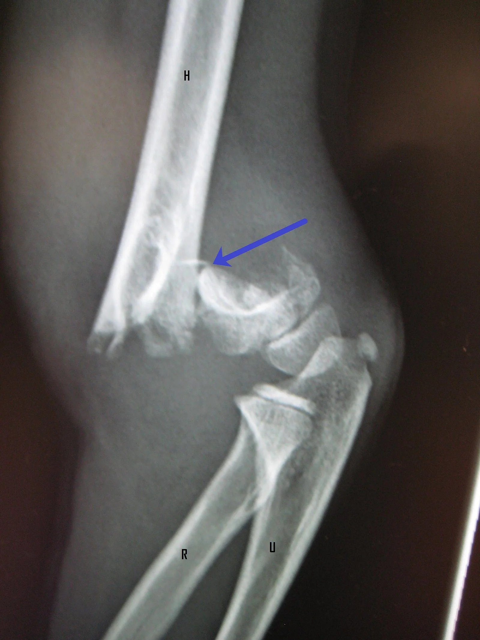
Long description
Elbow radiograph of a supracondylar fracture of the humerus in a young child.

Plain radiographs remain the first-line imaging for any suspected fracture. [1] Oblique views and imaging of the joint above and below the injury reduce missed lesions. Growth-plate injuries may look normal on the first film, particularly type I and type V. [1]
Ultrasound is useful in infants (unossified epiphyses), for guide reduction, and for assessing perfusion.
MRI is the most sensitive tool for occult physeal bars, early growth-plate damage, and marrow injury. The optimal sequence for identifying a physeal bar is a fat-suppressed three-dimensional gradient recalled echo image, in which normal physis is bright and bone is dark. [1]
CT helps define physeal bar size and complex fracture geometry when surgery is planned.
In suspected abuse, a full skeletal survey (standard series of radiographs including skull, chest, spine, pelvis and all limbs) is indicated when abuse is suspected in a young child. Skeletal surveys should be interpreted by a radiologist experienced in paediatric trauma. When clinically suspected, MRI of the brain and spine, and ophthalmology review for retinal haemorrhages, are part of the abuse evaluation. [7]
| Type | Mechanism / description | Frequency | Risk of growth disturbance |
|---|---|---|---|
| I | Transverse line through the physis from a longitudinal or shearing force; epiphysis separates from metaphysis | Around 5% | Usually low if reduced gently; high if not recognised |
| II | Fracture along the physis exiting through the metaphysis (Thurston-Holland fragment) | About 75% | Low-moderate |
| III | Intra-articular fracture entering the physis and exiting the epiphysis | About 10% | High (articular surface and growth) |
| IV | Fracture crossing epiphysis, physis and metaphysis | About 10% | High; anatomic reduction essential |
| V | Crush/compression of the growth plate | Least common; often diagnosed retrospectively | Highest: often leads to growth arrest |
Based on the review by Lou and colleagues, lower-numbered types are less severe and more subtle, while higher-numbered types are more severe and carry greater risk of complications and growth disturbance. [1] Type III and type IV involve the joint surface and risk both growth disturbance and traumatic osteoarthritis. [1]
The Peterson modification (renaming and adding patterns) and Ogden's expanded system exist, but the original Salter-Harris system remains the most used clinically because of its simplicity. [1]
Growth disturbance depends on both fracture type and anatomic location. Fractures of the distal femur, proximal tibia and distal tibia have disproportionately high rates of physeal arrest, so both morphology and location must be considered when estimating long-term risk. [1]
| Type | Description | Typical management |
|---|---|---|
| I | Undisplaced | Long-arm cast; orthopaedic follow-up |
| II | Displaced but posterior cortex intact (hinge) | Closed reduction and Kirschner-wire fixation for most |
| III | Completely displaced | Urgent closed reduction and Kirschner-wire fixation; open if vascular compromise |
| IV | Multidirectionally unstable fracture | Operative fixation; more complex construct |
Displaced Gartland type III and IV fractures carry the highest risk of neurovascular compromise from arterial entrapment, intimal injury, thrombosis or vasospasm. [2]
A forearm bow measured at more than approximately 10% of radial length on standard radiographs suggests pathological plastic deformation rather than normal curvature. [4]
Two bedside categories matter:
- Pink pulseless hand: good colour and warmth, absent pulses. Perfusion is preserved. Some patients can be observed closely after reduction.
- Pale pulseless hand: pale, cold, delayed capillary refill. This is critical ischaemia and calls for urgent reduction and vascular exploration. [2]
The general principle is to respect the child's remodelling capacity. Most paediatric fractures unite and realign without surgery, so the treating clinician must know the acceptable thresholds of angulation, rotation, shortening and displacement, which vary by region, age and site. [8] Salter-Harris injuries need the opposite instinct: growth plates do not remodel angular deformity well, particularly near the knee and ankle.
Immobilise the limb in the position found, provide analgesia, obtain radiographs including the joint above and below, and document neurovascular status. Splint rather than reduce on the ward when the deformity is complex, when there is swelling, or when vascular status is abnormal. Escalate any abnormal perfusion immediately.
- Salter-Harris I and II: gentle closed reduction if displaced, immobilisation in a cast. Reassess for signs of persistent displacement; fix percutaneously if unstable. Avoid repeated attempts, which can convert a benign injury into a physeal bar.
- Salter-Harris III and IV: anatomic reduction is required because the joint surface is involved. Open reduction with internal fixation is commonly needed. [1]
- Salter-Harris V (crush): often not visible initially. Immobilise and arrange close follow-up, since growth arrest may present months later. [1]
- Physeal bar excision is reserved for bars involving less than 50% of the physis, in patients with at least two years of growth remaining; bars affecting 50% or more usually need completion epiphysiodesis. [1]
- Guided growth with tension-band plates can correct angular deformity in children with substantial growth remaining. [1]
Undisplaced Gartland type I fractures are treated in a long-arm cast. [2] Displaced (type II to IV) fractures require reduction and Kirschner-wire fixation in most cases. [2]
Where vascular compromise is present, the priority is restoration of perfusion rather than a particular implant. Closed reduction with percutaneous pinning is standard for most displaced fractures; external fixation may be chosen where vascular exploration is needed, to allow unrestricted access without repeated manipulation. [2]
For a pale pulseless hand, reduce and explore urgently. For a pink pulseless hand, a more selective approach with reduction and close observation may be used, because evidence shows both strategies can achieve good outcomes. Advanced imaging should not delay surgery where limb-threatening ischaemia is clinically evident. [2]
Neurovascular assessment in a displaced supracondylar humerus fracture
- Displaced supracondylar fractureImmobilise in the position found, provide analgesia, document baseline neurovascular status
- Assess pulses, colour, warmth, capillary refill, sensation and movement
- Question: Hand status?
Warm and pink with pulses
- Closed reduction and Kirschner-wire fixation
Pink but pulseless
- Reduce, reassess, then close observation or exploration depending on protocol
Pale, cold, pulseless
- Urgent reduction and vascular exploration; imaging must not delay surgery
Reduction is simple and does not require imaging when the history and examination are typical. Two techniques exist:
- Hyperpronation (or forced pronation): turning the forearm into full pronation while pressing over the radial head.
- Supination-flexion: supinating the forearm, then flexing the elbow while applying pressure over the radial head.
In a randomised clinical study of 116 patients, hyperpronation was more successful than supination-flexion on first attempt (85% versus 53%), on second attempt (50% versus 28%), and as a crossover technique when supination-flexion failed (100% versus 50%). [5] The study concluded that hyperpronation, a single-movement technique, should be used first. [5]
After reduction the child typically begins using the arm within minutes. No radiograph is needed in the classic presentation, but image the elbow when the mechanism is unclear, there is swelling or bony tenderness, or reduction fails.
Treatment is nonoperative. [3] Swathe the arm gently in a soft bandage or pin the sleeve to the shirt when movement is painful; often even immobilisation is unnecessary. Prognosis is universally excellent, with full remodelling and no functional deficit. [3] Explain to parents that a lump (callus) may appear in the first weeks and remodel over time.
These are usually undisplaced spiral or torus-type fractures of the distal tibial metaphysis in a newly mobile child. They can be subtle on the first radiograph and are easily mistaken for transient synovitis of the hip. [9] Management is protected weight-bearing in a short leg walking cast or boot for a few weeks, with repeat radiographs if the diagnosis was missed the first time. Always keep non-accidental trauma and (rarely) a tumour on the differential list when the presentation is atypical.
Management accepts that the bone is bent but intact. If the bowing is minor and forearm rotation is largely preserved, observation with analgesia and repeat radiographs may be sufficient. Significant bowing (over about 10% of length) with restricted pronation and supination, or progressive deformity, requires closed reduction under adequate analgesia or anaesthesia, sometimes with an assistant providing counter-pressure. Because intact bone is deformed plastically, closed reduction can be difficult and recurrence is possible; a cast is applied after correction. [4]
There is no single fracture that proves abuse; the diagnosis rests on the whole picture. [7] The immediate pathway is:
- Treat the injury.
- Arrange a full skeletal survey in children under two years, with review by a radiologist who reads paediatric studies.
- Involve the paediatric or child-safeguarding team before discussing the suspicion with parents.
- Consider bone fragility work-up (including genetic evaluation for osteogenesis imperfecta) where the history or examination suggests it. [7]
- Document carefully and without accusation.
Classic metaphyseal lesions, once thought specific for abuse, can also be seen in osteogenesis imperfecta, so they are not pathognomonic. [7]
| Pattern | Typical mechanism | Treatment and red flag |
|---|---|---|
| Greenstick | Bending force producing an incomplete fracture with one cortex intact as a hinge | Reduce if angulation exceeds the accepted limit for age and site; cast. Remodels well in the young child |
| Buckle (torus) | Axial compression of the metaphysis, often distal radius | Simple splint or soft cast for a few weeks; excellent remodelling and low complication rate |
| Plastic deformation | Longitudinal compression beyond the elastic limit, causing permanent bowing without a break | Reduce if bowing exceeds about 10% of length or forearm rotation is lost; cast after reduction |
| Salter-Harris I-II | Shearing or avulsion through the physis | Gentle reduction, immobilisation; repeat attempts can create a physeal bar |
| Salter-Harris III-IV | Intra-articular extension across the growth plate | Anatomic reduction, usually open reduction and internal fixation |
| Toddler tibia | Twisting of the foot on a planted leg in a newly mobile child | Short leg walking boot or cast; repeat or oblique radiographs if initially missed |
| Supracondylar humerus | Fall on an outstretched hand with the elbow extended | Reduction and Kirschner-wire fixation; urgent vascular exploration if the hand is pale and pulseless |
| Nursemaid's elbow | Traction with the forearm pronated, deceleration from being lifted | Hyperpronation or supination-flexion reduction; no imaging if classical |
| Birth clavicle | Shoulder dystocia, macrosomia or instrument-assisted delivery | Nonoperative swathing, no reduction; expect excellent remodelling |
| Fracture patterns of concern | Injuries inconsistent with development or history | Treat, skeletal survey, safeguarding review, and exclusion of osteogenesis imperfecta |
- Growth disturbance: 5 to 10% of physeal fractures result in clinically significant growth arrest, limb-length discrepancy or angular deformity. [1]
- Physeal bar formation develops months to years after injury and is more common in the lower extremity than the upper. [1]
- Cubitus varus is the most common residual deformity after a distal humeral physeal injury. [3]
- Neurovascular injury in supracondylar fractures includes brachial artery occlusion or entrapment and nerve stretch or transection, with compartment syndrome as a limb-threatening consequence.
- Plastic deformation may cause persistent deformity and loss of forearm rotation if it is missed. [4]
- Missed toddler or birth fracture can turn a simple injury into a late callus, limp and family anxiety.
The outlook is excellent for most injuries on this page. Paediatric bones remodel strongly; birth clavicle fractures unite and remodel completely; toddler fractures heal without long-term disability; nursemaid's elbow resolves immediately with reduction. The exceptions are growth-plate injuries involving germinal cells, high-energy crush injuries, and injuries complicated by vascular compromise or infection.
Prevention is behavioural and environmental: safe trampolines (one child at a time), soft playground surfaces, no swinging by the hands, car seats appropriate to age, and education of new parents on how to lift and support an infant. Clinically, early recognition of abuse and immediate engagement of safeguarding teams prevent repeated injury.
Refer urgently on the same day for a pale or pulseless hand, an open fracture, suspected compartment syndrome, suspected non-accidental trauma, or any injury in which the neurovascular examination or the radiograph cannot be reconciled with the history. Refer to a paediatric orthopaedic service for Salter-Harris III or IV injuries, displaced supracondylar fractures, significant plastic deformation, early physeal bar on follow-up, and any child in whom the expected healing or remodelling is not occurring. Families of disabled children, infants with repeated fractures, and adolescents with sports-related physeal stress injuries benefit from multidisciplinary care.
- Neonates and preterm infants: epiphyses are unossified, so radiographs may be misleading; ultrasound or MRI is often decisive. Birth fractures remain rare but are easily missed, with reports of late presentation from swelling or callus. [3]
- Osteogenesis imperfecta: recurrent fractures, blue sclera, family history and atypical bone density. Distinguish from abuse before accusation. [7]
- Children with developmental delay: may not bear weight or describe pain; examine carefully and obtain imaging early.
- Plain radiographs are usually available and are the first-line investigation. Use oblique views of the distal tibia and comparison films of the uninjured wrist when resources allow.
- Splints and padded bandages suffice for many injuries. Plaster may not be available; a removable brace or even firm bandaging can be used for buckle and toddler fractures in stable circumstances.
- Ultrasound performed by a trained clinician is a valuable substitute for MRI in infants.
- Growth-plate bar excision and guided growth need specialist centres; prioritise early recognition and timely referral rather than attempting complex procedures locally.
- Safeguarding systems vary: even without a formal child-protection pathway, a clinician who suspects abuse should involve paediatrics, social services and legal authorities according to local arrangements.
- Paediatric bone bends, buckles and fractures incompletely because the periosteum is thick and the growth plate is weaker than cortex.
- Roughly 15 to 30% of paediatric fractures involve the physis; Salter-Harris type II is the commonest (about 75%), while types III, IV and V carry higher growth risk. [1]
- Growth-plate (Salter-Harris) type I and V can look normal on the first radiograph.
- Supracondylar humerus fractures are the most common paediatric elbow fractures (about 60% of elbow injuries, ages 5-8); displaced (Gartland III-IV) types cause brachial artery and nerve injury.
- A pale pulseless hand is an emergency; a pink pulseless hand may be observed after reduction.
- Nursemaid's elbow is the commonest condition in paediatric orthopaedic clinics; hyperpronation succeeds more often than supination-flexion (85% versus 53% first attempt). [5]
- Birth clavicle fractures are the most common birth fracture (0.05 to 3.2%) and are treated nonoperatively with excellent remodelling.
- Toddler fractures are subtle and often misdiagnosed as transient synovitis.
- Plastic forearm bowing exceeding approximately 10% of radial length is pathological and may need reduction.
- Classical fracture patterns are not pathognomonic for abuse; osteogenesis imperfecta and other fragility disorders must be excluded.
- Children's fractures generally remodel well, but physeal injuries can cause permanent growth disturbance in 5 to 10% of cases.
Answer each one in your head first, then open the answer.
A 7-year-old boy falls from a climbing frame onto an outstretched hand and arrives with a swollen, painful elbow. The hand is pale, cold, with capillary refill over 3 seconds and no palpable radial pulse. What is the most likely fracture and the immediate priority?
Show answerHide answer
Gartland type III (or IV) supracondylar humerus fracture with vascular compromise. The immediate priority is urgent fracture reduction to restore distal perfusion, with vascular exploration if the hand remains pale and pulseless afterwards. Advanced imaging should not delay surgery when limb-threatening ischaemia is clinically evident. Do not perform repeated manipulation attempts.
Which Salter-Harris type is most common, and which types are most likely to cause growth arrest and joint surface damage?
Show answerHide answer
Salter-Harris type II is most common (about 75% of physeal fractures). Types III, IV and V carry disproportionate risk of growth disturbance, and types III and IV additionally involve the articular surface, risking osteoarthritis. Growth arrest also depends on anatomic site: distal femur, proximal tibia and distal tibia are highest-risk locations. [1]
A toddler of 18 months refuses to use one arm after her mother pulled her up by the hand. There is no swelling and the radiograph is normal. What is the diagnosis and best first reduction?
Show answerHide answer
Nursemaid's elbow (radial head subluxation). Reduce by hyperpronation (forced pronation with thumb pressure over the radial head), which succeeded more often than supination-flexion in a randomised study (85% versus 53% on first attempt). Expect the child to use the arm within minutes.
A neonate's parents are told their baby has a clavicle fracture from delivery. What treatment and prognosis should you discuss?
Show answerHide answer
Treatment is nonoperative: gentle swathing in a bandage or pinning the sleeve to the shirt only if movement is painful; often no immobilisation is needed. Prognosis is excellent, with full remodelling and no functional deficit, although a palpable callus may appear in the first weeks. Most are right-sided.
An 11-month-old who has just started to walk has a limp. Radiographs are reported as normal. What two diagnoses must not be missed, and what can you do?
Show answerHide answer
Toddler (tibial) fracture, which may be invisible initially and is easily mistaken for transient synovitis; and a hip or systemic process. Repeat or oblique radiographs, close follow-up, and safeguarding awareness if the presentation is atypical are the next steps.
A 6-month-old non-ambulatory infant is found to have a classic metaphyseal lesion. Does this prove non-accidental trauma?
Show answerHide answer
No. Classic metaphyseal lesions are often associated with non-accidental trauma but are not specific; they can also occur in osteogenesis imperfecta. The whole clinical picture, including a skeletal survey, family and medical history, bone-fragility work-up and safeguarding input, is needed. [7]
What measurement distinguishes pathological plastic bowing from normal forearm curvature?
Show answerHide answer
Bowing of more than approximately 10% of radial length on radiography is suggestive of pathological plastic deformation, particularly when accompanied by limited pronation-supination.
Common paediatric orthopaedic injuries (this page); fractures in osteogenesis imperfecta; transient synovitis of the hip; brachial plexus birth injury; developmental dysplasia of the hip; child safeguarding and suspected physical abuse (see your local paediatric and safeguarding services); uncomplicated falls and accidental injury in childhood.
- Lou M, Knebel A, Eberson J, Eberson C. Pediatric physeal fractures and growth disturbance: current concepts and surgical approaches. World Journal of Pediatric Surgery 2026;9(4):e001187. Accessed .
- Kazemtash M, Hecking C, Hoeflich C, Stefanidou S, Schmandra T. Supracondylar Humerus Fractures in Children With Vascular Compromise: A Report of Two Cases and Review of Literature. Cureus 2026;18(5):e109999. Accessed .
- McDonald TC, Higdon CL, Cutchen WA. Neonatal Birth Fractures. Journal of the Pediatric Orthopaedic Society of North America 2024;9:100131. Accessed .
- Wetzel C, Rohl M, Manes T, Kadado A. Plastic Deformation of the Pediatric Forearm: Recognition, Radiographic Measurement, and Management. Journal of the Pediatric Orthopaedic Society of North America 2026;15:100372. Accessed .
- Ulici A, Herdea A, Carp M, Nahoi CA, Tevanov I. Nursemaid's Elbow - Supination-flexion Technique Versus Hyperpronation/forced Pronation: Randomized Clinical Study. Indian Journal of Orthopaedics 2019;53(1):117-121. Accessed .
- Flaherty A, Ghandour S, Mirochnik K, Lucaciu A, Nassour N, Kwon JY, Harris MB, Ashkani-Esfahani S. Identifying Risk Factors of Children Who Suffered Physical Abuse: A Systematic Review. Journal of the American Academy of Orthopaedic Surgeons Global Research and Reviews 2025;9(1). Accessed .
- Taha O, Weintraub M, Elfilali MM, Bassetti JA, Collins L, Fry CW, Russo CM. Osteogenesis Imperfecta or Non-accidental Trauma? The Diagnostic Dilemma in Pediatric Fractures. Journal of the Pediatric Orthopaedic Society of North America 2025;13:100224. Accessed .
- Baskovic M, Buzuk J, Dujic B, Juric D, Jurkovic K, Pehar K, Vukovic S. Conservative Treatment of Pediatric Fractures: Narrative Review of Acceptable Deformity and Remodeling Potential. Medical Sciences (Basel) 2026;14(4):432. Accessed .
- Seyahi A, Uludag S, Altintas B, Demirhan M. Tibial torus and toddler's fractures misdiagnosed as transient synovitis: a case series. Journal of Medical Case Reports 2011;5:305. Accessed .
Figure credits
Educational content for medical students and doctors. It is not a substitute for clinical judgement or local guidelines.